- Physical Examination
- Surgical Examination
- Ophthalmology
- Clinical Skills
- Orthopedics
- Surgery Videos
- Laparoscopy
- Pediatrics
- Funny Videos
- Cardiothoracic Surgery
- Nursing Videos
- Plastic Surgery
- Otorhinolaryngology
- Histology and Histopathology
- Neurosurgery
- Dermatology
- Pediatric Surgery
- Urology
- Dentistry
- Oncology and Cancers
- Anatomy Videos
- Health and Fitness
- Radiology
- Anaesthesia
- Physical Therapy
- Pharmacology
- Interventional Radiology
- Cardiology
- Endocrinology
- Gynecology
- Emergency Medicine
- Psychiatry and Psychology
- Childbirth Videos
- General Medical Videos
- Nephrology
- Physiology
- Diet and Food Health
- Diabetes Mellitus
- Neurology
- Women Health
- Osteoporosis
- Gastroenterology
- Pulmonology
- Hematology
- Rheumatology
- Toxicology
- Nuclear Medicine
- Infectious Diseases
- Vascular Disease
- Reproductive Health
- Burns and Wound Healing
- Other
Top videos

Must Watch Very Special New Funny Video 2023 Doctor Funny Video Injection Wala Funny Video | Comedy Video Episode 124 By Fun Comedy Ltd
@funcomedyltd
#funcomedyltd
#doctor
#comedy
#wala
Hello Dear Viewers,
If We have any mistake. please comment and tell us, what is our mistake? We will try to solve this mistake next. please watch our videos and give us confidence to trying best. Thank you for watching this video.
IMPORTANT NOTE:-
This video are no any kind of risk. This video are totally acting no risk no Dangerous act no Physical Harm or Death its ok for viewers.
injection wala comedy video injection wala video injection funny video injection injection wala injection injection doctor doctor doctor sui wala wala suji wala suji wala cartoon doctor cartoon funny video tui tui injection cartoon 22 cartoon video injection video cartoon cartoon comedy video doctor video wala cartoon busy fun ltd my family our fun tv fun tv 24 fun tv 420 funny day funny family ding dong bidik fun tv roma fun tv
#cartoon
#comedyvideo
#doctor_doctor
#busyfunltd
#newfunnyvideo2022
#newfunniestcomedy
#injectionfunnyvideo
#sui_wala
#myfamily
#busyfunltd
#funnyday
#bidikfuntv
#mohafuntv
#dingdong

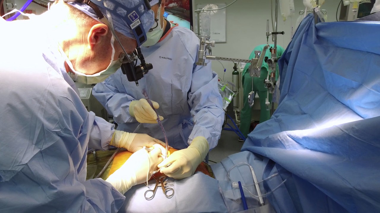
Dr. Ailawadi, M.D., the Chair of Cardiac Surgery at Michigan Medicine, specializes in minimally invasive valve surgery as well as complex cardiac operations. This video shows step by step footage of a Coronary Artery Bypass Graft (CABG) in a complex patient. In this case, CABG was performed through a sternotomy (through the breast bone) using the internal thoracic artery and saphenous leg veins to bypass obstructed coronary arteries. In this video, Dr. Ailawadi will perform a triple vessel bypass (CABG) which has been shown to minimize the risk of future heart attack and help patients live longer in the setting of complex coronary artery disease.
To learn more about cardiac surgery at Michigan Medicine, visit: https://medicine.umich.edu/dept/cardiac-surgery
To learn more about Frankel Cardiovascular Center, visit: https://www.umcvc.org/
To watch the full playlist, visit: https://www.youtube.com/playli....st?list=PLNxqP-XbH8B
-------------------------------------------------------
Subscribe to Michigan Medicine’s YouTube channel for upcoming videos and future live streams featuring our experts answering your questions.
-------------------------------------------------------
Follow Michigan Medicine on Social:
Twitter: https://twitter.com/umichmedicine
Instagram: https://www.instagram.com/umichmedicine/
Facebook: https://www.facebook.com/MichiganMedicine/
Follow the U-M Frankel Cardiovascular Center on Social:
Twitter: https://twitter.com/umichcvc
Facebook: https://www.facebook.com/Unive....rsityofMichiganCardi
#MichiganMedicine #MedEd #CardiacSurgery #UniversityOfMichiganHealth #FrankelCardiovascularCenter #Cardiology #CardiacSurgeon
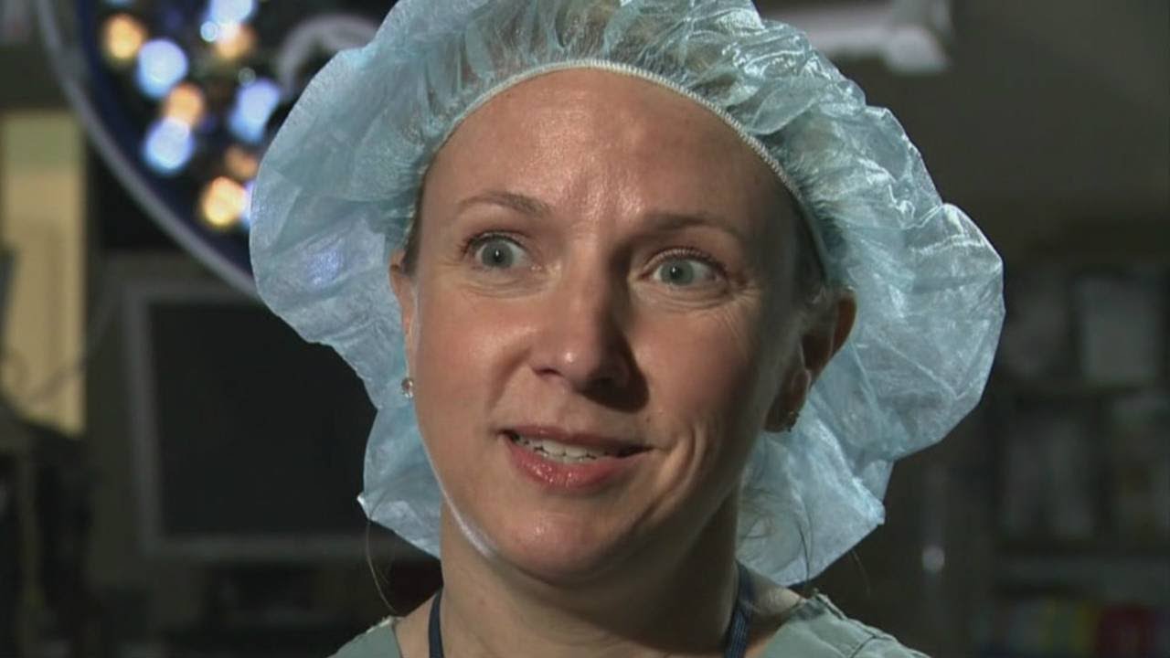
Female heart surgeons are rare, but pediatric female surgeons are even more so.

Cardiac anesthesiology is a subspecialty of anesthesiology that entails caring for patients undergoing major heart surgeries, including those that require cardiopulmonary bypass. I made this video to show a cardiac anesthesiologist's typical setup for surgery.
0:00 Start
0:28 IV pole #1
1:18 Perfusionist equipment
1:47 Anesthesia machine
3:01 Medications
3:36 Pacemaker
4:10 Echocardiography
4:34 IV pole #2
4:55 Arterial line
5:25 Defibrillators
5:40 OR table
---------
Find Max Feinstein, MD online:
Instagram: @MaxMFeinstein
Twitter: @MaxMFeinstein
Website: http://www.MaxFeinsteinMD.com
---------
The information in this video is not intended nor implied to be a substitute for professional medical advice, diagnosis or treatment. All content, including text, graphics, images, and information, contained in this video is for general information purposes only and does not replace a consultation with your own doctor/health professional.
---------
Music
Subtle Swagger by Ron Gelinas Chillout Lounge | https://soundcloud.com/atmospheric-music-portal
Music promoted by https://www.free-stock-music.com
Creative Commons Attribution 3.0 Unported License
https://creativecommons.org/li....censes/by/3.0/deed.e
---------
#Anesthesiology #Residency #MedicalSchool
![So You Want to Be a CARDIOTHORACIC SURGEON [Ep. 13]](https://i.ytimg.com/vi/sdxz242qDFA/maxresdefault.jpg)
So you want to be a cardiothoracic surgeon. You like the idea of open heart surgery and the glory that comes with being a CT surgeon. Let’s debunk the public perception myths of what it means to be a cardiothoracic surgeon, and give it to you straight. This is the reality of cardiothoracic surgery.
✒️ Accompanying Blog Post: https://medschoolinsiders.com/....medical-student/so-y
💌 Sign up for my weekly newsletter - https://medschoolinsiders.com/newsletter
🌍 Website & blog - https://medschoolinsiders.com
📸 Instagram - https://instagram.com/medschoolinsiders
🐦 Twitter - https://twitter.com/medinsiders
🗣️ Facebook - https://facebook.com/medschoolinsiders
🎥 My Youtube Gear: https://kit.co/kevinjubbalmd/
👀 Hand Picked Productivity Tools: https://www.amazon.com/shop/medschoolinsiders
🎵My Study Playlist: https://open.spotify.com/user/....1231934998/playlist/
TIME STAMPS:
00:41 - What is Cardiothoracic Surgery?
04:08 - How to Become a Cardiothoracic Surgeon
06:29 - Subspecialties within Cardiothoracic Surgery
07:49 - What You’ll Love About Cardiothoracic Surgery
09:10 - What You Won’t Love About Cardiothoracic Surgery
10:04 - Should You Become a Cardiothoracic Surgeon?
LINKS FROM VIDEO:
So You Want to Be Playlist: https://www.youtube.com/playli....st?list=PL2ADAFpTg5a
Day in the Life Playlist: https://www.youtube.com/playli....st?list=PLTCN43UFAlB
#medicalschool #cardiothoracicsurgery #soyouwanttobe
====================
Disclaimer: Content of this video is my opinion and does not constitute medical advice. The content and associated links provide general information for general educational purposes only. Use of this information is strictly at your own risk. Kevin Jubbal, M.D. and Med School Insiders LLC will not assume any liability for direct or indirect losses or damages that may result from the use of information contained in this video including but not limited to economic loss, injury, illness or death. May include affiliate links to Amazon. As an Amazon Associate, I may earn a commission on qualifying purchases made through them (at no extra cost to you).

Emory has one of the few heart and vascular centers nationally performing robotic cardiac surgery using the daVinci Surgical System. Emory's robotic surgeons have completed numerous cases and are recognized in Atlanta, the Southeast and across the country for their expertise in cardiac surgery. Some of the cardiac and thoracic conditions treated by Emory cardiac surgeons include mitral valve repair and replacement, atrial septal defect repair, atrial myxoma and thrombi, coronary bypass (LIMA to LAD), mediastinal mass excision, thymectomy, epicardial lead placement and pericardial window.
Dr. Erik Beyer, Florida Medical Center's chief of cardiac surgery, discusses performed a procedure called a micro-thoracotomy.
Dr. Joseph McGinn explains minimally invasive bypass, the procedure he pioneered as an alternative to open heart surgery.

To learn more about coronary artery bypass grafting (CABG), please visit https://cle.clinic/3b7dqpE
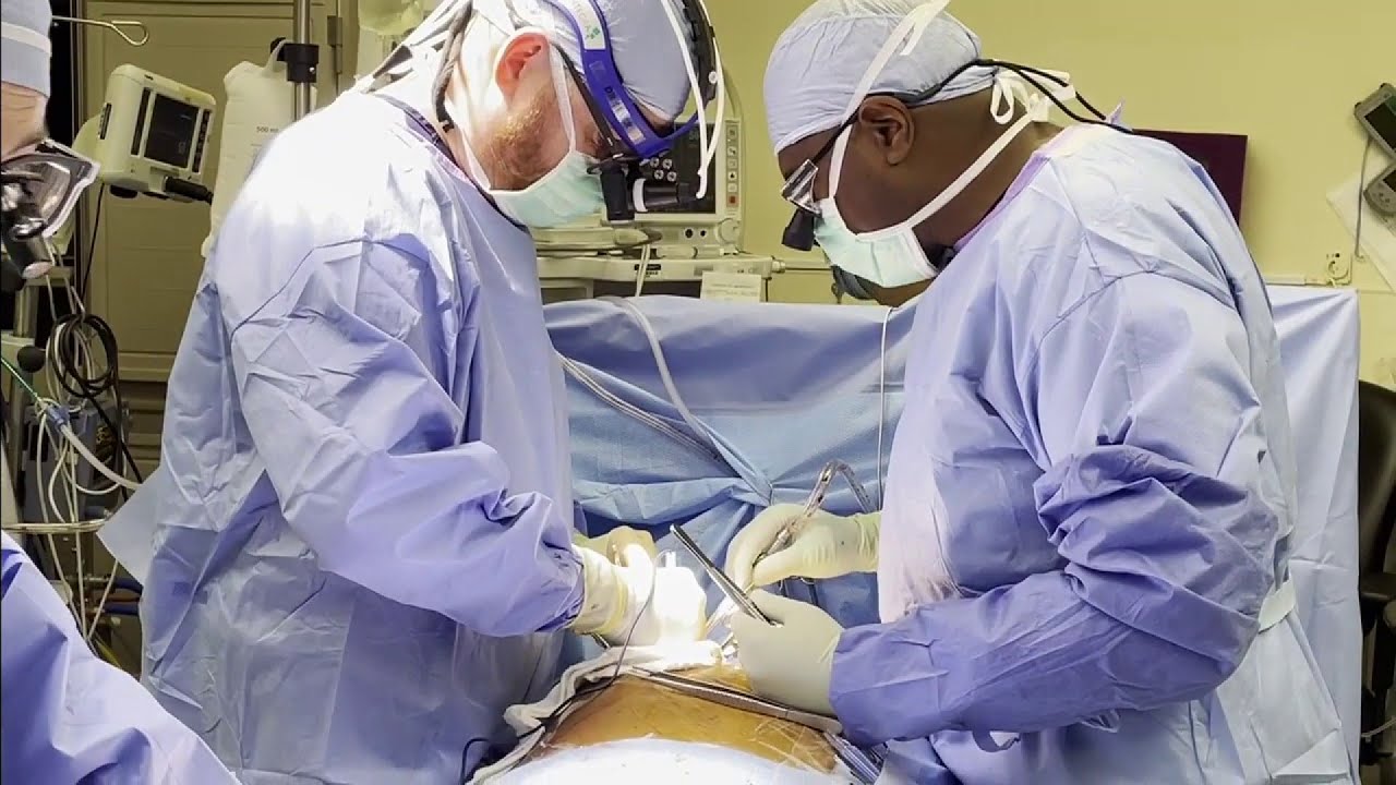
Cardiac surgeons Faisal Baaeen, MD and Edward Soltesz, MD talk about coronary artery bypass graft (CABG) surgery in this informative video.
They describe:
• how blockages are bypassed
• how vessels are used for the bypass graft
• reoperation experiences
• the importance of complete revascularization
• and options such as off pump bypass surgery and minimally invasive surgery
Dr. Bakaeen is the Director of the Coronary Artery Bypass Center at Cleveland Clinic. To learn more about him, please visit https://cle.clinic/2INN9AV
Dr. Soltesz is a cardiovascular and heart transplant surgeon. To learn more about him, please visit https://cle.clinic/3o86RMt
▶Share this video with others: https://youtu.be/Cp59BCMVHHc
▶Subscribe to learn more about @clevelandclinic
#clevelandclinic #coronaryartery #bypasssurgery #heartsurgery #heartcare #cardiacsurgery

Ellis demonstrates how to clean a reusable inner cannula, care for a tracheostomy site, and suction a tracheostomy.
Our Critical Nursing Skills video tutorial series is taught by Ellis Parker MSN, RN-BC, CNE, CHS and intended to help RN and PN nursing students study for your nursing school exams, including the ATI, HESI and NCLEX.
#ClinicalSkills #NCLEX #tracheostomy #patientcare #ATI #Kaplan #LVN #PN #RN #nurseeducator #nurse #nursingstudent #murse #clinicals #clinicalnursingskills
00:00 What to expect Tracheostomy Care and Suctioning
0:33 Explaining the process Tracheostomy Care and Suctioning
1:10 Positioning patient for a Tracheostomy Care and Suctioning
1:33 Opening tray
1:46 Pouring saline
1:58 Removing inner cannula
2:14 Removing clean gloves
2:25 Donning sterile gloves
3:16 Showing tray contents
3:53 Removing previous dressing
4:06 Pouring saline
4:27 Cleaning stoma
5:10 Cleaning faceplate
5:20 Drying site
5:30 Cleaning inner cannula
6:00 Drying inner cannula
6:20 Reinserting inner cannula
6:40 Placing new gauze
7:00 Replacing ties
8:00 Replacing oxygen
8:13 Preparing for suction
8:58 Checking suction
9:30 Opening saline
9:42 Opening kit
9:58 Donning sterile gloves
11:04 Setting up saline container
11:20 Pouring saline
11:52 Connecting catheter to suction
12:46 Inserting catheter
13:10 Removing catheter
13:24 Rinsing catheter
13:40 Reoxyginating
14:05 Reinserting catheter
14:17 Removing catheter
14:29 Rinsing catheter
14:44 Reoxyginating
14:55 Cleaning up
15:09 Chatting about sterility
17:00 Checking a tie
🚨 Reminder: shipping deadlines are looming 👀
🎁 Regular Shipping: Order by Friday, December 15
🚀 Expedited Shipping: Order by Monday, December 18
🔍 Still searching for last-minute gifts? Consider a Level Up RN Gift Card! 💌 It’s not only a thoughtful present but also the perfect way to share treasures like Pharmacology Flashcards OR digital treasures like Flashables Digital Nursing Flashcards & the Level Up RN membership. Give the gift of knowledge this holiday season! 🧠⚡️💖 bit.ly/LevelUpRNGC
🚪 Access our Cram Courses, Quizzes and Videos all in one ad free space with Level Up RN Membership https://bit.ly/LevelUpRNMembership
Want more ways to MASTER Clinical Skills? Check out our flashcards & videos!
👇👇👇👇👇👇👇👇👇👇
👉 https://bit.ly/clinicalnursingskills 👈
☝️👆☝️👆☝️👆☝️👆☝️👆
This is your one-stop-shop for materials to help you LEARN & REVIEW so you can PASS Nursing School.
🤔🤔🤔 DO YOU WANT TO PASS your classes, proctored exams and the NCLEX? 🤔🤔🤔 Our resources are the best you can buy. They are built with a single goal: help you pass with no fluff. Everything you need, and nothing you don’t. Don’t take our word for it, though! Check out our hundreds of ⭐️⭐️⭐️⭐️⭐️ reviews from nurses who passed their exams and the NCLEX with Level Up RN.
🗂️ Our Ultimate Nursing School Survival kit is your number 1 resource to get through nursing school and to pass the NCLEX. Whether you're just starting school or you’re already prepping for the NCLEX, this bundle of flashcards is the best you can buy. It covers all the information you need to know to pass all your exams and it has FREE shipping!
➡️ https://bit.ly/TUNSSK ⬅️
L👀king for EVEN MORE resources to survive Nursing School? Make your Nursing School experience your own! Life’s difficult enough—learning shouldn’t be.
🪅 Games https://nursesquad.com
💻 Digital resources https://bit.ly/NursingStudyCourses
📅 Organizational tools https://bit.ly/OrganizingSchool
✨Want perks? Join our channel!
https://youtube.com/leveluprn/join
🏷 Head to https://leveluprn.com/specials for all our latest deals!🥳️
📧 LOOKING FOR FREE RESOURCES TO HELP WITH YOUR EXAMS? Get exclusive tips, latest video releases and more delivered to your email!
➡️ https://leveluprn.com/signup ⬅️
⚕ 👩 LEVEL UP NURSE SQUAD 👩⚕️
All of the nurses at Level Up RN are here to help! Cathy Parkes started helping her fellow classmates back when she was in nursing school, tutoring so they could pass their exams and graduate. After she got her BSN and started working as an RN at Scripps Encinitas Hospital, she started this YouTube channel to help nursing students around the world. Since then she has built a team of top-notch dedicated nurses and nurse educators who are focused on improving nursing education and supporting career advancement for nurses everywhere. With flashcards, videos, courses, organizational tools and more, we are singularly focused on helping students and nurses Level Up on their exams and nursing careers.

Ellis demonstrates how to administer an intradermal, subcutaneous, and intramuscular injection.
Our Critical Nursing Skills video tutorial series is taught by Ellis Parker MSN, RN-BC, CNE, CHS and intended to help RN and PN nursing students study for your nursing school exams, including the ATI, HESI and NCLEX.
#NCLEX #ClinicalSkills #injections #HESI #Kaplan #ATI #NursingSchool #NursingStudent #Nurse #RN #PN #Education #LVN #LPN #nurseeducator
00:00 What to expect
00:20 Intradermal injections
00:35 Cleaning site
00:54 Explaining bevel up
1:40 Inserting needle
2:00 Injecting medication
2:16 Withdrawing needle
2:29 Subcutaneous Injections
2:39 Selecting site for subcutaneous injections
3:08 Cleaning subcutaneous injections site
3:18 Pinching subcutaneous injections site
3:30 Inserting needle subcutaneous injections
4:13 Injecting medication subcutaneous injections
4:23 Post injection
4:36 Intramuscular injection
4:54 Locating intramuscular injection site
5:18 Cleaning intramuscular injection site
5:38 Inserting needle intramuscular injection
6:28 Anchoring needle intramuscular injection
6:44 Injecting medication intramuscular injection
6:55 Withdrawing needle intramuscular injection
7:05 Disposing of needle
7:43 Cleaning site
8:00 Displacing with Z-track
8:10 Inserting needle
8:23 Releasing tissue
🚨 Reminder: shipping deadlines are looming 👀
🎁 Regular Shipping: Order by Friday, December 15
🚀 Expedited Shipping: Order by Monday, December 18
🔍 Still searching for last-minute gifts? Consider a Level Up RN Gift Card! 💌 It’s not only a thoughtful present but also the perfect way to share treasures like Pharmacology Flashcards OR digital treasures like Flashables Digital Nursing Flashcards & the Level Up RN membership. Give the gift of knowledge this holiday season! 🧠⚡️💖 bit.ly/LevelUpRNGC
🚪 Access our Cram Courses, Quizzes and Videos all in one ad free space with Level Up RN Membership https://bit.ly/LevelUpRNMembership
Want more ways to MASTER Clinical Skills? Check out our flashcards & videos!
👇👇👇👇👇👇👇👇👇👇
👉 https://bit.ly/clinicalnursingskills 👈
☝️👆☝️👆☝️👆☝️👆☝️👆
This is your one-stop-shop for materials to help you LEARN & REVIEW so you can PASS Nursing School.
🤔🤔🤔 DO YOU WANT TO PASS your classes, proctored exams and the NCLEX? 🤔🤔🤔 Our resources are the best you can buy. They are built with a single goal: help you pass with no fluff. Everything you need, and nothing you don’t. Don’t take our word for it, though! Check out our hundreds of ⭐️⭐️⭐️⭐️⭐️ reviews from nurses who passed their exams and the NCLEX with Level Up RN.
🗂️ Our Ultimate Nursing School Survival kit is your number 1 resource to get through nursing school and to pass the NCLEX. Whether you're just starting school or you’re already prepping for the NCLEX, this bundle of flashcards is the best you can buy. It covers all the information you need to know to pass all your exams and it has FREE shipping!
➡️ https://bit.ly/TUNSSK ⬅️
L👀king for EVEN MORE resources to survive Nursing School? Make your Nursing School experience your own! Life’s difficult enough—learning shouldn’t be.
🪅 Games https://nursesquad.com
💻 Digital resources https://bit.ly/NursingStudyCourses
📅 Organizational tools https://bit.ly/OrganizingSchool
✨Want perks? Join our channel!
https://youtube.com/leveluprn/join
🏷 Head to https://leveluprn.com/specials for all our latest deals!🥳️
📧 LOOKING FOR FREE RESOURCES TO HELP WITH YOUR EXAMS? Get exclusive tips, latest video releases and more delivered to your email!
➡️ https://leveluprn.com/signup ⬅️
⚕ 👩 LEVEL UP NURSE SQUAD 👩⚕️
All of the nurses at Level Up RN are here to help! Cathy Parkes started helping her fellow classmates back when she was in nursing school, tutoring so they could pass their exams and graduate. After she got her BSN and started working as an RN at Scripps Encinitas Hospital, she started this YouTube channel to help nursing students around the world. Since then she has built a team of top-notch dedicated nurses and nurse educators who are focused on improving nursing education and supporting career advancement for nurses everywhere. With flashcards, videos, courses, organizational tools and more, we are singularly focused on helping students and nurses Level Up on their exams and nursing careers.

Ellis Parker MSN, RN-BC, CNE, CHSE covers Incentive Spirometry. The Critical Nursing Skills - Shorts series is intended to help RN and PN nursing students study for nursing school exams, including the ATI, HESI and NCLEX.
#NCLEX #HESI #Kaplan #ATI #NursingSchool #NursingStudent #Nurse #RN #PN #Education #LVN #LPN #clinicalskills #safety
Comments? Suggestions? Please share! Your feedback can help inform our future videos and study resources. 🙂
🤔🤔🤔 DO YOU WANT TO PASS your classes, proctored exams and the NCLEX? 🤔🤔🤔 Our flashcards are the best you can buy. They are built with a single goal: help you pass with no fluff. Everything you need, and nothing you don’t. Don’t take our word for it, though! Check out our hundreds of 5-star reviews from nurses who passed their exams and the NCLEX with Level Up RN.
Our #Clinical Nursing Skills Flashcards are available at
➡️ https://bit.ly/clinicalnursingskills
👇SHOP ALL OUR FLASHCARDS👇
http://bit.ly/allstudycards
🗂️ Our Ultimate Nursing School Survival kit is your number 1 resource to get through nursing school and to pass the NCLEX. Whether you're just starting school or you’re already prepping for the NCLEX, this bundle of flashcards is the best you can buy. It covers all the information you need to know to pass all your exams and it has FREE shipping!
➡️ https://bit.ly/TUNSSK ⬅️
📧 LOOKING FOR FREE RESOURCES TO HELP WITH YOUR EXAMS? Get exclusive tips, latest video releases and more delivered to your email!
➡️ https://www.leveluprn.com/signup ⬅️
Want perks? Join our channel!
➡️ https://www.youtube.com/leveluprn/join ⬅️
👩⚕️ LEVEL UP NURSE SQUAD 👩⚕️
All of the nurses at Level Up RN are here to help! Cathy Parkes started helping her fellow classmates back when she was in nursing school, tutoring so they could pass their exams and graduate. After she got her BSN and started working as an RN at Scripps Encinitas Hospital, she started this YouTube channel to help nursing students around the world. Since then she has built a team of top-notch dedicated nurses and nurse educators who are focused on improving nursing education and supporting career advancement for nurses everywhere. With flashcards, videos, courses, organizational tools and more, we are singularly focused on helping students and nurses Level Up on their exams and nursing careers.
👋 STAY CONNECTED 👋
TikTok: https://tiktok.com/@leveluprn
Instagram: https://www.instagram.com/leveluprn/
Facebook: https://fb.me/LevelUpRN
Pinterest: https://www.pinterest.com/leveluprn/
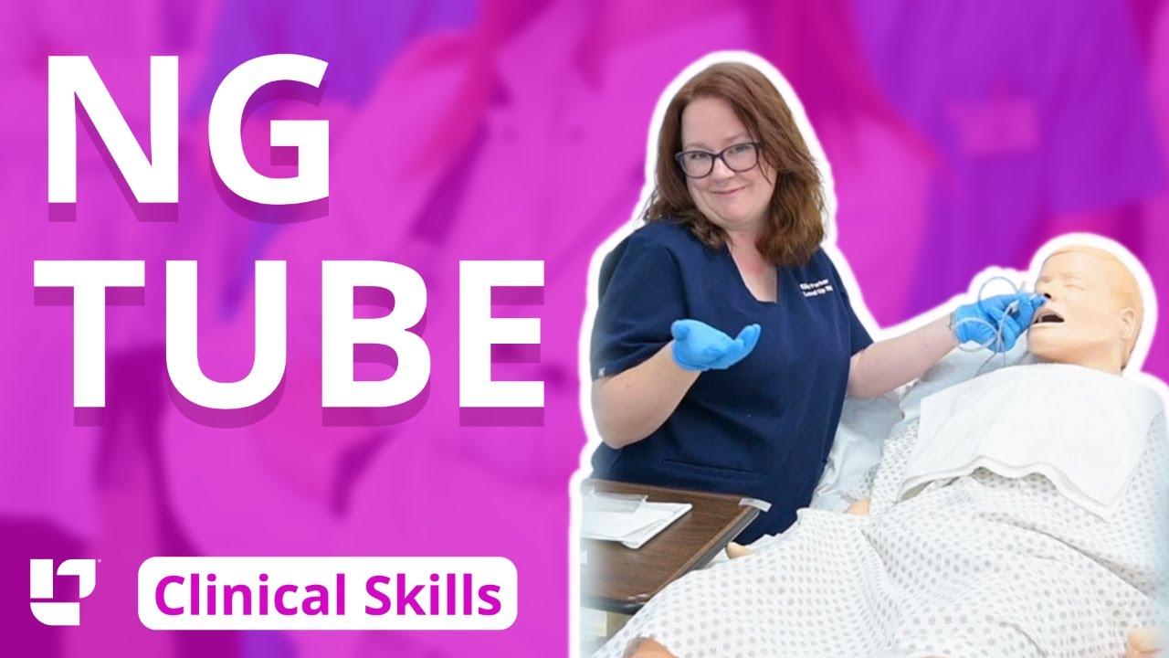
Ellis demonstrates how to insert and then remove an NG tube. This includes drawing gastric residual and checking the pH. After the demonstration, Ellis provides additional tips about clamping the NG tube and using the blue pigtail.
Our Critical Nursing Skills video tutorial series is taught by Ellis Parker MSN, RN-BC, CNE, CHS and intended to help RN and PN nursing students study for your nursing school exams, including the ATI, HESI and NCLEX.
#NCLEX #HESI #Kaplan #ATI #NursingSchool #NursingStudent #Nurse #RN #PN #Education #LVN #LPN #ClinicalSkills #NGTube #nurseeducator
00:00 What to expect
00:30 Preparing NG tube patient
00:56 Preparing NG tube equipment
1:29 Measuring the NG tube
2:02 Preparing for NG tube insertion
2:28 Inserting the NG tube
3:17 Checking placement with pH
4:23 Anchoring with split-tape
5:32 Connecting to suction
6:05 Disconnecting from suction
6:17 What to do before removal?
7:03 Removing NG tube
7:40 Additional tips on clamping
8:31 The blue pigtail
🚨 Reminder: shipping deadlines are looming 👀
🎁 Regular Shipping: Order by Friday, December 15
🚀 Expedited Shipping: Order by Monday, December 18
🔍 Still searching for last-minute gifts? Consider a Level Up RN Gift Card! 💌 It’s not only a thoughtful present but also the perfect way to share treasures like Pharmacology Flashcards OR digital treasures like Flashables Digital Nursing Flashcards & the Level Up RN membership. Give the gift of knowledge this holiday season! 🧠⚡️💖 bit.ly/LevelUpRNGC
🚪 Access our Cram Courses, Quizzes and Videos all in one ad free space with Level Up RN Membership https://bit.ly/LevelUpRNMembership
Want more ways to MASTER Clinical Skills? Check out our flashcards & videos!
👇👇👇👇👇👇👇👇👇👇
👉 https://bit.ly/clinicalnursingskills 👈
☝️👆☝️👆☝️👆☝️👆☝️👆
This is your one-stop-shop for materials to help you LEARN & REVIEW so you can PASS Nursing School.
🤔🤔🤔 DO YOU WANT TO PASS your classes, proctored exams and the NCLEX? 🤔🤔🤔 Our resources are the best you can buy. They are built with a single goal: help you pass with no fluff. Everything you need, and nothing you don’t. Don’t take our word for it, though! Check out our hundreds of ⭐️⭐️⭐️⭐️⭐️ reviews from nurses who passed their exams and the NCLEX with Level Up RN.
🗂️ Our Ultimate Nursing School Survival kit is your number 1 resource to get through nursing school and to pass the NCLEX. Whether you're just starting school or you’re already prepping for the NCLEX, this bundle of flashcards is the best you can buy. It covers all the information you need to know to pass all your exams and it has FREE shipping!
➡️ https://bit.ly/TUNSSK ⬅️
L👀king for EVEN MORE resources to survive Nursing School? Make your Nursing School experience your own! Life’s difficult enough—learning shouldn’t be.
🪅 Games https://nursesquad.com
💻 Digital resources https://bit.ly/NursingStudyCourses
📅 Organizational tools https://bit.ly/OrganizingSchool
✨Want perks? Join our channel!
https://youtube.com/leveluprn/join
🏷 Head to https://leveluprn.com/specials for all our latest deals!🥳️
📧 LOOKING FOR FREE RESOURCES TO HELP WITH YOUR EXAMS? Get exclusive tips, latest video releases and more delivered to your email!
➡️ https://leveluprn.com/signup ⬅️
⚕ 👩 LEVEL UP NURSE SQUAD 👩⚕️
All of the nurses at Level Up RN are here to help! Cathy Parkes started helping her fellow classmates back when she was in nursing school, tutoring so they could pass their exams and graduate. After she got her BSN and started working as an RN at Scripps Encinitas Hospital, she started this YouTube channel to help nursing students around the world. Since then she has built a team of top-notch dedicated nurses and nurse educators who are focused on improving nursing education and supporting career advancement for nurses everywhere. With flashcards, videos, courses, organizational tools and more, we are singularly focused on helping students and nurses Level Up on their exams and nursing careers.

Thank you so much for watching❤
If you enjoyed this video ▶Please leave a LIKE👍 ▶SHARE this video ▶【SUBSCRIBE】my channel for more new videos And click the BELL 🔔so you don't miss any of my videos HERE
https://www.youtube.com/c/nurs....eminder?sub_confirma
You can support my work by purchasing your NurseMinder Merch https://teespring.com/stores/nurseminder-nation (or click on merch pics under the video)
Or simply do your Amazon shopping after clicking on one of the links below
-------------------------------------------------------------------------
Thank you so much! I appreciate you!♥♥
------------------------------------------------------------------------
Nurses often prime IV lines with the hopes that there are no air bubbles. In this video, I will share a couple of tips to help reduce the risk or frequency of air bubbles during line priming. I will also talk about how to troubleshoot the air bubbles when they appear during an infusion
Providing patient care and influencing safe patient outcomes requires that registered nurses and licensed practice nurses maintain air free IV lines. Learn the strategies and tips to decrease the risk of air bubbles appearing in your primary or secondary medication line as well as troubleshooting tips to remove those alarming bubbles. Your patients will thank you!
Whether you are providing normal saline, a medication, or a combination, ensure that all fluids are compatible.
Supplies used in this video include the Alaris Primary Infusion line, alcohol swabs and a sterile 10 cc syringe ... and a nail in the wall :)
------------------------------------------------------------------------
❤️ ~ You may also be interested in watching ~ ❤️
PICC line assessment https://youtu.be/tnKClpU-J1g
How To Access a PICC line https://youtu.be/SCF6bmk8KWc
Putting on Sterile Gloves https://youtu.be/xNwkKLqDJn4
Organizational Plans for Nursing https://youtu.be/_NATxwPwHzc
Medication Conversions https://youtu.be/TCPBXg2TYCs
------------------------------------------------------------------------
💻COMMENT in the description box below and share your ideas
👍 LIKE the video
🗣 SHARE with your friends
📥 SUBSCRIBE ... hit the BELL 🔔
Subscribe to NurseMinder https://www.youtube.com/c/nurs....eminder?sub_confirma
------------------------------------------------------------------------
Amazon Affiliate Links
------------------------------------------------------------------------
Want to support me in another way? Enter Amazon through my links and continue to do your shopping. Simple and Easy Way to support the work I do.
The following list is the equipment I use (or if my version is no longer sold, a close replica).
📱 Phone 11 Cell Phone https://amzn.to/2WpOJfz
💻 MacBook Pro https://amzn.to/2YyxQC1
👉 Final Cut Video Editing software https://amzn.to/3fqlAd9
🎙️ Rode NT USB microphone (Audio Recording) for post-production voiceover https://amzn.to/2W2RJj1
👉 Neewer Professional Recording Stand – mount microphone and adjust positioning to keep it close but out of the camera’s view: https://amzn.to/3fjB4zs
👉 Manfrotto Tripod (hold cell phone) https://amzn.to/2YKGYUz
💡 Neewer Ring Light to reduce shadows and improve lighting. https://amzn.to/3dk5OP5
Disclaimer: I recommend only products that I know and trust to be of high quality. Links are provided for quick access. Some of the links contained in this checklist are affiliate links and I may receive a commission if make a purchase from the affiliate. This helps me to keep creating and offering free content.

Learn what's working for other Nursing Students! Check out our Top 10 Most Popular Lessons Here: https://bit.ly/3nda5u3
Central Line Dressing Change- Nursing Skills
FREE Nursing School Cheat Sheets at: http://www.NURSING.com
Get the full PPE Donning & Doffing lesson here:
https://nursing.com/lesson/cen....tral-line-dressing-c
Welcome to the NURSING Family, we call it the most supportive nursing cohort on the planet.
At NURSING.com, we want to help you remove the stress and overwhelm of nursing school so that you can focus on becoming an amazing nurse.
Check out our freebies and learn more at: (http://www.nursing.com)
Central Line Dressing Change - Nursing Skills:
In this video we’re going to talk about central line dressing changes. In this particular video, we’re going to look at a PICC Line, but the same strategy is also used for a Central Line. Remember the dressing should be changed every 7 days or as needed for peeling or soiling
This includes PICC lines. Sterile technique must be maintained to prevent Central-Line Associated Bloodstream Infections (CLABSI)
We love you guys! Go out and be your best selves today! And, as always, happy nursing!
Bookmarks:
0.05 Introduction
0.22 Mask application
0:36 Patient positioning
0:48 Dressing removal
1:20 Sterilization
1:26 Dressing change kit
2:14 Sterile gloves (Lesson link below)
https://nursing.com/lesson/ski....lls-01-04-sterile-gl
2:50 Cleaning the site
3:30 Bio patch application
4:20 Changing infusion caps
4:41 Labeling the dressing
5:00 Outro
Visit us at https://nursing.com/medical-disclaimer/ for disclaimer information.
NCLEX®, NCLEX-RN® are registered trademarks of the National Council of State Boards of Nursing, INC. and hold no affiliation with NURSING.com.

Full Tummy Tuck 3D Video - http://drlandsman.com
Look great... feel great
•Smart Liposuction + Liposculpture
•Abdominplasty (Tummy Tuck)
+ Full Mini Modified
•Brazilian Lift with Fat Transfer
•Vaginal Aesthetics & Rejuvenation
•Laser Hair Removal
•Full Body Lift
•Thigh lift
•Brachioplasty (Arm Lift) + Short Scar
Expertise in Body Contouring
Board Certified Plastic Surgeon
Expertise in body contouring combines skin excision techniques and advanced fat contouring technology
Weight control personalized training and smoking cessation results in a healthier lifestyle improved shape and longer lasting results
With over 2 decades of experience Dr Lloyd Landsman provides state of the art cosmetic and plastic surgery
Dr Landsman integrates the finest and safest products with the newest procedures
A customized treatment plan is created for each patient utilizing classic surgical and minimally invasive techniques for optimal results
Call for your complimentary consultation to learn how Dr Landsman can help you look your very best
Visit http://drlandsman.com Call 631 864 4111
Main Office 994 W Jericho Tpke Smithtown NY 11787
Affiliates East Islip • Westbury • Jackson Heights • Manhattan

Dr.Young Cho explains what happens during a tummy tuck procedure, and what he does to get that hourglass shape.

If you’ve lost a significant amount of weight, either after pregnancy or through exercise and dietary changes, excess skin and weakened abdominal muscles can leave you self-conscious about your appearance. In this video, Dr. Catherine Hannan and Dr. Lauren Patrick, two of our Board-Certified Plastic Surgeons, are performing a Tummy Tuck (Abdominoplasty) surgery. Tummy Tuck surgery gets rid of the excess skin, as well as tightens your abdominal muscles, resulting in a flatter and smoother abdomen. The results of the surgery are permanent except in cases of large weight gain or pregnancy after surgery.
We are so excited to have taken a part in our patient's body transformation journey!
Before & After Gallery:
https://www.westendplasticsurg....ery.com/surgical/bod
To learn more, visit our website or call (202) 785-4187
http://www.westendplasticsurgery.com
~~~~~~~~~~~~~~~~~~~
Social Media:
✨ Instagram: http://www.instagram.com/westendplasticsurgery
✨ Facebook: http://www.facebook.com/westendplasticsurgery
✨ Twitter: http://www.twitter.com/weplasticsurg
✨ Blog: https://www.westendplasticsurgery.com/blog
✨ Business Inquiries: info@westendplasticsurgery.com
~~~~~~~~~~~~~~~~~~~
#TummyTuck #Abdominoplasty
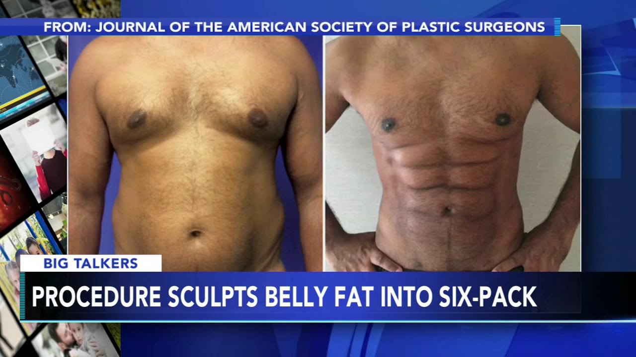
If you've always wanted six-pack abs, but can't seem to get to the gym - there's now a short-cut for that. Researchers at the University of Miami have developed a new plastic surgery technique called abdominal etching. It can reshape belly fat to make you look like you spend a lot of time at the gym.
READ MORE: https://6abc.cm/2Vv5Tu4
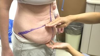
After MacKenzie Walker lost 100 pounds, her "after" picture remained elusive. So she asked plastic surgeon Dr. Anthony Youn to perform an abdominoplasty.
