- Physical Examination
- Surgical Examination
- Ophthalmology
- Clinical Skills
- Orthopedics
- Surgery Videos
- Laparoscopy
- Pediatrics
- Funny Videos
- Cardiothoracic Surgery
- Nursing Videos
- Plastic Surgery
- Otorhinolaryngology
- Histology and Histopathology
- Neurosurgery
- Dermatology
- Pediatric Surgery
- Urology
- Dentistry
- Oncology and Cancers
- Anatomy Videos
- Health and Fitness
- Radiology
- Anaesthesia
- Physical Therapy
- Pharmacology
- Interventional Radiology
- Cardiology
- Endocrinology
- Gynecology
- Emergency Medicine
- Psychiatry and Psychology
- Childbirth Videos
- General Medical Videos
- Nephrology
- Physiology
- Diet and Food Health
- Diabetes Mellitus
- Neurology
- Women Health
- Osteoporosis
- Gastroenterology
- Pulmonology
- Hematology
- Rheumatology
- Toxicology
- Nuclear Medicine
- Infectious Diseases
- Vascular Disease
- Reproductive Health
- Burns and Wound Healing
- Other
Top videos

Intestinal obstruction.....
This video is only educational purposes and this is not for entertainment....this is surgery time

This video provides a guide peforming a respiratory examination in an OSCE station, including real-time auscultation sounds of common pathology such as coarse crackles, fine crackles, wheeze and stridor.
You can access our step-by-step OSCE guide to accompany this video here: https://geekymedics.com/respiratory-examination-2/
Check out our other awesome clinical skills resources including:
• 🔥 Geeky Medics Bundles (discounted products): https://app.geekymedics.com/purchase/bundles/
• ✨ 1000+ OSCE Stations: https://app.geekymedics.com/pu....rchase/osce-stations
• 🏥 Geeky Medics OSCE Revision Book: https://app.geekymedics.com/purchase/book/
• 📝 150+ PDF OSCE Checklists: https://geekymedics.com/pdf-osce-checklists/
• 🗂️ 3000+ OSCE Flashcards: https://app.geekymedics.com/pu....rchase/flashcard-col
• 📱 Geeky Medics OSCE App: https://geekymedics.com/geeky-medics-app/
• 🩺 Medical Finals SBA Question Pack: https://app.geekymedics.com/pu....rchase/medical-stude
• 💊 PSA Question Pack: https://app.geekymedics.com/pu....rchase/prescribing-s
Chapters:
- Introduction 00:00
- General inspection 00:40
- Inspection of the hands 00:50
- Schamroth's window test 01:09
- Heart rate and respiratory rate 01:50
- Jugular venous pressure 02:02
- Face, eyes and mouth 02:13
- Anterior chest inspection 02:36
- Trachea and cricosternal distance 03:01
- Palpation of apex beat 03:16
- Chest expansion 03:28
- Lung percussion 03:50
- Auscultation of lungs 04:21
- Vocal resonance 05:03
- Lymph node palpation 05:32
- Inspection of posterior chest 06:04
- Posterior chest expansion 06:10
- Percussion of posterior chest 06:32
- Auscultation of posterior chest 06:55
- Sacral and pedal oedema 08:04
- Summary of findings 08:39
Subscribe to our newsletter to be the first to know about our latest content: https://geekymedics.com/newsletter/ ✉️
Join the Geeky Medics community: 👩👩👧👧
Twitter: http://www.twitter.com/geekymedics
Instagram: https://instagram.com/geekymedics
Facebook: http://www.facebook.com/geekymedics
Always adhere to your medical school/local hospital guidelines when performing examinations or clinical procedures. DO NOT perform any examination or procedure on patients based purely upon the content of these videos. Geeky Medics accepts no liability for loss of any kind incurred as a result of reliance upon the information provided in this video.
Some people have found this video useful for ASMR purposes.
Special thanks to www.easyauscultation.com and Andy Howes for providing some of the respiratory sounds.

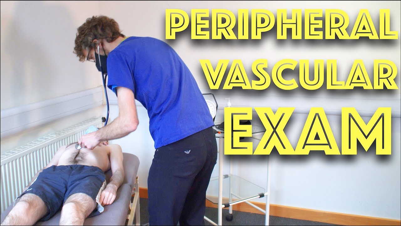
Examination of Peripheral Vascular System - Clinical Skills OSCE Revision - Dr Gill
In this video, we demonstrate the peripheral vascular examination - a less common examination, but still vitally important, particularly amongst the older population
Starting with the examination of the hands looking for clinical signs of vascular compromise, we then check the pulses of the major arteries of the upper body - the radial, brachial and carotid arteries, before moving down to assess for an abdominal aortic aneurysm.
At this point, I feel it's a practical step to check the femoral pulses before doing the overview of the legs.
After visually assessing we must examine the major vascular areas of leg.- namely the popliteal pulses, before wrapping up around the ankle with the posterior tibial and dorsalis pedis pulses
For completeness, the cardiovascular examination is demonstrated here
https://www.youtube.com/watch?v=ECs9O5zl6XQ&t=2s
#PeripheralVascular #ClinicalSkills #DrGill

Watch this clinical examination video to learn how to diagnose cervical spine pathology.
This video clip is part of the FIFA Diploma in Football Medicine and the FIFA Medical Network. To enrol or to find our more click on the following link http://www.fifamedicalnetwork.com
The Diploma is a free online course designed to help clinicians learn how to diagnose and manage common football-related injuries and illnesses. There are a total of 42 modules created by football medicine experts. Visit a single page, complete individual modules or finish the entire course.
The network provides the opportunity for clinicians around the world to meet and share ideas relating to football medicine. Ask about an interesting case, debate current practice and discuss treatment strategies. Create a profile and log on to interact with other health professionals from around the globe.
This is not medical advice. The content is intended as educational content for health care professionals and students. If you are a patient, seek care of a health care professional.

Ear Examination ENT is often a challenging examination, crossing over with the cranial nerve examination of the vestibular cochlear exam as well at other neurological assessments of balance
Here we will review the ear examination, looking both at the use of the otoscope, but also the Dix-Hallpike Manoeuvre, along with HINTS assessment. the Webers and Rinne's test is also included to determine types of hearing loss.
Often these ear examination techniques are performed separately, depending on the patients presenting complaint
#EARExamination #DrGill #ClinicalSkills

The most reliable clinical sign to detect ascites is checking for bilateral flank dullness. If a patient with ascites is lying supine, fluid accumulates in the flank regions, leading to dullness on percussion. At the same time, the air-filled bowel loops are forced upwards by the free fluid due to buoyancy, resulting in tympanitic percussion. To locate specifically where dullness shifts to tympany, or the air-fluid level, percussion should be performed from the sides towards the middle. To confirm that the dullness is caused by ascites, ask the patient to switch to a lateral decubitus position. If ascites is present, the air-filled bowel loops will shift accordingly and remain at the surface of the fluid. As a result, the air-fluid level will shift as well. This is known as shifting dullness.
Subscribe to AMBOSS YouTube for the latest clinical examination videos, medical student interviews, study tips and tricks, and live webinars!
Free 5 Day Trial: https://go.amboss.com/amboss-YT
Instagram: https://www.instagram.com/amboss_med/
Facebook: https://www.facebook.com/AMBOSS.Med/
Twitter: https://twitter.com/ambossmed
Blog: https://blog.amboss.com/us
#AMBOSSMed #ClinicalExamination
Peripheral Vascular Examination OSCE - Clinical Skills - Dr Gill
In the cardiovascular examination, particularly in the case of an OSCE station, we conclude the examination often by stating that the examiner would want to perform:
- An ECG
- Check full blood count
- and "do a peripheral vascular examination
In this video, we demonstrate that oft-talked about, but comparatively less common examination.
Starting off, with the examination of the hands, the radial, brachial and carotid pulses. before moving down to assess for a AAA, checking the femoral and popliteal pulses, before wrapping up around the ankle with the posterior tibial and dorsalis pedis pulses
For completeness, the cardiovascular examination is demonstrated here
https://www.youtube.com/watch?v=ECs9O5zl6XQ&t=2s
#PeripheralVascular #ClinicalSkills #DrGill

Watch this clinical examination video to learn how to diagnose inguinal related groin pain.
This video clip is part of the FIFA Diploma in Football Medicine and the FIFA Medical Network. To enrol or to find our more click on the following link http://www.fifamedicalnetwork.com
The Diploma is a free online course designed to help clinicians learn how to diagnose and manage common football-related injuries and illnesses. There are a total of 42 modules created by football medicine experts. Visit a single page, complete individual modules or finish the entire course.
The network provides the opportunity for clinicians around the world to meet and share ideas relating to football medicine. Ask about an interesting case, debate current practice and discuss treatment strategies. Create a profile and log on to interact with other health professionals from around the globe.
This is not medical advice. The content is intended as educational content for health care professionals and students. If you are a patient, seek care of a health care professional.
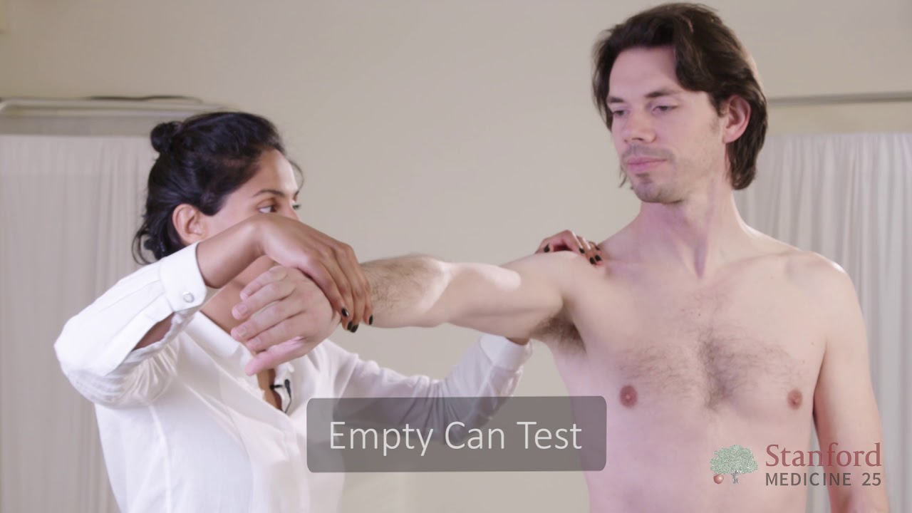
This video is brought to you by the Stanford Medicine 25 to teach you the common causes of shoulder pain and how to diagnose them by the physical exam.
The Stanford Medicine 25 program for bedside medicine at the Stanford School of Medicine aims to promote the culture of bedside medicine to make current and future clinicians and other healthcare provides better at the art of physical diagnosis and more confident at the bedside of their patients.
Visit us:
Website: http://stanfordmedicine25.stanford.edu/
Blog: http://stanfordmedicine25.stanford.edu/blog.html
Facebook: https://www.facebook.com/StanfordMedicine25
Twitter: https://twitter.com/StanfordMed25
Diagnoses covered in this video:
Rotator Cuff Pathology
Impingement Syndrome
Biceps Tendinopathy
Adhesive Capsulitis (Frozen Shoulder)
Acromioclavicular (AC) Joint Disease
Shoulder Instability
Labral Tears (SLAP Lesions)

Lesson on clinical examination of a scaphoid fracture and assessment of the anatomic snuffbox. The scaphoid bone is one of the carpal bones of the wrist. A scaphoid fracture is important to rule out due to risk of avascular necrosis, which is a compromise of bone vasculature leading to death of the bone. Scaphoid fractures can occur with a FOOSH injury. In this lesson, we discuss the clinical assessment to rule out a scaphoid fracture, including assessing and localizing the anatomic snuffbox.
If you find this lesson helpful, please consider liking, subscribing, and clicking the notification bell to help support this channel and stay up-to-date on future lessons.
*Subscribe for more free medical lessons* https://www.youtube.com/channe....l/UCFPvnkCZbHfBvV8Ap
-------------------------------------------------------------------------------------------------------------
For books and more information on these topics
https://www.amazon.com/shop/jjmedicine
Support future lessons (and get other cool stuff) ➜ https://www.patreon.com/jjmedicine
Follow me on Twitter! ➜ https://twitter.com/JJ_Medicine
Come join me on Facebook! ➜ https://www.facebook.com/JJ-Me....dicine-1006426481611
Start your own website with BlueHost ➜ https://www.bluehost.com/track/jjmedicine/
Check out the best tool to help grow your YouTube channel (it’s helped me!)
https://www.tubebuddy.com/jjmedicine
-------------------------------------------------------------------------------------------------------------
Check out some of my other lessons.
Medical Terminology - The Basics - Lesson 1:
https://www.youtube.com/watch?v=04Wh2E9oNug
Fatty Acid Synthesis Pathway:
https://www.youtube.com/watch?v=WuQS_LpNMzo
Wnt/B Catenin Signaling Pathway:
https://www.youtube.com/watch?v=NGVP4J9jpgs
Upper vs. Lower Motor Neuron Lesions:
https://www.youtube.com/watch?v=itNd74V53ng
Lesson on the Purine Synthesis and Salvage Pathway:
https://www.youtube.com/watch?v=e2KFVvI8Akk
Gastrulation | Formation of Germ Layers:
https://www.youtube.com/watch?v=d6Kkn0SECJ4
Introductory lesson on Autophagy (Macroautophagy):
https://www.youtube.com/watch?v=UmSVKzHc5yA
Infectious Disease Playlist
https://www.youtube.com/playli....st?list=PLRjNoiRtdFw
Dermatology Playlist
https://www.youtube.com/playli....st?list=PLRjNoiRtdFw
Pharmacology Playlist
https://www.youtube.com/playli....st?list=PLRjNoiRtdFw
Hematology Playlist
https://www.youtube.com/playli....st?list=PLRjNoiRtdFw
Rheumatology Playlist
https://www.youtube.com/playli....st?list=PLRjNoiRtdFw
Endocrinology Playlist
https://www.youtube.com/playli....st?list=PLRjNoiRtdFw
Nephrology Playlist
https://www.youtube.com/playli....st?list=PLRjNoiRtdFw
----------------------------------------------------------------------------------------------------
**MEDICAL DISCLAIMER**: JJ Medicine does not provide medical advice, and the information available on this channel does not offer a diagnosis or advice regarding treatment. Information presented in these lessons is for educational purposes ONLY, and information presented here is not to be used as an alternative to a healthcare professional’s diagnosis and treatment of any person/animal.
Only a physician or other licensed healthcare professional are able to determine the requirement for medical assistance to be given to a patient. Please seek the advice of your physician or other licensed healthcare provider if you have any questions regarding a medical condition.
----------------------------------------------------------------------------------------------------
*Although I try my best to present accurate information, there may be mistakes in this video. If you do see any mistakes with information in this lesson, please comment and let me know.*
I am always looking for ways to improve my lessons! Please don't hesitate to leave me feedback and comments - all of your feedback is greatly appreciated! :) And please don't hesitate to send me any messages if you need any help - I will try my best to be here to help you guys :)
Thanks for watching! If you found this video helpful, please like and subscribe!
JJ
Dr. Mohan Rao, Senior General & Laparoscopic consultant at Apollo Spectra Hospitals, MRC Nagar explains How can one self-examination of Hernia be done

#final #fumc #mbbs #medicalstudents #mbbsabroad #doctor #fcps #fcpspart #surgeryeducation #surgeryreview #trainee #exampreparation

Inguinal or groin hernias are the most common type of hernias and most of the time occur in men. We talked with CU Medicine surgeon, Dr. Sam Phinney, about groin hernias and how they are treated. https://www.cumedicine.us/abou....t-cu-medicine/health


The anatomy of the direct and indirect inguinal hernia.
Music:
Berries and Lime by Gregory David
https://www.epidemicsound.com/track/z6iCiiyCPm/
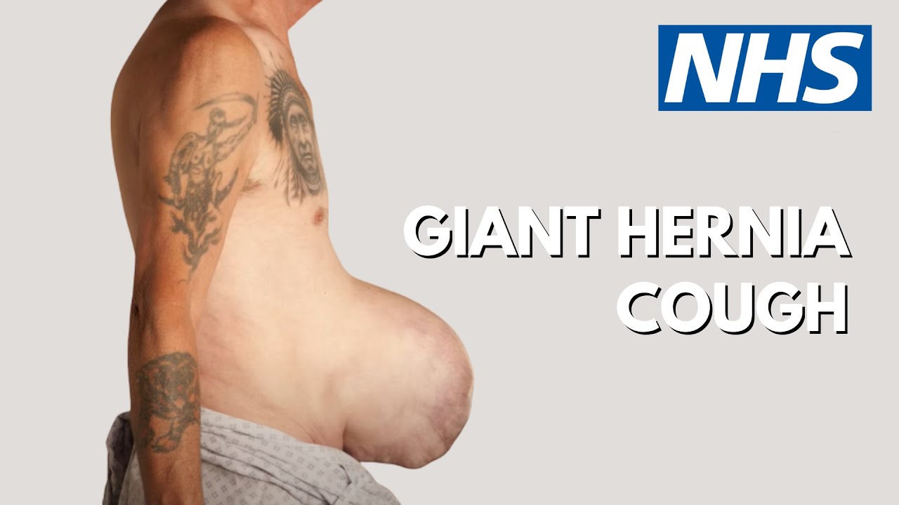
Patient Glenn Williams had a hernia measuring 20cm x 30cm. Consultant Graham Offer has performed ground breaking surgery to help Glenn.

Contact us to find out more http://www.londonvisionclinic.com/contact-us/ Mr Carp explains the risks involved in losing sight as being extremely rare. Only 1 in 5 million may lose sight in one eye.
