أهم مقاطع الفيديو

Full Tummy Tuck 3D Video - http://drlandsman.com
Look great... feel great
•Smart Liposuction + Liposculpture
•Abdominplasty (Tummy Tuck)
+ Full Mini Modified
•Brazilian Lift with Fat Transfer
•Vaginal Aesthetics & Rejuvenation
•Laser Hair Removal
•Full Body Lift
•Thigh lift
•Brachioplasty (Arm Lift) + Short Scar
Expertise in Body Contouring
Board Certified Plastic Surgeon
Expertise in body contouring combines skin excision techniques and advanced fat contouring technology
Weight control personalized training and smoking cessation results in a healthier lifestyle improved shape and longer lasting results
With over 2 decades of experience Dr Lloyd Landsman provides state of the art cosmetic and plastic surgery
Dr Landsman integrates the finest and safest products with the newest procedures
A customized treatment plan is created for each patient utilizing classic surgical and minimally invasive techniques for optimal results
Call for your complimentary consultation to learn how Dr Landsman can help you look your very best
Visit http://drlandsman.com Call 631 864 4111
Main Office 994 W Jericho Tpke Smithtown NY 11787
Affiliates East Islip • Westbury • Jackson Heights • Manhattan

This animation shows you how a tummy tuck is performed at Boerhaave Medical Centre. Curious? Watch the video!
Boerhaave Medical Centre sets itself the goal of providing the highest quality care. Quality not just in terms of treatment, but also in terms of our staff and the before and after care given. By providing thorough information and clear advice in advance, carefully supporting our patients through the procedure and caring for them afterwards, we believe this quality is assured.
Although we are one of the largest clinics in the Netherlands and have built up many years of experience, we continuously strive to improve. After all, the Boerhaave Medical Centre intends to remain a pioneer in the healthcare sector, by working in accordance with the latest medical findings and techniques both now and in the future.
We offer the highest standard of plastic surgery in our cosmetic care department. For 365 days a year, you can also come to us for non-surgical treatments, such as injectables, permanent hair removal and gastric balloons.
We have been awarded the ZKN quality mark and are certified to ISO 9001-2008 for giving advice and carrying out plastic surgery, including after care.
Visit our website for more information: https://www.boerhaave.com/all-....treatments/upperbody
Follow us:
Facebook: https://www.facebook.com/boerhaavemc
Google+: https://plus.google.com/+BoerhaaveNl-Kliniek
Pinterest: https://nl.pinterest.com/BoerhaaveMC/
Instagram: https://www.instagram.com/BoerhaaveMC/

#anatomy #histology #biology #bytesizemed
✨If you would like my help studying the structure of bones, check out my long-form video on it.
🔅Structure of Bone : https://youtu.be/MYInVEnnS_I
💫 For more videos like this, subscribe to my channel!
Byte Size Med: https://youtube.com/channel/UC....ZghvlgylH3r_CWfA18eF
📚Factual References & for Further Reading:
- DiFiore's Atlas of Histology
- Junqueira's Basic Histology
- Gartner's Concise Histology
- Openstax Anatomy and Physiology
https://openstax.org/details/b....ooks/anatomy-and-phy
- Openstax Biology
https://openstax.org/details/books/biology-2e
(The last two are links to open-source references. They are NOT affiliate links)
🌤 Note:
These are just a collection of my notes. So use them the way you would use borrowed notes from a friend. 📝
The images in this video are hand-drawn for illustration and explanation only.✍️ Hence, they may not be anatomically accurate. I am just one person making these videos. If there are any errors, that is unintentional. I try super hard to avoid them. Please let me know if you find any, so it gets clarified for other viewers. Science constantly evolves and changes. New discoveries are made everyday. So some of the information in these videos may become outdated. If you notice that, please let me know so I can update them.
⚡️Disclaimer:
These videos are NOT a substitute for a medical textbook. Textbooks are written by experts (which I do not claim to be), edited, proofread and referenced. Please use them.
The information has been sourced from multiple references as mentioned above. I draw all the pictures myself. But if I have inadvertently infringed on any copyright, that is completely unintentional. I only make these videos to impart education. If I have accidentally violated copyright in any way, do let me know so I can make the necessary changes or give credit to anyone who is owed the same.
These videos are NOT intended for patient education. They are NOT a substitute for diagnosis and treatment by a licensed medical professional. Always seek the advice of a qualified health care provider for any questions you may have regarding any medical condition, so that they can address your individual needs.
🔅They are ONLY meant to help students of medicine and health sciences with studying, and should be used for just that purpose and absolutely nothing else.
Byte Size Med. All Rights Reserved.
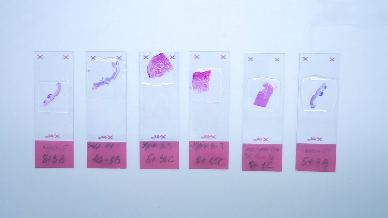
The most popular and one of the principal stains in histology is hematoxylin and eosin stain. It gives us an overview of the tissue and its structure. Hematoxylin binds with basophilic structures – for example DNA and RNA. So we can observe nuclei stained in blue or purple color. Eosin binds to acidophilic substances such as positively charged amino acid side chains. So as the result cytoplasm is pink or orange. All samples in laboratory are stained with H&E. There are several different types of hematoxylins and eosins used in histology which will give us different results.
In this video you will see, how we stain slides with different types of hematoxylins and eosins. Finally, we will compare the results.
• Subscribe to our channel: https://www.youtube.com/c/BioVitrumEN
• Watch other videos about histological process: https://www.youtube.com/playli....st?list=PLw4LQHit0MU
• Our website: http://en.biovitrum.ru/

Join the Amoeba Sisters a they explore different muscle tissues and then focus on the sliding filament theory in skeletal muscle! This video also briefly talks about muscle naming, some vocabulary (such as agonists and antagonists) before focusing on the sliding filament model. Video also mentions general roles of tropomyosin and troponin.
---------------------------------------------------------
Table of Contents:
00:00 Intro
0:39 Muscle Tissue Types
1:58 Muscle Characteristics
2:33 Skeletal Muscle Naming and Arrangement
3:26 Actin Myosin and Sarcomere
4:32 Sliding Filament Model
6:55 Tropomyosin an Troponin
---------------------------------------------------------
Factual References:
Betts, J. Gordon, et al. “10.3 Muscle Fiber Contraction and Relaxation - Anatomy and Physiology 2e | OpenStax.” Openstax.org, 20 Apr. 2022, openstax.org/books/anatomy-and-physiology-2e/pages/10-3-muscle-fiber-contraction-and-relaxation.
Urry, Lisa A, et al. Campbell Biology. 11th ed., New York, Ny, Pearson Education, Inc, 2017.
---------------------------------------------------------
Further Reading Recommendations:
What about I and A bands? What actually initiates the power stroke? How does calcium get released and from where? Remember, there is a lot more detail! We recommend this page from Openstax to learn more:
https://openstax.org/books/bio....logy-2e/pages/38-4-m
-----------------------------------------------
The Amoeba Sisters videos demystify science with humor and relevance. The videos center on Pinky's certification and experience in teaching biology at the high school level. Amoeba Sisters videos only cover concepts that Pinky is certified to teach, and they focus on her specialty: secondary life science. Learn more about our videos here: https://www.amoebasisters.com/our-videos
Support Us? https://www.amoebasisters.com/support-us
Our Resources and Handouts: https://www.amoebasisters.com/handouts
Biology Video Playlist: https://www.youtube.com/playli....st?list=PLwL0Myd7Dk1
GIFs: https://www.amoebasisters.com/gifs.html
Comics: https://www.amoebasisters.com/....parameciumparlorcomi
Unlectured Series: https://www.amoebasisters.com/unlectured
Connect with us!
Website: https://www.AmoebaSisters.com
Twitter: https://www.twitter.com/AmoebaSisters
Facebook: https://www.facebook.com/AmoebaSisters
Tumblr: https://www.amoebasisters.tumblr.com
Pinterest: https://www.pinterest.com/AmoebaSisters
Webtoon: https://www.webtoons.com/en/challenge/amoeba-sisters-sisterhood/list?title_no=289479&page=1
Instagram: https://www.instagram.com/amoebasistersofficial/
TikTok: https://www.tiktok.com/@amoebasistersofficial
Visit our Redbubble store at https://www.amoebasisters.com/store
TIPS FOR VIEWING EDU YOUTUBE VIDEOS:
Want to learn tips for viewing edu YouTube videos including changing the speed, language, viewing the transcript, etc? https://www.amoebasisters.com/....pinkys-ed-tech-favor
MUSIC:
Our intro music designed and performed by Jeremiah Cheshire.
End music in this video is listed free to use/no attribution required from the YouTube audio library.
COMMUNITY:
We take pride in our AWESOME community, and we welcome feedback and discussion. However, please remember that this is an education channel. See YouTube's community guidelines and how YouTube handles comments that are reported by the community. We also reserve the right to remove comments.
TRANSLATIONS:
Spanish Subtitles Translated by Jeremy García
Hindi Subtitles: Translated by Alisha Aggarwal
We gladly accept subtitle translations from our community. Learn more here: https://www.amoebasisters.com/....pinkys-ed-tech-favor We want to thank our amazing community for the generosity of their time in continuing to create translated subtitles.
We also have videos dubbed in Spanish and Portuguese using an artificial voice via https://aloud.area120.google.com to increase accessibility. See our Amoeba Sisters en Español channel https://www.youtube.com/channe....l/UC1Njo3LBy53cOPngz and Amoeba Sisters em Português https://www.youtube.com/channe....l/UCYTQPX2X_mXe0ZMPi

► Sign up here and try our FREE content: http://lectur.io/freecontentyt
► If you’re an medical educator or faculty member, visit: http://lectur.io/medytb2u
This video “Respiratory Histology” is part of the Lecturio course “Histology” ► WATCH the complete course on http://lectur.io/respiratoryhistology
► LEARN ABOUT:
- The cellular components of epithelium
- Structure and function of the conchae
- The cellular components of olfactory epithelium
- Components of the true vocal cord
- Function of the epiglottis
- The difference between bronchus, bronchiole, respiratory bronchiole
- Alveolar duct and Alveolar sac
- Components that make up the interalveolar septum
- Type I and Type II of alveolar cells, macrophages and endothelium
- Two separate blood supplies to the lungs and their functions
- Summary of the functions and the system of the respiratory system
► THE PROF: Your lecturer is Professor Geoff Meyer. He is currently teaching at the School of Anatomy, Physiology and Human Biology at the University of Western Australia (UWA). As a leading anatomy and histology expert he is also coordinating the Federative International Program for Anatomical Terminologies (FIPAT) of the International Federation of Associations of Anatomists (IFAA). Besides medical research on the ovarian function, steroidogenesis, corpus luteum, angiogenesis, and microcirculation, Geoff Meyer’s research activities also focus on developing innovative, computer-aided learning and teaching tools. For his inventiveness, Geoff Meyer has received a number of awards, including the Australian University Teaching Award.
► LECTURIO is your single-point resource for medical school:
Study for your classes, USMLE Step 1, USMLE Step 2, MCAT or MBBS with video lectures by world-class professors, recall & USMLE-style questions and textbook articles. Create your free account now: http://lectur.io/respiratoryhistology
► INSTALL our free Lecturio app
iTunes Store: https://app.adjust.com/z21zrf
Play Store: https://app.adjust.com/b01fak
► READ TEXTBOOK ARTICLES related to this video:
Histology of the Airways
http://lectur.io/respiratoryhistoloyarticle
► SUBSCRIBE to our YouTube channel: http://lectur.io/subscribe
► WATCH MORE ON YOUTUBE: http://lectur.io/playlists
► LET’S CONNECT:
• Facebook: https://www.facebook.com/lectu....rio.medical.educatio
• Instagram: https://www.instagram.com/lecturio_medical_videos
• Twitter: https://twitter.com/LecturioMed
• Pinterest: https://www.pinterest.de/lecturiomedical
• LinkedIn: https://www.linkedin.com/company/lecturio-medical/
0:00 Introduction
2:00 Respiratory system Summary of structure and function
3:32 Conducting portion
7:49 The Nasal cavities
11:27 The nasal cavity
13:06 Respiratory mucosa
15:26 Olfactory mucosa
17:13 Olfactory receptor
21:16 The larynx (and the epiglottis)
25:44 The trachea
29:16 The bronchi
31:35 Bronchiole
38:50 Alveolus
40:45 Air blood barrier
43:36 Alveolar macrophages
45:35 Blood supply and lymphatic drainage of the pulmonary lobule

#anatomy #histology #bytesizemed
✨If you would like my help studying about cartilage, you can check out my long-form video linked at the bottom of the screen.
💫 For more videos like this, subscribe to my channel, Byte Size Med.
📚Factual References & for Further Reading:
- DiFiore's Atlas of Histology
- Junqueira's Basic Histology
- Gartner's Concise Histology
- Openstax Anatomy and Physiology
https://openstax.org/details/b....ooks/anatomy-and-phy
- Openstax Biology
https://openstax.org/details/books/biology-2e
(The last two are links to open-source references. They are NOT affiliate links)
🌤 Note:
These are just a collection of my notes. So use them the way you would use borrowed notes from a friend. 📝
The images in this video are hand-drawn for illustration and explanation only.✍️ Hence, they may not be anatomically accurate. I am just one person making these videos. If there are any errors, that is unintentional. I try super hard to avoid them. Please let me know if you find any, so it gets clarified for other viewers. Science constantly evolves and changes. New discoveries are made everyday. So some of the information in these videos may become outdated. If you notice that, please let me know so I can update them.
⚡️Disclaimer:
These videos are NOT a substitute for a medical textbook. Textbooks are written by experts (which I do not claim to be), edited, proofread and referenced. Please use them.
The information has been sourced from multiple references as mentioned above. I draw all the pictures myself. But if I have inadvertently infringed on any copyright, that is completely unintentional. I only make these videos to impart education. If I have accidentally violated copyright in any way, do let me know so I can make the necessary changes or give credit to anyone who is owed the same.
These videos are NOT intended for patient education. They are NOT a substitute for diagnosis and treatment by a licensed medical professional. Always seek the advice of a qualified health care provider for any questions you may have regarding any medical condition, so that they can address your individual needs.
🔅They are ONLY meant to help students of medicine and health sciences with studying, and should be used for just that purpose and absolutely nothing else.
Byte Size Med. All Rights Reserved.

Classification of epithelium, discussion on lining epithelium of 3 major system (GIT, Urogenital and Respiratory system


Website : https://www.udemy.com/course/h....istology/?referralCo
Human Histology is one of the basic subject in a Medical Student career. By learning Histology in a proper way, this will help you to get a Visual memory of the Human body. Using this Visual memory, you can Learn any other subjects with little effort.
This Course is very well organized with lot of Histology images, Line diagrams, simple presentations and clear Explanations. This course has 33 videos, 19 chapters, 6 hours long covering all topics. Every topic is made Simple and Complete. Dr Ram has a great teaching style and has a good experience in teaching medical subjects to students.
After finishing this course, you will be better in your basics, with ability to visualize the human body and this will create an intense thirst to learn more. We give 100% guarantee that you will have a complete and in-depth understanding in short time, You will start to enjoy Learning Medicine because of the visualization of human body you get from this course and you will be ready to face any Medical exams in world.
Course features:
- Complete Histology lectures covering all chapters
- 19 chapters | 33 Videos | 6 Hours
- Clear Histology images
- Line diagrams for easy understanding
- Lot of memory tips
- High quality audio and Videos
- Can be viewed in Pc, or Phones or TV
Course content: ( 19 Chapters, 33 videos )
I The Cell - 3 Lessons
1. Nucleus
2. Cytoplasm
3. Cell Junctions
II Tissues - 11 Lessons
4. Epithelial tissues
5. Connective tissues
6. Muscular tissues
7. Nervous tissues
8. Bones
9. Cartilage
10. Lymphoid tissues
III Organ systems - 19 Lessons
11. Cardiovascular system
12. Respiratory system
13. Gastrointestinal system
14. Liver and Exocrine pancreas
15. Endocrine system
16. Urinary system
17. male reproductive system
18. Female reproductive system
19. The skin
Instructor : Dr Ram , Med Madness

Learn about the structural unit of compact bone (the osteon) and it's four basic parts: central canal, lamellae, lacunae, and canaliculi

✨This video is on the structure and functions of the three types of cartilage (Hyaline, Elastic and Fibrocartilage). I hope it helps! ☀️
🌟What's in this video?
0:00 - Intro
0:07 - Connective Tissue Recap
1:16 - Structure of Connective Tissue
1:57 - Structural Components of Cartilage
3:38 - Types of Cartilage
3:49 - Hyaline Cartilage
8:05 - Elastic Cartilage
8:55 - Fibrocartilage
✨ Other videos you may need:
🔅 Connective Tissue : https://youtu.be/xw_ALdt5n-A
🔅 Collagen : https://youtu.be/3e2JYMNS_W4
🔅 Ossification: https://youtu.be/86V9SNWD_No
🔅Histology: https://www.youtube.com/playli....st?list=PL1rG930trF2
💫 For more videos like this, subscribe to my channel!
Byte Size Med: https://youtube.com/channel/UC....ZghvlgylH3r_CWfA18eF
📚Factual References & for Further Reading:
- DiFiore's Atlas of Histology
- Junqueira's Basic Histology
- Gartner's Concise Histology
- Openstax Anatomy and Physiology
https://openstax.org/details/b....ooks/anatomy-and-phy
- Openstax Biology
https://openstax.org/details/books/biology-2e
(The last two are links to open-source references. They are NOT affiliate links)
🌤 Note:
These are just a collection of my notes. So use them the way you would use borrowed notes from a friend. 📝
The images in this video are hand-drawn for illustration and explanation only.✍️ Hence, they may not be anatomically accurate. I am just one person making these videos. If there are any errors, that is unintentional. I try super hard to avoid them. Please let me know if you find any, so it gets clarified for other viewers. Science constantly evolves and changes. New discoveries are made everyday. So some of the information in these videos may become outdated. If you notice that, please let me know so I can update them.
⚡️Disclaimer:
These videos are NOT a substitute for a medical textbook. Textbooks are written by experts (which I do not claim to be), edited, proofread and referenced. Please use them.
The information has been sourced from multiple references as mentioned above. I draw all the pictures myself. But if I have inadvertently infringed on any copyright, that is completely unintentional. I only make these videos to impart education. If I have accidentally violated copyright in any way, do let me know so I can make the necessary changes or give credit to anyone who is owed the same.
These videos are NOT intended for patient education. They are NOT a substitute for diagnosis and treatment by a licensed medical professional. Always seek the advice of a qualified health care provider for any questions you may have regarding any medical condition, so that they can address your individual needs.
🔅They are ONLY meant to help students of medicine and health sciences with studying, and should be used for just that purpose and absolutely nothing else.
Byte Size Med. All Rights Reserved.
![Histology of Exocrine Gland [Epithelium 7 of 7]](https://i.ytimg.com/vi/NkU7YJ7eYd0/maxresdefault.jpg)
Histological features and cellular biology of exocrine glands. This video is a part of our Histology Video Course (https://youtube.com/playlist?l....ist=PLnr1l7WuQdDynxT
Additional YouTube Content
Biochemistry videos: https://youtube.com/playlist?l....ist=PLnr1l7WuQdDzCUC
Anatomy Videos: https://youtube.com/playlist?l....ist=PLnr1l7WuQdDz2dK
DaVinci Cases Videos: https://youtube.com/playlist?l....ist=PLnr1l7WuQdDyJUl
The DaVinci Hour Podcast: https://youtube.com/playlist?l....ist=PLnr1l7WuQdDwSm9
DaVinci Academy Website: https://www.dviacademy.com/

**PLEASE READ FULLY
Purpose of the video is to help Esthetician’s review chapters in their text book to better prepare for State Bord testing, by simply reading and going over some of the material, it’s not intended to replace any teaching from any Beauty College. Every instructor does things different, Keep in mind I am in the state of Texas, also keep in mind that when in school students are to follow guidelines and might be required to do things a bit different, I teach my students the text book because that is where the state board questions come from and the goal is for them to pass their board exams. I also teach them and go over real working situations they might come across in the salon or spa.
* I am not affiliated with TDLR or PSI in any way
PSI Bulletin Link
https://candidate.psiexams.com/bulletin/display_bulletin.jsp?ro=yes&actionname=83&bulletinid=173&bulletinurl=.pdf
Glymed store: https://glymedplus.io/home/index?store=0011298
email: glamandbeyondinfo@gmail.com

Brain tumor survivor Robert Alvarez and neurosurgeon Sujit Prabhu, M.D., explain why and how Robert played the guitar during his surgery for a grade II astrocytoma. It was the first time a brain tumor patient played a musical instrument during an awake craniotomy at MD Anderson.
Read Robert Alvarez's story: https://www.mdanderson.org/pub....lications/cancerwise
Learn about awake craniotomy for brain tumors: https://www.mdanderson.org/pub....lications/cancerwise
Request an appointment at MD Anderson by calling 1-877-632-6789 or online at: https://my.mdanderson.org/Requ....estAppointment?cmpid

http://www.highimpact.com - This brain surgery animation was used to demonstrate a young girl's craniotomy, cranioplasty, and reconstructive skull surgery after her vehicle was struck by a tractor-trailer. The procedures included the evacuation of a large epidural hematoma, the draining of the epidural space, and the reassembly of bone fragments to repair the skull.
More Brain Surgery Animations: https://tinyurl.com/y6m4lkdf
WHAT HAPPENED
A teenage girl was riding home with her parents and boyfriend from a Wednesday night church service when a tractor-trailer struck the back driver’s side of their car as they were traveling through an intersection. The impact sent the car spinning into oncoming traffic where it struck another vehicle. When paramedics arrived, the 17-year-old was unresponsive with bleeding from her left ear and a laceration from behind her left ear.
She was rushed to the hospital where she underwent a series of CT scans that showed a severely comminuted open skull fracture with an underlying 1.1 cm subdural hematoma. She was taken to the operating room where an emergency craniotomy was performed to evacuate the hematoma and reassemble the skull fragments. The patient gradually began to wake up and was discharged six days later, after she showed she could maneuver up and down the hallway.
The biggest challenge in a traumatic brain injury case like this - where most of the damages are deeply underlying and undetectable on the surface - is that the only visual evidence is in the form of 2D black-and-white radiographic films. This can look ambiguous to the typical juror because it’s often difficult to discern where these snapshots are located inside the person’s skull. Tony Seaton, Esq., and Robert Bates, Esq., needed to reinforce this 2D radiographic evidence with maximum 3D context.
We equipped them with a custom Diagnostic Slice Chooser: an interactive presentation that presents radiographic slides within a three-dimensional model of the patient’s head. We also designed the model accurately to the patient’s likeness and colorized the films to highlight key areas of damage. The attorneys could show the complete depth and magnitude of his client’s injuries at every level both before and after the surgery. After establishing the full extent of damages, we also created an animation to walk viewers through the surgical experience the patient would undergo as a result of her injuries.
The visual presentation helped jurors understand the destructive impact this collision had on this young teenager’s life, and Mr. Seaton and Mr. Bates, Esq., were able to acquire a $4.5M settlement for his client.
Read the Full Case Study: https://tinyurl.com/yy4v2dyh

http://www.nucleushealth.com/ - This 3D medical animation depicts two operations, called craniotomy and craniectomy, in which the skull is opened to access the brain. The normal anatomy of the skull and tissues surrounding the brain are shown, including arteries and veins. The animation lists the common reasons for these procedures, and briefly introduces intracranial pressure.
Video ID: ANH13109
Transcript:
Your doctor may recommend a craniotomy or a craniectomy procedure to treat a number of different brain diseases, injuries, or conditions.
Your skull is made of bone and serves as a hard, protective covering for your brain. Just inside your skull, three layers of tissue, called meninges, surround your brain. The thick, outermost layer is the dura mater. The middle tissue layer is the arachnoid mater and the innermost layer is the pia mater. Between the arachnoid mater and the pia mater is the subarachnoid space, which contains blood vessels and a clear fluid called cerebrospinal fluid. Blood vessels, called bridging veins, connect the surface of your brain with the dura mater. Other blood vessels, called cerebral arteries, bring blood to your brain.
Inside your skull, normal brain function requires a delicate balance of pressure between the blood in your blood vessels, the cerebrospinal fluid that surrounds your brain, and your brain tissue. This is called normal intracranial pressure. Increased intracranial pressure may result from: brain tumors, head injuries, problems with your blood vessels, or infections in your brain or spinal cord. These conditions put pressure on your brain and may cause it to swell or change shape inside your skull, which can lead to serious brain injury.
Your doctor may recommend a craniotomy to remove: abnormal brain tissue, such as a brain tumor, a sample of tissue by biopsy, a blood clot, called a hematoma, excess cerebrospinal fluid, or pus from an infection, called an abscess.
A craniotomy may also be done to: relieve brain swelling,
stop bleeding, called a hemorrhage, repair abnormal blood vessels, repair skull fractures, or repair damaged meninges.
Finally, a craniotomy may also be done to: treat brain conditions, such as epilepsy, deliver medication to your brain, or implant a medical device, such as a deep brain stimulator.
The most common reason for a craniotomy is to remove a brain tumor.
#Craniotomy #Craniectomy #BrainSurgery
Dr. Akshay Syal takes us to NYU Langone Health where new A.I. technology is diagnosing brain tumors in record time, which opens the doors to possible new life-saving treatments.
» Subscribe to NBC News: http://nbcnews.to/SubscribeToNBC
» Watch more NBC video: http://bit.ly/MoreNBCNews
NBC News Digital is a collection of innovative and powerful news brands that deliver compelling, diverse and engaging news stories. NBC News Digital features NBCNews.com, MSNBC.com, TODAY.com, Nightly News, Meet the Press, Dateline, and the existing apps and digital extensions of these respective properties. We deliver the best in breaking news, live video coverage, original journalism and segments from your favorite NBC News Shows.
Connect with NBC News Online!
NBC News App: https://smart.link/5d0cd9df61b80
Breaking News Alerts: https://link.nbcnews.com/join/....5cj/breaking-news-si
Visit NBCNews.Com: http://nbcnews.to/ReadNBC
Find NBC News on Facebook: http://nbcnews.to/LikeNBC
Follow NBC News on Twitter: http://nbcnews.to/FollowNBC
Get more of NBC News delivered to your inbox: nbcnews.com/newsletters
#NBCNews #AI #Cancer

It’s called gamma knife surgery, but there’s no cutting involved.
It’s been used at Mayo Clinic for 30 years as an alternative to open brain surgery.
The patient’s head is held still during the procedure with a headframe, which also serves as a map for the radiation. Using 3D imaging — typically an MRI — as a guide, the gamma knife is targeted directly at the tumor.
And with no hospital stay and minimal side effects, it’s a procedure that is efficient and can be lifesaving.
More health and medical news on the Mayo Clinic News Network. https://newsnetwork.mayoclinic.org/
Journalists: Clean and nat sound versions of this pkg available for download at https://newsnetwork.mayoclinic.org/
Register (free) at https://newsnetwork.mayoclinic.org/request-account/

Brain port surgery is a minimally invasive surgical technique performed through a specially designed tube about the size of a dime. Using neuronavigation GPS-like guidance, the brain port is inserted into the brain with millimeter accuracy and is used as a channel to guide the surgeon and his/her instruments to various regions of the brain. Colloid cysts, metastatic tumors, and a variety of tumors within the ventricles are often candidates for this approach.
