- Physical Examination
- Surgical Examination
- Ophthalmology
- Clinical Skills
- Orthopedics
- Surgery Videos
- Laparoscopy
- Pediatrics
- Funny Videos
- Cardiothoracic Surgery
- Nursing Videos
- Plastic Surgery
- Otorhinolaryngology
- Histology and Histopathology
- Neurosurgery
- Dermatology
- Pediatric Surgery
- Urology
- Dentistry
- Oncology and Cancers
- Anatomy Videos
- Health and Fitness
- Radiology
- Anaesthesia
- Physical Therapy
- Pharmacology
- Interventional Radiology
- Cardiology
- Endocrinology
- Gynecology
- Emergency Medicine
- Psychiatry and Psychology
- Childbirth Videos
- General Medical Videos
- Nephrology
- Physiology
- Diet and Food Health
- Diabetes Mellitus
- Neurology
- Women Health
- Osteoporosis
- Gastroenterology
- Pulmonology
- Hematology
- Rheumatology
- Toxicology
- Nuclear Medicine
- Infectious Diseases
- Vascular Disease
- Reproductive Health
- Burns and Wound Healing
- Other
Top videos
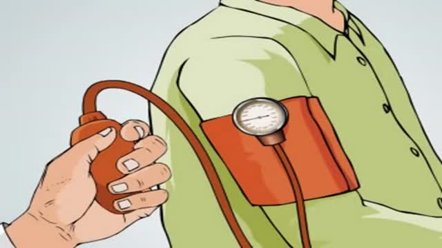
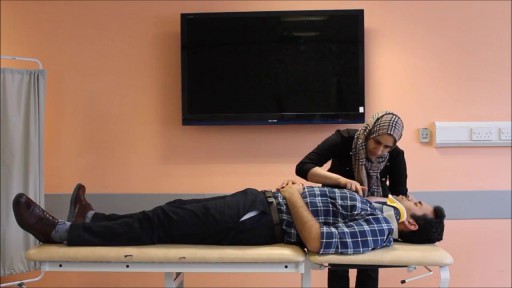
A simple video showing the small tips to be followed by patients which a clinician should provide. The video is simple, easy to understand and can be provided to the patient for their reference.
Improve blood sugar control in adults with type 2 diabetes through new advancements
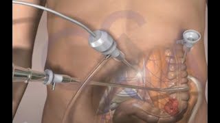
This surgical animation is for patient education and describes a laparoscopic colectomy, which is a type of minimally invasive surgery for colon cancer. Laparoscopic colectomy, also called minimally invasive colectomy, involves several small incisions in your abdomen. Instead of a big incision, the surgeon makes a few small cuts (0.5-1 centimeters) in the abdominal cavity to insert a surgical camera and instruments and perform the operation. A slightly bigger incision, about 3.5 centimeters wide, is made to remove the tumor.
When compared to traditional open surgery, laparoscopic colectomy can result in much less pain and swifter recovery. Depending on the procedure, most laparoscopic colectomy patients leave the hospital and return to normal activities more quickly than patients recovering from open surgery.
Colorectal cancer is the second leading cause of cancer death in the United States.
For more information about 3d animation videos, please visit https://www.amerra.com
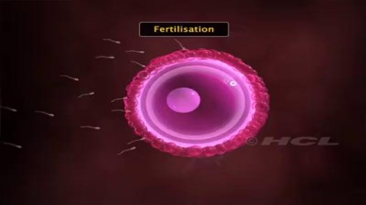
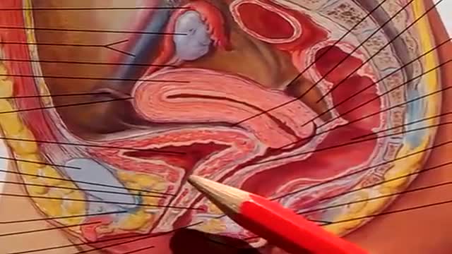
Millions of sperms are deposited into the vagina during sexual intercourse. The sperms make their way through the cervix into the uterus and then on to the fallopian tubes. As they swim along this way their numbers decline. Only a few hundred sperm will get close to the egg. During the trip, sperm prepare themselves to meet the egg by subtle alterations of their heads and movement patterns. Once inside the fallopian tube, the sperm attracts the egg by releasing a chemical. The egg is surrounded by a protective covering called the zona pellucida, which allows only one sperm to penetrate it. Once inside the egg, the head of the fertilizing sperm releases its genetic contents, which fuses with the nucleus of the egg. Fertilisation is now complete. Sperm are able to survive for 2-3 days within the female's reproductive tract. The length of the time that a woman's egg can be fertilized by a man's sperm ranges from 12-24 hours.
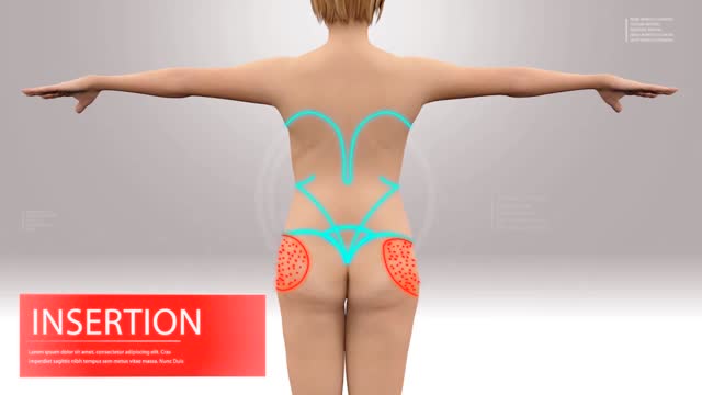
Liposuction & Facelift
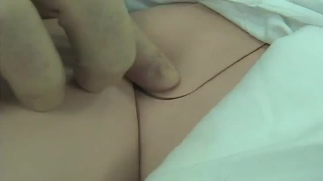
Femoral Venous Line Placement
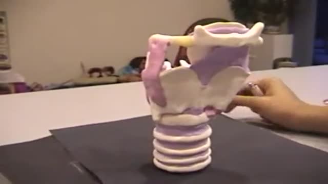
Larynx Anatomy and Physiology
The urinary bladder is a hollow muscular organ that collects urine from the kidneys before disposal by urination. A hollow muscular, and distensible (or elastic) organ, the bladder sits on the pelvic floor. Urine enters the bladder via the ureters and exits via the urethra.
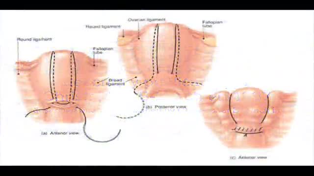
B-Lynch suture for uterine atony technique described
It sounds like you're questioning whether or not your water may have broken, and this can actually be a hard thing for a lot of women to tell. Usually if your water breaks, it's just a trickle of fluid, and you're afraid to admit it to anyone because you think you peed your pants. And it is normal to pee your pants when you're pregnant because the bladder is right below the uterus, and if the baby moves just right, it might kick out a little bit of urine. So if you feel a trickle or a little tiny gush of fluid, what you want to do is put a pad or a pantie-liner on after going to the bathroom and emptying your bladder, and wait an hour and see if fluid continues to come out. And if it does, then you're not having bladder leakage issues - your water is probably broken.

To save humanity, a dietitian travels to the past. A lot.
Subscribe now: https://www.youtube.com/c/funn....yordie?sub_confirmat
CREDITS:
Director: Elliot Dickerhoof
Producers: Chuck Armstrong, Charlie Stockman, Elliot Dickerhoof
Writers: Chuck Armstrong & Charlie Stockman
Actors: Chuck Armstrong, Charlie Stockman, Kelly Vrooman
Executive Producer: Darren Miller
DP: Cody Jacobs
Gaffer: Jordan Holtane
AC: Giselle Gonzalez
Sound Mixer: Marcos Castro
Costume Designer: Kate Bergh
Hair and Makeup Artist: Jessica Leigh Schwartz
PA: Elyssa Phillips
Get more Funny Or Die
-------------------------------
Like FOD on Facebook: https://www.facebook.com/funnyordie
Follow FOD on Twitter: https://twitter.com/funnyordie
Follow FOD on Tumblr: http://funnyordie.tumblr.com/
Follow FOD on Instagram: http://instagram.com/funnyordie
Follow FOD on Vine: https://vine.co/funnyordie
Follow FOD on Pinterest: http://www.pinterest.com/funnyordie
Follow FOD on Google+: https://plus.google.com/+funnyordie
See the original at: http://www.funnyordie.com/videos/74dd9afee2
Immunomodulating effect of autohaemotherapy (a literature review). PMID 3534085 [PubMed indexed for MEDLINE]
J Hyg Epidemiol Microbiol Immunol. 1986;30(3):331-6.
Immunomodulating effect of autohaemotherapy (a literature review).
Klemparskaya NN, Shalnova GA, Ulanova AM, Kuzmina TD, Chuhrov AD.
Abstract
An analysis is presented of experimental and clinical data from different authors on the stimulating effect of autohaemotherapy with regard to the immunological reactivity of humans and animals as well as in vitro experiments with lymphocytes. Erythrolysate has been found to exert a more powerful effect than intact erythrocytes. The stimulating effect of autohaemotherapy on both irradiated and non-irradiated animals manifests itself in an increase in resistance to infection (increased LD50 in experimental infection), enhanced production of antibodies to microbial and tissue antigens and activated functioning of cell-mediated immune defence mechanisms. The favourable influences on radioresistance and the antitumour effect of authohaemotherapy are described. Induced desensitization plays an important part in the mechanism of action of autohaemotherapy. The administration of large doses of erythrocytes or of erythrolysate results in immunosuppression. Autohaemotherapy does not cause side effects and is feasible both on an in-and out-patient basis.
PMID: 3534085
[PubMed - indexed for MEDLINE]
http://www.ncbi.nlm.nih.gov/pubmed/3534085
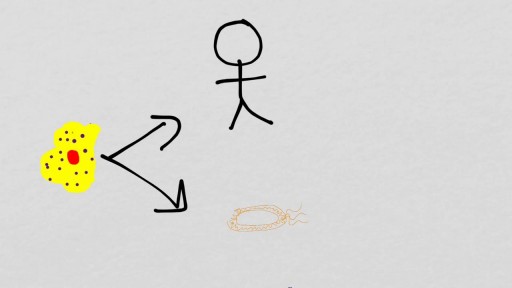
Autohemotherapy: an immunization with our own blood
http://www.geocities.ws/autohemoterapiabr/
http://autohemoterapia.fortunecity.com/
http://www.geocities.ws/autohemoterapiabr/aht_english.htm
http://autohemoterapia.fortunecity.com/aht_english.htm
-
Auto-hemotherapy PDF files in GOOGLE sites:
https://sites.google.com/site/autohemotherapy/
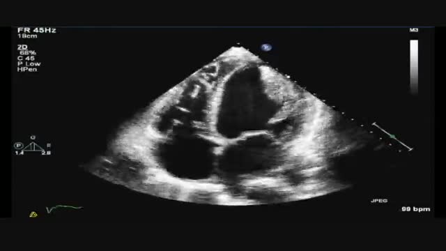
TEE of a giant LV thrombus as well as other, smaller thrombi in a 51 year-old man who came to the hospital complaining of chest pain. He was subsequently taken to the cardiac cath lab and was found to have clean coronary arteries. Surprisingly, he was clinically stable despite his TEE findings and an EF of 20%.

Dr. Joanna Chikwe, explains how patients may feel after heart surgery.
Learn more about the Smidt Heart Institute at Cedars-Sinai at https://ceda.rs/3meIA2A
Learn more about Dr. Joanna Chikwe at https://ceda.rs/3ul6I6t
Connect with us:
https://twitter.com/CedarsSinai
https://www.facebook.com/CedarsSinai
https://www.instagram.com/CedarsSinai
Cedars-Sinai is a leader in providing high-quality healthcare encompassing primary care, specialized medicine and research. Since 1902, Cedars-Sinai has evolved to meet the needs of one of the most diverse regions in the nation, setting standards in quality and innovative patient care, research, teaching and community service. Today, Cedars- Sinai is known for its national leadership in transforming healthcare for the benefit of patients. Cedars-Sinai impacts the future of healthcare by developing new approaches to treatment and educating tomorrow’s health professionals. Additionally, Cedars-Sinai demonstrates a commitment to the community through programs that improve the health of its most vulnerable residents.
http://www.landging.com/skeletal-system-animation-knee-surgery.html
This skeletal system animation demonstrates the new concept of knee surgery procedure.

An ectopic pregnancy results when a fertilized egg implants outside the uterus. Unfortunately, there's no way to transplant an ectopic pregnancy into your uterus, so ending the pregnancy is the only option. About 2 percent of pregnancies are ectopic. Because ectopic pregnancy is potentially dangerous for you, it's important to recognize the early signs and get treatment as soon as possible.
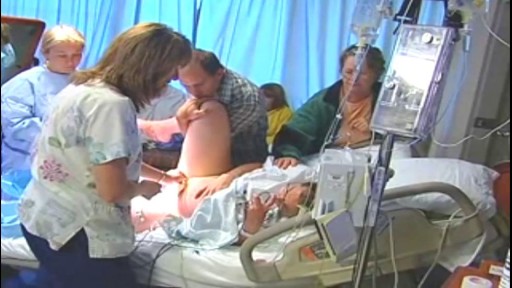
How to push a baby out video how to prevent tearing during labor and delivery
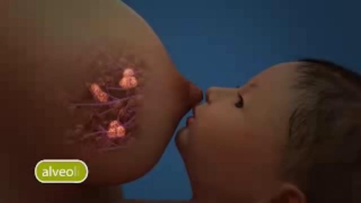
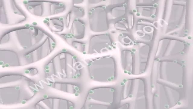
Prompted by the hormone prolactin, the alveoli take proteins, sugars, and fat from your blood supply and make breast milk. A network of cells surrounding the alveoli squeeze the glands and push the milk out into the ductules, which lead to a bigger duct.
