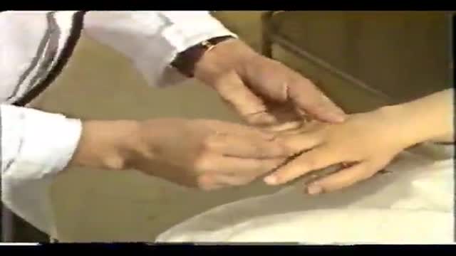
- Physical Examination
- Surgical Examination
- Ophthalmology
- Clinical Skills
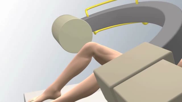
- Orthopedics
- Surgery Videos
- Laparoscopy
- Pediatrics
- Funny Videos
- Cardiothoracic Surgery
- Nursing Videos
- Plastic Surgery
- Otorhinolaryngology
- Histology and Histopathology
- Neurosurgery
- Dermatology
- Pediatric Surgery
- Urology
- Dentistry
- Oncology and Cancers
- Anatomy Videos
- Health and Fitness
- Radiology
- Anaesthesia
- Physical Therapy
- Pharmacology
- Interventional Radiology
- Cardiology
- Endocrinology
- Gynecology
- Emergency Medicine
- Psychiatry and Psychology
- Childbirth Videos
- General Medical Videos
- Nephrology
- Physiology
- Diet and Food Health
- Diabetes Mellitus
- Neurology
- Women Health
- Osteoporosis
- Gastroenterology
- Pulmonology
- Hematology
- Rheumatology
- Toxicology
- Nuclear Medicine
- Infectious Diseases
- Vascular Disease
- Reproductive Health
- Burns and Wound Healing
- Other
Top videos
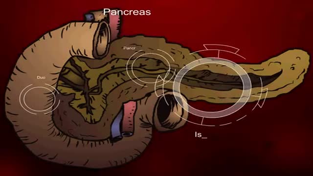
Insulin Processes Mechanism Animation 3D

This patented device replaces cumbersome metal retractors for a variety of surgical procedures. The surgeon has maximum unobstructed exposure and the size of the required incision is minimized.
This brief exam will help you to quickly detect major risks and prompt you to refer patients to appropriate specialists.

Liposuction for weight loss with Abdominoplasty and Body Lifting

At Children's Hospital, Dr. Mary Bedard and the NICU nursing staff save the life of a tiny infant struggling from a serious intestinal infection. ~ Detroit Medical Center
Integrative Physical Examination Lecture
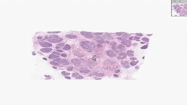
Histology of Active Breast
This new surgical technique provide good stability for all type of fracture even severe comminution. Each fragment are reduced and several pin sleeves are inserted circumferentially and tighten by braded cable through the sleeve box. The final features of surgery seems blooming sunflower 'Himwari in Jananese'.
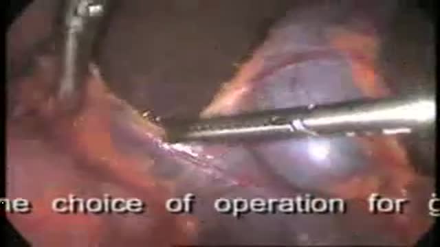
Gall Stones
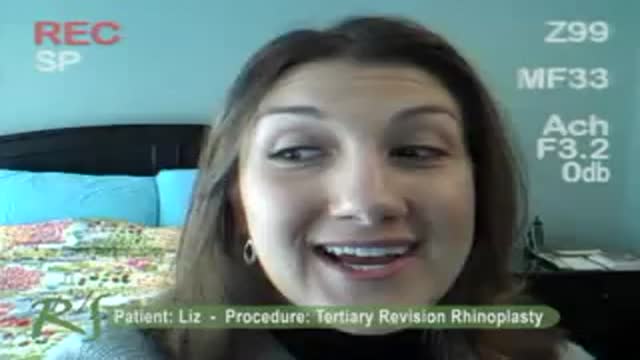
LIZ: The first time the doctor made my tip too narrow and I didnt look like myself. The second time the doctor made my tip too wide, and actually took out (removed) extra bone from the side of my nose. That didnt need to be taken out (removed)
My initial consultation with Dr. Nassif was fantastic! He treated me liker his own daughter, and was very caring and thorough. He went over everything!
DR. NASSIF: Liz came into me for a revision rhinoplasty. She told me that shes had two previous rhinoplasties. She was unhappy with the way her nose appeared on her face. She felt it was asymmetric, the tip was kind of bulbous, or large appearing, especially when she looked up, this view, it was very asymmetric. And so, her whole goal was to make it look better, hopefully make it her LAST surgery, and also to help with her breathing.
One of the things thats very important about revision rhinoplasty that you always have to consider is; What are you going to find in there? Even though you can feel the nose, you can palpate it, you can look at it, and you can guess what the other doctors have performed; your first up-hill battle is to see how much scar tissue youre going to be able to identify with. So when you have to open up the nose, you have to remove the scar tissue, identify it: whats there, whats present, whats been removed. Then after you do that, and you have cartilage now ready for grafting, or fascia, or perichondrium, you have to start rebuilding it. Rebuilding it (cartilage) is the second big stage after weve already carved everything; weve carved the cartilage. In that scenario when Im playing with the nose, in regards to staring at the profile, staring at the front of the nose, I go back and forth and look inside and outside of the nose to make sure its as symmetric as possible. That takes a long time One of Lizs main complaints was that on her profile, that her tip stuck out too far. And so one of the things I had to do in surgery is called a medial cura tuck-up, I had to push the tip back, by pushing the tip back, it can make the tip look a little bit wider. But in this situation, I was able to bring everything in as much as I can. After Im finished with everything, and Im happy, then we go ahead and we start to close the nose. Thats putting every little small stitch in perfectly, so that the scar will be minimally visible.
Chinese Complete Physical Clinical Exam
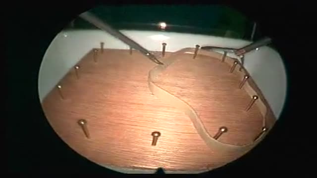
This task requires streching a rubber band around 16 nails on a wooden board. A penalty is calculated when the rubber band is not streched around a nail at the end of the task. Score = time (seconds) + number of missed nails x 10. Performance standard: Score = 62 sec [Kolkman 2008]
This shows an animated procedure for Interventional Cardiologists in injecting stemcells.
bilateral tubal ligation as modified Pomeroy technique during a C-Section
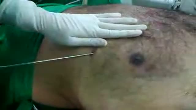
A central venous catheter, also called a central line, is a long, thin, flexible tube used to give medicines, fluids, nutrients, or blood products over a long period of time, usually several weeks or more. A catheter is often inserted in the arm or chest through the skin into a large vein.
Male Breast Liposuction Reduction
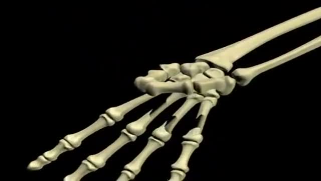
An animated illustration showing muscles of the forearm
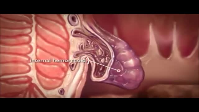
The veins around your anus tend to stretch under pressure and may bulge or swell. Swollen veins (hemorrhoids) can develop from an increase in pressure in the lower rectum. Factors that might cause increased pressure include: Straining during bowel movements.

Watch that video to know What is Trypophobia? Do You Have it ?

Toxin is a protein produced by the bacterium Clostridium botulinum, and is extremely neurotoxic.
