- Physical Examination
- Surgical Examination
- Ophthalmology
- Clinical Skills
- Orthopedics
- Surgery Videos
- Laparoscopy
- Pediatrics
- Funny Videos
- Cardiothoracic Surgery
- Nursing Videos
- Plastic Surgery
- Otorhinolaryngology
- Histology and Histopathology
- Neurosurgery
- Dermatology
- Pediatric Surgery
- Urology
- Dentistry
- Oncology and Cancers
- Anatomy Videos
- Health and Fitness
- Radiology
- Anaesthesia
- Physical Therapy
- Pharmacology
- Interventional Radiology
- Cardiology
- Endocrinology
- Gynecology
- Emergency Medicine
- Psychiatry and Psychology
- Childbirth Videos
- General Medical Videos
- Nephrology
- Physiology
- Diet and Food Health
- Diabetes Mellitus
- Neurology
- Women Health
- Osteoporosis
- Gastroenterology
- Pulmonology
- Hematology
- Rheumatology
- Toxicology
- Nuclear Medicine
- Infectious Diseases
- Vascular Disease
- Reproductive Health
- Burns and Wound Healing
- Other
Top videos
Histology of Medium Artery and Vein
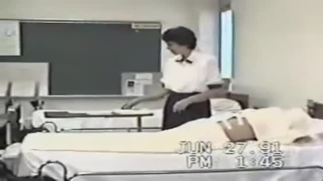
Changing Dressing Wounds
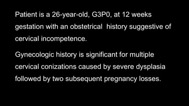
Cerclage In Pregnancy Laparoscopic HD
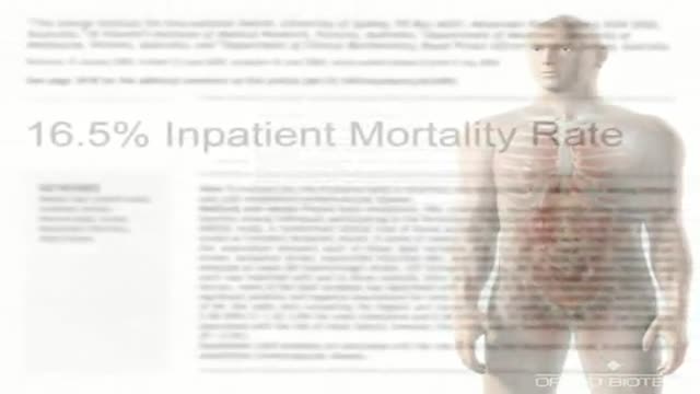
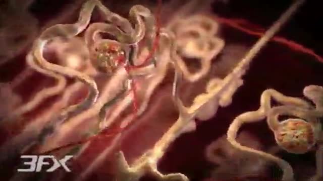
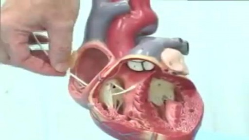
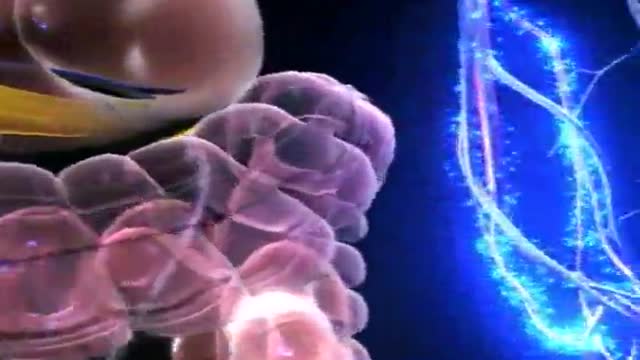
Congestive Heart Failure Animation
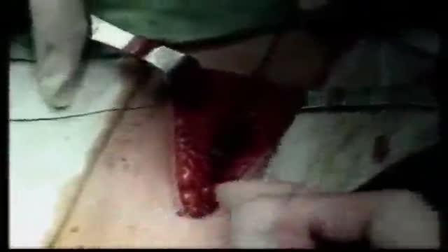
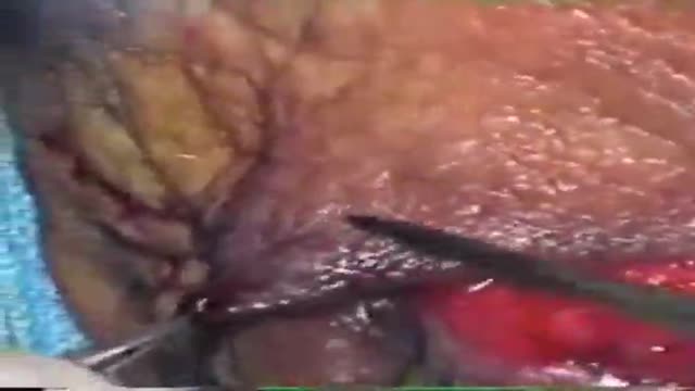
Abdominal Wall Closure
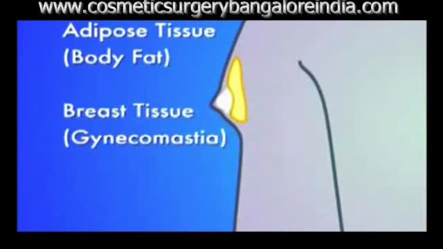
Before and After images following gynecomastia correction with surgical video and animation
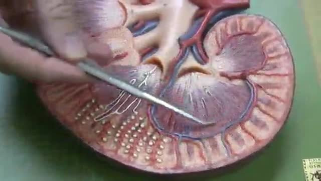
A great video showing dunctions and physiology of the Nephron
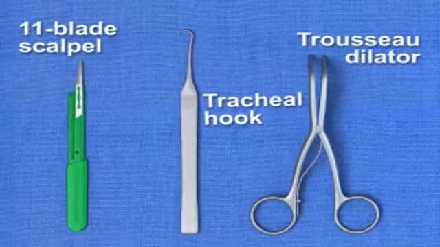
Traditional Surgical Cricothyrotomy
Diabetic Nephropathy Animation 3D
Pulmonary Artery Swan Ganz Catheter
Eye Lid Jones Procedure
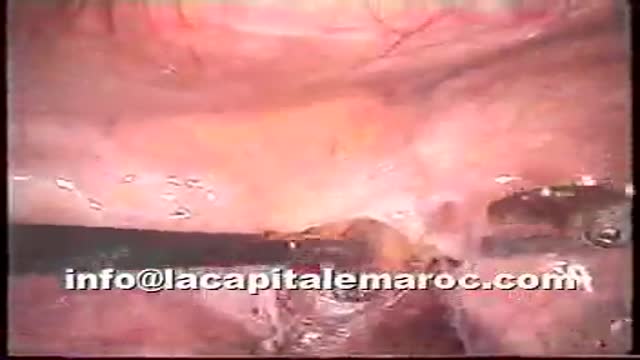
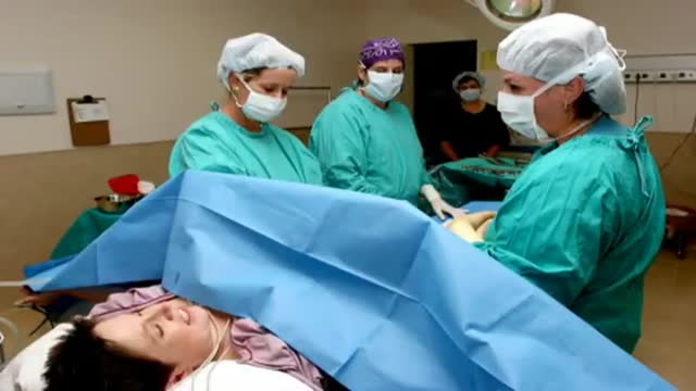
Pelvic lymphadenectomy
Lasic in 10 years old girl for Myopia
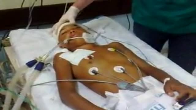
Male 19 y. age victim of penetrating brain injury. All the criteria for the encephalic death diagnosis were present. The presence of this complex spinal reflex doesn't exclude the brain death diagnosis and must be known and understood by the professionals involved in this very important diagnosis
Pain Management During Birth
Diabetes Animation 3D
Understanding narcolepsy symptoms to improve alertness.
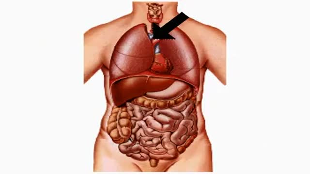
How Smoking and Drinking Affect Your Body
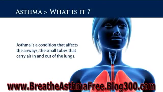
http://breatheasthmafree.blog300.com - Bronchitis Asthma Symptoms - Asthma Treatments For Adults
Asthma Sufferer?
Ex-Sufferer Reveals Own Natural
Remedy for Eliminating Asthma!...
http://breatheasthmafree.blog300.com
Bronchitis Asthma Symptoms - Asthma Treatments For Adults
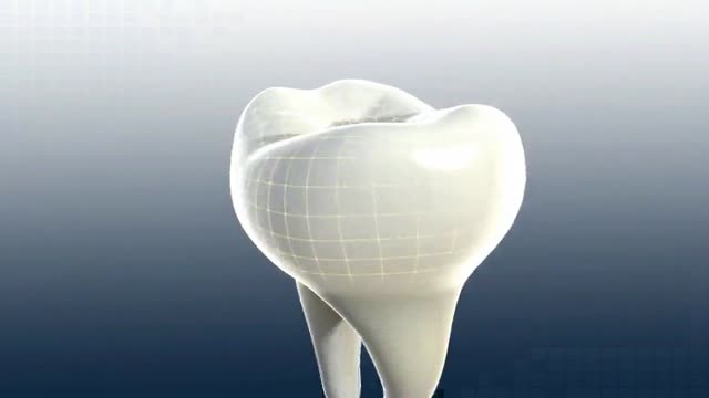
http://www.landging.com/dental-animation.html 3D teeth virtual reality animation with high quality textures, for patient education purpose.
