- Physical Examination
- Surgical Examination
- Ophthalmology
- Clinical Skills
- Orthopedics
- Surgery Videos
- Laparoscopy
- Pediatrics
- Funny Videos
- Cardiothoracic Surgery
- Nursing Videos
- Plastic Surgery
- Otorhinolaryngology
- Histology and Histopathology
- Neurosurgery
- Dermatology
- Pediatric Surgery
- Urology
- Dentistry
- Oncology and Cancers
- Anatomy Videos
- Health and Fitness
- Radiology
- Anaesthesia
- Physical Therapy
- Pharmacology
- Interventional Radiology
- Cardiology
- Endocrinology
- Gynecology
- Emergency Medicine
- Psychiatry and Psychology
- Childbirth Videos
- General Medical Videos
- Nephrology
- Physiology
- Diet and Food Health
- Diabetes Mellitus
- Neurology
- Women Health
- Osteoporosis
- Gastroenterology
- Pulmonology
- Hematology
- Rheumatology
- Toxicology
- Nuclear Medicine
- Infectious Diseases
- Vascular Disease
- Reproductive Health
- Burns and Wound Healing
- Other
Top videos
living with chronic fatigue syndrome
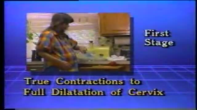
First stage of labour with its signs and symptoms like uterine contractions and the show

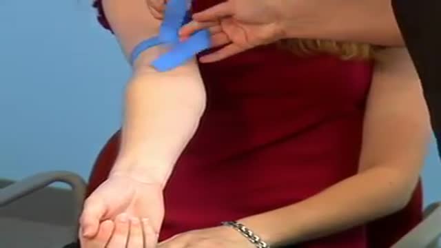
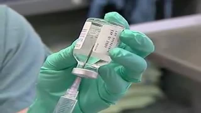
IM Injection instructions
Intra-Uterine Device IUD Insertion Demonstration
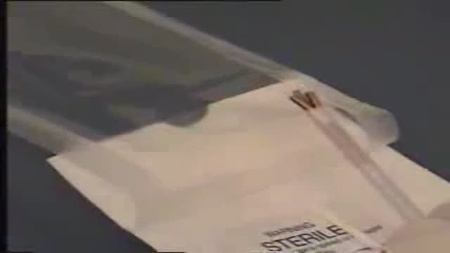
What is Venipuncture? While venipuncture can refer to a variety of procedures, including the insertion of IV tubes into a vein for the direct application of medicine to the blood stream, in phlebotomy venipuncture refers primarily to using a needle to create a blood evacuation point. As a phlebotomist, you must be prepared to perform venipuncture procedures on adults, children, and even infants while maintaining a supportive demeanor and procedural accuracy. Using a variety of blood extraction tools, you must be prepared to respond to numerous complications in order to minimize the risk to the patient while still drawing a clean sample. In its entirety, venipuncture includes every step in a blood draw procedure—from patient identification to puncturing the vein to labeling the sample. Patient information, needle placement, and emotional environment all play a part in the collection of a blood sample, and it's the fine details that can mean the difference between a definite result and a false positive. After placing the tourniquet and finding the vein, it's time for the phlebotomist to make the complex choice on what procedure will best suit the specific situation. Keeping this in mind, it should be noted that the following information is not an instructional guide on how to perform these phlebotomy procedures. Rather, the information below is intended to serve as an educational resource to inform you of the equipment and procedures you will use. Venipuncture Technqiues Venipuncture with an Evacuated or Vacuum Tube: This is the standard procedure for venipuncture testing. Using a needle and sheath system, this procedure allows multiple sample tubes to be filled through a single puncture. This procedure is ideal for reducing trauma to patients. After drawing the blood, the phlebotomist must make sure the test stopper is correctly coded and doesn't contact exposed blood between samples. Venipuncture with a Butterfly Needle : This is a specialized procedure that utilizes a flexible, butterfly needle adaptor. A butterfly needle has two plastic wings (one on either side of the needle) and is connected to a flexible tube, which is then attached to a reservoir for the blood. Due to the small gauge of the needle and the flexibility of the tube, this procedure is used most often in pediatric care, where the patients tend to have smaller veins and are more likely to move around during the procedure. After being inserted into a vein at a shallow angle, the butterfly needle is held in place by the wings, which allow the phlebotomist to grasp the needle very close to the skin. Phlebotomists should be careful to watch for blood clots in the flexible tubing. Venipuncture with a Syringe: This technique is typically only used when there is a supply shortage, or when a technician thinks it is the appropriate method. It uses the classic needle, tube, and plunger system, operating in a similar manner to the vacuum tube but requiring multiple punctures for multiple samples. Additionally, after the blood is drawn it must be transferred to the appropriate vacuum tube for testing purposes. If you choose to use this method, remember to check for a sterile seal, and use a safety device when transferring the sample. Fingerstick (or Fingerprick): This procedure uses a medical lance to make a small incision in the upper capillaries of a patient's finger in order to collect a tiny blood sample. It is typically used to test glucose and insulin levels. When performing a Fingerstick, the phlebotomist should remember to lance the third or fourth finger on the non-dominant arm. Never lance the tip or the center of the finger pad; instead, lance perpendicular to the fingerprint lines. Heelstick (or Heelprick): Similar to the Fingerstick procedure, this process is used on infants under six months of age. A medical lance is used to create a small incision on the side of an infant's heel in order to collect small amounts of blood for screening. As with a Fingerstick, the incision should be made perpendicular to the heel lines, and it should be made far enough to the left or right side of the heel to avoid patient agitation. Before performing a Heelstick, the infant's heel should be warmed to about 42 degrees Celsius in order to stimulate capillary blood and gas flow. Therapeutic Phlebotomy: This involves the actual letting of blood in order to relieve chemical and pressure imbalances within the blood stream. Making use of a butterfly needle, this therapy provides a slow removal of up to one pint of blood. Though the blood removed is not used for blood transfusions, the procedure and concerns are the same as with routine blood donation. As with any phlebotomy procedure, one should pay close attention to the patient in order to prevent a blood overdraw. Bleeding Time: A simple diagnostic test that is used to determine abnormalities in blood clotting and platelet production. A shallow laceration is made, followed by sterile swabbing of the wound every 30 seconds until the bleeding stops. Average bleed times range between one and nine minutes. As a phlebotomist, you should familiarize yourself with the application and cross-application of these procedures in order to recognize when a procedure is necessary, and what the risks are for each.
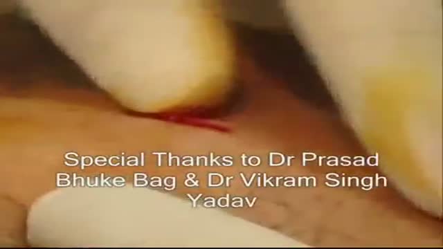
Watch that video to know How Does Circumcision Affect Your Sexual Functions ?
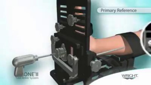
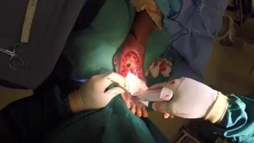
TAA is performed either under general anesthetic or nerve block. A tourniquet is used at the time of surgery to control bleeding and improve visualization during the surgery. The ankle is approached from the front or the side depending on the type of implant being used. Bone is then cut, allowing for placement of the metal and plastic components that re-create the ankle joint. Sometimes the patient will have a tight calf muscle or tight Achilles tendon that needs to be lengthened to improve range of motion of the ankle. The wounds are then closed using stitches or staples, and a splint is applied. A period of non-weightbearing in either a cast or cast boot is necessary to allow the implants to heal in place.
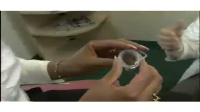
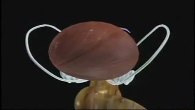
Cervical Cap for Birth Control
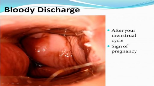
Watch that video to know the Types of Female Genital discharge
Watch that video of A man with one inch-wide hole in his face
Robotic Prostatectomy: Cornell Athermal Robotic Technique

Emboli Formation in Artery
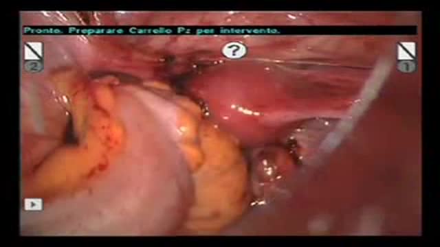
Robot-Assisted Laparoscopic Rectal resection for Endometriosis.Operation performed by D.Vitobello, director of divisione of Gynaecology, and G.Baldazzi,director of Surgical department. Abano Terme Hospital Padova (Italy)
Digital Local Anaesthesia
A video showing the clinical and physical medical examination of the back,the axilla and the lungs
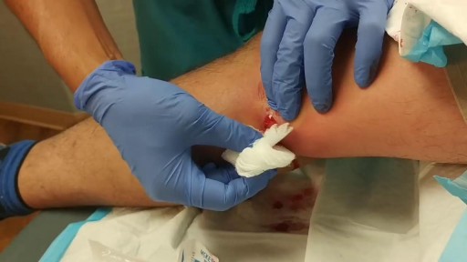
Popping and draining a leg abscess
Compromise of the blood supply from microvascular disease, often in association with lack of sensation because of neuropathy, predisposes persons with diabetes mellitus to foot infections. These infections span the spectrum from simple, superficial cellulitis to chronic osteomyelitis. Diabetic foot infections typically take one of the following forms: Cellulitis Deep-skin and soft-tissue infections Acute osteomyelitis Chronic osteomyelitis Cellulitis Tender, erythematous, nonraised skin lesions are present, sometimes with lymphangitis Lymphangitis suggests group A streptococcal infection Bullae are typical of Staphylococcus aureus infection, but occasionally occur with group A streptococci
The procedure of Suprapubic Cystostomy
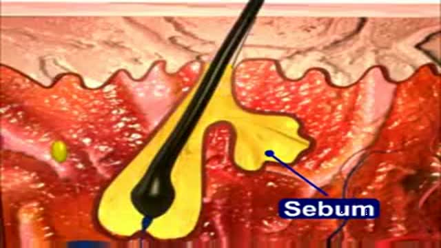
This 3D Animation video shows the process of acne formation and its treatment
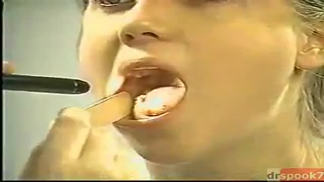
The exam should be performed in an orderly fashion as follows: 1. Have the patient stick out their tongue so that you can examine the posterior pharynx (i.e. the back of the throat). Ask the patient to say "Ah", which elevates the soft palate, giving you a better view. If you are still unable to see, place the tongue blade � way back on the tongue and press down while the patient again says "Ah," hopefully improving your view. This causes some people to gag, particularly when the blade is pushed onto the more proximal aspects of the tongue. It may occasionally be important to determine whether the gag reflex is functional (e.g. after a stroke that impairs CNs 9 or 10; or to determine if a patient with depressed level of consciousness is able to protect their airway from aspiration). This is done by touching a q-tip against the posterior pharynx, uvula or tongue. It is not necessary to do this during your routine exam as it can be quite noxious!
2. Note that the uvula hangs down from the roof of the mouth, directly in the mid-line. With an "Ah," the uvula rises up. Deviation to one side may be caused by CN 9 palsy (the uvula deviates away from the affected side), a tumor or an infection. CN9 Pasly Cranial Nerve 9 Dysfunction: Patient has suffered stroke, causing loss of function of left CN 9. As a result, uvula is pulled towards the normally functioning (ie right) side. 3. The normal pharynx has a dull red color. In the setting of infection, it can become quite red, frequently covered with a yellow or white exudate (e.g. with Strep. Throat or other types of pharyngitis).
4. The tonsils lie in an alcove created by arches on either side of the mouth. The apex of these arches are located lateral to and on a line with the uvula. Normal tonsils range from barely apparent to quite prominent. When infected, they become red, are frequently covered by whitish/yellow discharge. In the setting of a peritonsilar abscess, the tonsils appear asymmetric and the uvula may be pushed away from the affected side. When this occurs, the tonsil may actually compromise the size of the oral cavity, making breathing quite difficult.
5. Look carefully along the upper and lower gum lines and at the mucosa in general, which can appear quite dry if the patient is dehydrated.
6. Examine the teeth to get a sense of general dentition, particularly if the patient has a dental complaint. Pain produced by tapping on a tooth is commonly caused by a root abscess. Tooth Abscess: Tooth abscess involving left molar region. Associated inflammation of left face can clearly be seen. 7. Have the patient stick their tongue outside their mouth, which allows evaluation of CN 12. If there is nerve impairment, the tongue will deviate towards the affected side. Any obvious growths or abnormalities? Ask them to flip their tongue up so that you can look at the underside. If you see something abnormal, grasp the tongue with gauze so that you can get a better look. Left CN 12 Dysfunction: Stroke has resulted in L CN 12 Palsy. Tongue therefore deviates to the left.
8. Make note of any growths along the cheeks, hard palate (the roof of the mouth between the teeth), soft palate, or anywhere else. In particular, patients who smoke or chew tobacco are at risk for oral squamous cell cancer. Any areas which are painful or appear abnormal should also be palpated. Put on a pair of gloves to better explore these regions. What do they feel like? Are they hard? To what extent does a growth involve deeper structures? If the patient feels something that you cannot see, try to get someone else to hold the light source, freeing both your hands to explore the oral cavity with two tongue depressors.
