
- Physical Examination
- Surgical Examination
- Ophthalmology
- Clinical Skills
- Orthopedics
- Surgery Videos
- Laparoscopy
- Pediatrics
- Funny Videos
- Cardiothoracic Surgery
- Nursing Videos
- Plastic Surgery
- Otorhinolaryngology
- Histology and Histopathology
- Neurosurgery
- Dermatology
- Pediatric Surgery
- Urology
- Dentistry
- Oncology and Cancers
- Anatomy Videos
- Health and Fitness
- Radiology
- Anaesthesia
- Physical Therapy
- Pharmacology
- Interventional Radiology
- Cardiology
- Endocrinology
- Gynecology
- Emergency Medicine
- Psychiatry and Psychology
- Childbirth Videos
- General Medical Videos
- Nephrology
- Physiology
- Diet and Food Health
- Diabetes Mellitus
- Neurology
- Women Health
- Osteoporosis
- Gastroenterology
- Pulmonology
- Hematology
- Rheumatology
- Toxicology
- Nuclear Medicine
- Infectious Diseases
- Vascular Disease
- Reproductive Health
- Burns and Wound Healing
- Other
Top videos
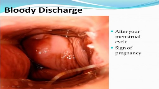

Watch that video to know the Types of Female Genital discharge
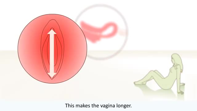
The big bang is the moment when the uterus, vagina, and anus contract simultaneously at 0.8-second intervals. A small orgasm may consist of three to five contractions; a biggie, 10 to 15. Many women report feeling different kinds of orgasms
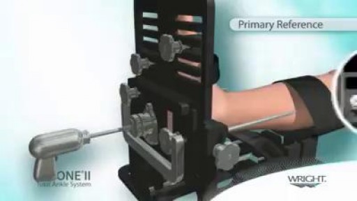
TAA is performed either under general anesthetic or nerve block. A tourniquet is used at the time of surgery to control bleeding and improve visualization during the surgery. The ankle is approached from the front or the side depending on the type of implant being used. Bone is then cut, allowing for placement of the metal and plastic components that re-create the ankle joint. Sometimes the patient will have a tight calf muscle or tight Achilles tendon that needs to be lengthened to improve range of motion of the ankle. The wounds are then closed using stitches or staples, and a splint is applied. A period of non-weightbearing in either a cast or cast boot is necessary to allow the implants to heal in place.
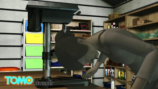
Watch that video of A man with one inch-wide hole in his face
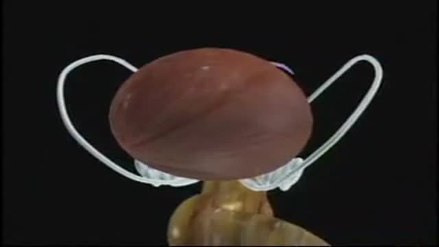
Robotic Prostatectomy: Cornell Athermal Robotic Technique

Emboli Formation in Artery
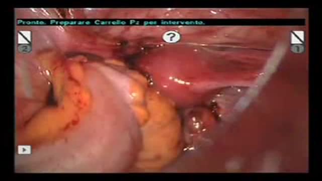
Robot-Assisted Laparoscopic Rectal resection for Endometriosis.Operation performed by D.Vitobello, director of divisione of Gynaecology, and G.Baldazzi,director of Surgical department. Abano Terme Hospital Padova (Italy)
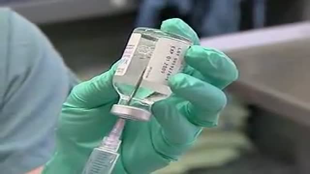
Digital Local Anaesthesia
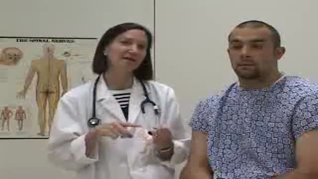
A video showing the clinical and physical medical examination of the back,the axilla and the lungs
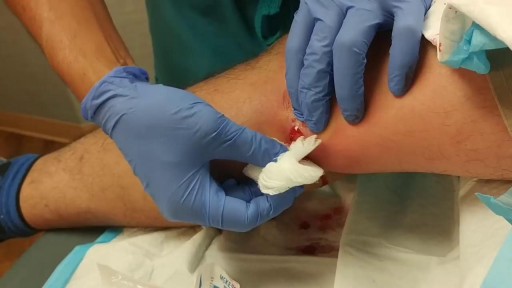
Popping and draining a leg abscess
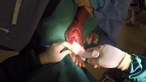
Compromise of the blood supply from microvascular disease, often in association with lack of sensation because of neuropathy, predisposes persons with diabetes mellitus to foot infections. These infections span the spectrum from simple, superficial cellulitis to chronic osteomyelitis. Diabetic foot infections typically take one of the following forms: Cellulitis Deep-skin and soft-tissue infections Acute osteomyelitis Chronic osteomyelitis Cellulitis Tender, erythematous, nonraised skin lesions are present, sometimes with lymphangitis Lymphangitis suggests group A streptococcal infection Bullae are typical of Staphylococcus aureus infection, but occasionally occur with group A streptococci

The exam should be performed in an orderly fashion as follows: 1. Have the patient stick out their tongue so that you can examine the posterior pharynx (i.e. the back of the throat). Ask the patient to say "Ah", which elevates the soft palate, giving you a better view. If you are still unable to see, place the tongue blade � way back on the tongue and press down while the patient again says "Ah," hopefully improving your view. This causes some people to gag, particularly when the blade is pushed onto the more proximal aspects of the tongue. It may occasionally be important to determine whether the gag reflex is functional (e.g. after a stroke that impairs CNs 9 or 10; or to determine if a patient with depressed level of consciousness is able to protect their airway from aspiration). This is done by touching a q-tip against the posterior pharynx, uvula or tongue. It is not necessary to do this during your routine exam as it can be quite noxious!
2. Note that the uvula hangs down from the roof of the mouth, directly in the mid-line. With an "Ah," the uvula rises up. Deviation to one side may be caused by CN 9 palsy (the uvula deviates away from the affected side), a tumor or an infection. CN9 Pasly Cranial Nerve 9 Dysfunction: Patient has suffered stroke, causing loss of function of left CN 9. As a result, uvula is pulled towards the normally functioning (ie right) side. 3. The normal pharynx has a dull red color. In the setting of infection, it can become quite red, frequently covered with a yellow or white exudate (e.g. with Strep. Throat or other types of pharyngitis).
4. The tonsils lie in an alcove created by arches on either side of the mouth. The apex of these arches are located lateral to and on a line with the uvula. Normal tonsils range from barely apparent to quite prominent. When infected, they become red, are frequently covered by whitish/yellow discharge. In the setting of a peritonsilar abscess, the tonsils appear asymmetric and the uvula may be pushed away from the affected side. When this occurs, the tonsil may actually compromise the size of the oral cavity, making breathing quite difficult.
5. Look carefully along the upper and lower gum lines and at the mucosa in general, which can appear quite dry if the patient is dehydrated.
6. Examine the teeth to get a sense of general dentition, particularly if the patient has a dental complaint. Pain produced by tapping on a tooth is commonly caused by a root abscess. Tooth Abscess: Tooth abscess involving left molar region. Associated inflammation of left face can clearly be seen. 7. Have the patient stick their tongue outside their mouth, which allows evaluation of CN 12. If there is nerve impairment, the tongue will deviate towards the affected side. Any obvious growths or abnormalities? Ask them to flip their tongue up so that you can look at the underside. If you see something abnormal, grasp the tongue with gauze so that you can get a better look. Left CN 12 Dysfunction: Stroke has resulted in L CN 12 Palsy. Tongue therefore deviates to the left.
8. Make note of any growths along the cheeks, hard palate (the roof of the mouth between the teeth), soft palate, or anywhere else. In particular, patients who smoke or chew tobacco are at risk for oral squamous cell cancer. Any areas which are painful or appear abnormal should also be palpated. Put on a pair of gloves to better explore these regions. What do they feel like? Are they hard? To what extent does a growth involve deeper structures? If the patient feels something that you cannot see, try to get someone else to hold the light source, freeing both your hands to explore the oral cavity with two tongue depressors.
The procedure of Suprapubic Cystostomy
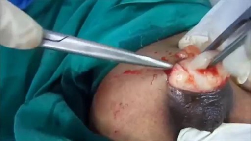
Watch that A Big Size Fibroadenoma Removal Under Local Anesthesia
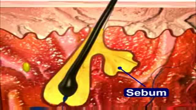
This 3D Animation video shows the process of acne formation and its treatment
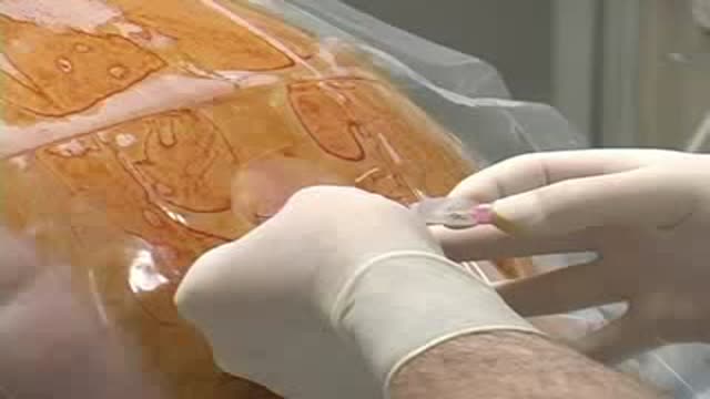
Thoracic Epidural Placement Paramedian Approach
With an Ophthalmoscope, light is shone into the eye and the retina and the optic nerve is examined. This is called as Examination of the Fundus. This is what the eye-doctor sees when he peeps into your eye! Through the transparent cornea, into the dark interior. The Fundus Exam When he looks into the eye with the Ophthalmoscope, he sees a orange glowing interior. That is the retina. The retina is actually transparent. It appears bright because of blood vessels in the choroid layer below. It is like looking at your ear against the bright sunlight. The yellow circle is the Optic Nerve, the cable of vision! A red, shiny dot attracts attention. That is the macula. If indicated, the exam of periphery of the retina is done with an Indirect ophthalmoscope. The ophthalmologist wears this instrument on the head and focuses the light into the eye with a lens held in his hand. This is usually done in a dark room.
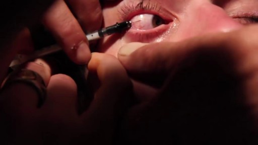
Eye Sclera Tattoo Procedure
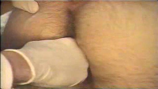
ectal exam is an internal examination of the rectum such as by a physician or other healthcare professional.
The digital rectal examination (DRE, Latin palpatio per anum or PPA) is a relatively simple procedure. The patient is placed in a position where the anus is accessible and relaxed (lying on the side, squatting on the examination table, bent over the examination table, etc). The physician inserts a gloved and lubricated finger into the rectum through the anus and palpates the insides.
The DRE is inadequate as a screening tool for colorectal cancer because it examines less than 10% of the colorectal mucosa; colonoscopy is preferred. However, it's an important part of a general examination, as many tumors or other diseases are made manifest in the distal part of the rectum.
This examination may be used: * for the diagnosis of rectal tumors and other forms of cancer; * in males, for the diagnosis of prostatic disorders, notably tumors and benign prostatic hyperplasia; * for the diagnosis of appendicitis or other examples of an acute abdomen (i.e. acute abdominal symptoms indicating a serious underlying disease); * for the estimation of the tonicity of the anal sphincter, which may be useful in case of fecal incontinence or neurologic diseases, including traumatic spinal cord injuries; * in females, for gynecological palpations of internal organs * for examination of the hardness and color of the feces (ie. in cases of constipation, and fecal impaction); * prior to a colonoscopy or proctoscopy. * to evaluate haemorrhoids
The DRE is frequently combined with an FOBT (fecal occult blood test), which may be useful for diagnosing the etiology of an anemia and/or confirming a gastrointestinal bleed.
Sometimes proctoscopy may also be part of a rectal examination.
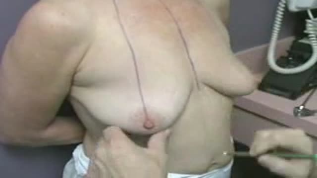
The Stevens Laser Bra works to create a more permanent breast lift for women with sagging breasts, and can be combined with other surgery at Marina Plastic Surgery in Los Angeles, including breast augmentation or reduction, to create a long-lasting, natural transformation of the breast area.
