- Physical Examination
- Surgical Examination
- Ophthalmology
- Clinical Skills
- Orthopedics
- Surgery Videos
- Laparoscopy
- Pediatrics
- Funny Videos
- Cardiothoracic Surgery
- Nursing Videos
- Plastic Surgery
- Otorhinolaryngology
- Histology and Histopathology
- Neurosurgery
- Dermatology
- Pediatric Surgery
- Urology
- Dentistry
- Oncology and Cancers
- Anatomy Videos
- Health and Fitness
- Radiology
- Anaesthesia
- Physical Therapy
- Pharmacology
- Interventional Radiology
- Cardiology
- Endocrinology
- Gynecology
- Emergency Medicine
- Psychiatry and Psychology
- Childbirth Videos
- General Medical Videos
- Nephrology
- Physiology
- Diet and Food Health
- Diabetes Mellitus
- Neurology
- Women Health
- Osteoporosis
- Gastroenterology
- Pulmonology
- Hematology
- Rheumatology
- Toxicology
- Nuclear Medicine
- Infectious Diseases
- Vascular Disease
- Reproductive Health
- Burns and Wound Healing
- Other
Top videos
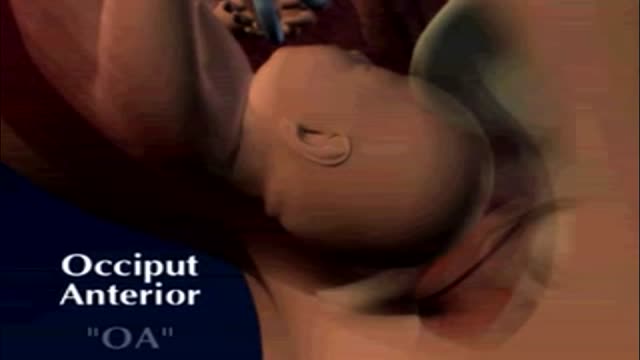
fetal position in womb at 34 weeks fetal position in womb week by week fetal position in womb at 19 weeksUnborn babies toss and turn and hold many different positions within the womb during the gestation period; pregnant women everywhere will attest to the fact that their children always start up the gymnastics at bedtime.

Colposcopy (kol-POS-kuh-pee) is a procedure to closely examine your cervix, vagina and vulva for signs of disease. During colposcopy, your doctor uses a special instrument called a colposcope. Your doctor may recommend colposcopy if your Pap test has shown abnormal results.

If you’re wondering ‘what’s the cause of my knee pain?’ or ‘what kind of knee pain do I have?’ the position of your knee pain can often tell you what type of knee pain you have.
You confirm this if you know the common symptoms an aggravations for each type of knee problem. So if you want to know ‘why my knee hurts’... here’s a quick look at the most common type of knee problems...
Patellofemoral Pain Syndrome (Or Runner’s Knee) (Old Name: Chondromalacia Patellae)
Infrapatellar Fat Pad Syndrome (Hoffa's Syndrome)
Patella Tendonitis (Jumper’s Knee)
Prepatellar Bursitis
Osgood-Schlatter Disease
Meniscus Tear
Medial Collateral Ligament Tear
Osteoarthritic Knee Pain
Pes Anserine Bursitis.
Iliotibial Band Syndrome
Quadriceps Tendinopathy
Popliteus Strain
Baker’s Cyst
ACL Or PCL Tear/Rupture
---------------------------------------
Check out my channel...
https://youtube.com/@BodyFixExercises
OTHER VIDEOS:
How To Fix Pain In The Front Of The Knee… (Runner's Knee) https://youtu.be/g0qmx_0enAA
Knee Strengthening Exercises To Prevent Knee Pain
https://youtu.be/Pk-ae_lyx7M
How To Treat Patellar Tendinopathy (Jumper’s Knee) & Quadriceps Tendinopathy
https://youtu.be/MkPwsb-rQwU
---------------------------------------
#bodyfixexercises #kneepainrelief #kneepain

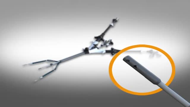
What is vascular access? What are the different types of accesses for hemodialysis? Does vascular access require surgery? Adina Voiculescu, M.D., FASDIN, General and Interventional Nephrologist at Brigham and Women's Hospital and Assistant Professor at Harvard Medical School, discusses the different types of vascular access, such as AV fistulas and AV grafts, and how to stay healthy while on hemodialysis.
Subscribe Link: https://www.youtube.com/channe....l/UCYrLjATd88gPwIKnt
0:00 - Intro
0:29 - Peritoneal dialysis & Hemodialysis
0:44 - Types of access to perform dialysis
1:48 - Recommendations
About Mass General Brigham:
Mass General Brigham combines the strength of two world-class academic medical centers, five nationally ranked specialty hospitals, 11 community hospitals, and dozens of health centers. Our doctors and researchers accelerate medical breakthroughs and drive innovations in patient care. They are leaders in medical education, serving as Harvard Medical School faculty and training the next generation of physicians. Mass General Brigham’s mission is to deliver the best, affordable health care to patients everywhere. Together, we transform the health of our communities and beyond.
#MassGeneralBrigham #MGB #Hemodialysis
Visit Mass General Brigham: https://www.massgeneralbrigham.org/
Find us on social:
Twitter: https://twitter.com/MassGenBrigham
Instagram: https://www.instagram.com/massgeneralbrigham/
Facebook: https://www.facebook.com/MassGeneralBrigham/
LinkedIn: https://www.linkedin.com/compa....ny/mass-general-brig
Mass General Brigham:
https://www.youtube.com/massgeneralbrigham
Hemodialysis: Types of Accesses for Kidney Dialysis and How to Stay Healthy | Mass General Brigham
https://youtu.be/_bxLpudpqnc

Time Management and Work Organization
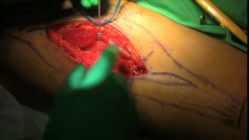
Infected Hernia Mesh Repair Surgery Video

For more information:
http://www.7activestudio.com
info@7activestudio.com
http://www.7activemedical.com/
info@7activemedical.com
7activestudio@gmail.com
Contact: +91- 9700061777,
+91- 9100061777
7 Active Technology Solutions Pvt.Ltd. is an educational 3D digital content provider for K-12. We also customize the content as per your requirement for companies platform providers colleges etc . 7 Active driving force "The Joy of Happy Learning" -- is what makes difference from other digital content providers. We consider Student needs, Lecturer needs and College needs in designing the 3D & 2D Animated Video Lectures. We are carrying a huge 3D Digital Library ready to use.
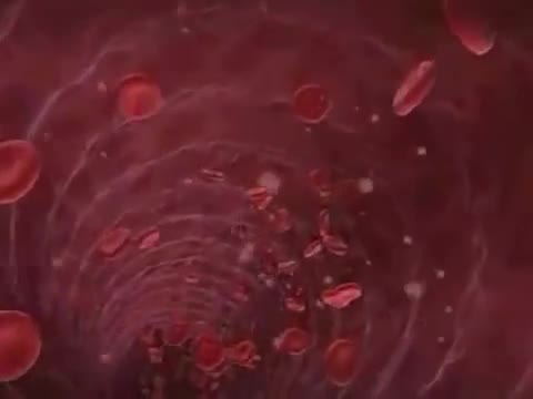
Kidney is most essential organ to remove nitrogenous waste materials from the body. Kidney was damaged by several human activities leads to kidney failure. Once it is damaged it cannot perform basic functions. To overcome this problem one of the best method we follows called hemodialysis. Hemodialysis is a process of removing of nitrogenous waste materials and excess fluids from the blood (collecting from arteries) through tubes containing semi permeable linings in the dialyzer and sending purified blood to the patient's body through veins. It covers the process of hemodialysis in step wise manner. Hemodialysis only performs some basic functions not all those which are performed by natural kidney like reabsorption etc..
Stop Nose Bleeds by Cautery
SPIDER Surgery-- Single Incision Gallbladder Removal

Intestinal obstruction.....
This video is only educational purposes and this is not for entertainment....this is surgery time
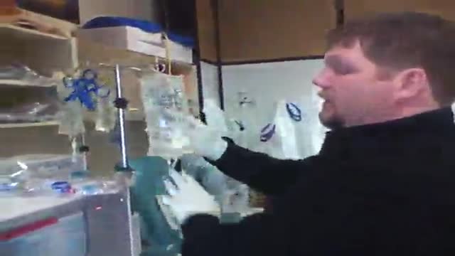
Hemodialysis Machine Setup

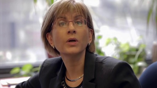
The most reliable clinical sign to detect ascites is checking for bilateral flank dullness. If a patient with ascites is lying supine, fluid accumulates in the flank regions, leading to dullness on percussion. At the same time, the air-filled bowel loops are forced upwards by the free fluid due to buoyancy, resulting in tympanitic percussion. To locate specifically where dullness shifts to tympany, or the air-fluid level, percussion should be performed from the sides towards the middle. To confirm that the dullness is caused by ascites, ask the patient to switch to a lateral decubitus position. If ascites is present, the air-filled bowel loops will shift accordingly and remain at the surface of the fluid. As a result, the air-fluid level will shift as well. This is known as shifting dullness.
Subscribe to AMBOSS YouTube for the latest clinical examination videos, medical student interviews, study tips and tricks, and live webinars!
Free 5 Day Trial: https://go.amboss.com/amboss-YT
Instagram: https://www.instagram.com/amboss_med/
Facebook: https://www.facebook.com/AMBOSS.Med/
Twitter: https://twitter.com/ambossmed
Blog: https://blog.amboss.com/us
#AMBOSSMed #ClinicalExamination

https://bit.ly/3HIStRc #shorts
Coloscopy | Colon Polyp Resection | Polypectomy
Colonoscopies are essential for detecting colorectal abnormalities, including colon polyps. Polypectomy, the surgical removal of these growths, can prevent them from becoming cancerous. This article offers a brief overview of colonoscopies, colon polyps, and polypectomy procedures.
A colonoscopy is an endoscopic examination allowing healthcare providers to visualize the colon and rectum using a colonoscope. The colonoscope, a flexible tube with a camera and light source, helps detect abnormalities, including polyps or tumors.
Colon polyps are abnormal growths arising from the colon's inner lining. While most polyps are benign, some can become malignant. Adenomatous polyps have a higher potential to become cancerous, whereas hyperplastic and inflammatory polyps pose a lower risk.
Polypectomy involves removing colon polyps during a colonoscopy. Two primary techniques include snare polypectomy, using a wire loop to cut the polyp, and cold forceps polypectomy, which employs forceps to grasp and remove smaller polyps.
Following a polypectomy, patients may experience mild discomfort or bleeding. Regular surveillance is crucial to minimize colorectal cancer risk. The frequency of surveillance colonoscopies depends on the number, size, and type of polyps found, as well as the patient's overall risk factors.
Colonoscopies and polypectomies play vital roles in detecting and removing colon polyps, reducing the risk of colorectal cancer, and maintaining optimal colon health.
Do you want to learn more about colon polyps and colonoscopy? check our:
Article @ https://bit.ly/41w5Ooq
For more free resources, find us on Pinterest & Facebook pages:
https://www.pinterest.ca/medicalartsofficial/
https://www.facebook.com/Medicalartsofficial
https://www.youtube.com/@medic....alarts?sub_confirmat
https://www.instagram.com/medicalartsofficial/
https://www.tiktok.com/@medicalarts
#endoscopicsurgery #digestivesystem
coloscopy
polyp
colon polyp
polypectomy
colonoscopy
colon polyp animation
gi endoscopy
@MedicalArts , 2023.

LSD is one of the most potent, mood-changing chemicals. It is manufactured from lysergic acid, which is found in the ergot fungus that grows on rye and other grains. It is produced in crystal form in illegal laboratories, mainly in the United States. These crystals are converted to a liquid for distribution. It is odorless, colorless, and has a slightly bitter taste.

Elbow Exam - Orthopaedic OSCE - Clinical Skills - Dr Gill
The elbow examination is a core skill - in this video, we demonstrate how to perform an elbow EXAM for an Orthopaedic Clinical Skills OSCE, which should be one of the more accessible examination stations for medical students.
For a passing grade in your Clinical Skills OSCE, an elbow assessment should follow the LOOK, FEEL, MOVE approach
Initially looking for erythema, scars, swelling and position
Palpating the elbow - specifically the olecranon, medial and lateral epicondyles, and radial head for heat, oedema and crepitus
Finally assess range of movement with flexion and extension at the elbow, before determining for tennis and golfers' elbows
Watch further orthopaedic examinations for your OSCE revision:
The Elbow - Deep Dive
https://youtu.be/SX5buhtCVDw
The Spine Examination:
https://youtu.be/pJxMHa6SCgU
The Knee examination
https://youtu.be/oyKH4EYfJDM
The Hip examination
https://youtu.be/JC9GKq5nSdQ
The GALS examination
https://youtu.be/5qJaf7gW-B0 - Gait, Arms, Legs, Spine - GALS screen
------------
Please note that there is no ABSOLUTE way to perform a clinical examination. Different institutions and even clinicians will have differing degrees of variations - the aim is the effectively identify medically relevant signs.
However during OSCE assessments. Different medical schools, nursing colleges and other health professional courses will have their own preferred approach to a clinical assessment - you should concentrate on THEIR marks schemes for your assessments.
The examination demonstrated here is derived from Macleods Clinical Examination - a recognised standard textbook for clinical skills.
Some people viewing this medical examination video may experience an ASMR effect
#clinicalskills #Elbow #DrGill

MRI of Fetal Brain Development
Sickle cell anemia (sickle cell disease) is a disorder of the blood caused by an inherited abnormal hemoglobin (the oxygen-carrying protein within the red blood cells). The abnormal hemoglobin causes distorted (sickled) red blood cells.

Parasitic twins: boy carrying dead twin inside him, giant tumor removed - tumors compilation

Click here to subscribe to Dr. Pimple Popper: https://www.youtube.com/@DrPimplePopper/
Join All Access Memberships here:
https://www.youtube.com/channe....l/UCgrsF4TYwmrV0QsXb
Click here to see my favorite POPS:
Most Popular Pops: https://www.youtube.com/playli....st?list=PLJZ_ok3xiAi
Blackheads: https://www.youtube.com/playli....st?list=PLJZ_ok3xiAi
DPOW’s: https://www.youtube.com/playli....st?list=PLJZ_ok3xiAi
Steatocystomas: https://www.youtube.com/playli....st?list=PLJZ_ok3xiAi
Cysts: https://www.youtube.com/playli....st?list=PLJZ_ok3xiAi
Lipomas: https://www.youtube.com/playli....st?list=PLJZ_ok3xiAi
Soft Pops: https://www.youtube.com/playli....st?list=PLJZ_ok3xiAi
Poppin Off' Show with Dr Pimple Popper: https://youtube.com/playlist?l....ist=PLJZ_ok3xiAi_XPY
__
Connect with Dr. Pimple Popper on Social Media:
Follow Dr. Pimple Popper on Instagram: https://bit.ly/3smTOED
Follow Dr. Pimple Popper on TikTok: https://bit.ly/2MXWDM9
Follow Dr. Pimple Popper on Facebook: https://bit.ly/3oU3Rz7
Follow Dr. Pimple Popper on Twitter: https://bit.ly/2MXWDM9
Follow Dr. Sandra Lee on Instagram: https://bit.ly/2LscBO8
Subscribe to SLMD YouTube to never miss a skincare video:
https://www.youtube.com/@SLMDskincare
More ways to connect with me:
Download the Dr. Pimple Popper All Access App:
https://apps.apple.com/us/app/....dr-pimple-popper/id1
Click here to begin free trial of All-Access: https://allaccess.drpimplepopper.com/
Welcome to the world of Dr. Pimple Popper, the one and only Sandra Lee, MD! As a board certified dermatologist, skin cancer surgeon, and cosmetic surgeon, I am a highly sought-after expert in the field of dermatology.
On this channel, you'll find a treasure trove of videos that offer a window into my world.
Hopefully you'll learn about various skin conditions, hair and nail issues, and cutting-edge cosmetic surgery techniques. Whether you're struggling with blackheads, acne, cysts, warts, or looking for Botox, fillers, or liposuction, you'll find helpful advice and information here.
But this channel isn't just about skin care - it's about the amazing people I encounter every day. You'll get to know some of my incredible patients and their stories, and maybe even fall in love with dermatology just as much as I have!
Disclaimer: This video may contain dermatologic surgical and/or procedural content. The content seen in this video is provided only for medical education purposes and is not intended to be a substitute for professional medical advice, diagnosis, or treatment.
#DrPimplePopper #DrSandraLee #Dermatology #SLMD #Skincare