
- Physical Examination
- Surgical Examination
- Ophthalmology
- Clinical Skills
- Orthopedics
- Surgery Videos
- Laparoscopy
- Pediatrics
- Funny Videos
- Cardiothoracic Surgery
- Nursing Videos
- Plastic Surgery
- Otorhinolaryngology
- Histology and Histopathology
- Neurosurgery
- Dermatology
- Pediatric Surgery
- Urology
- Dentistry
- Oncology and Cancers
- Anatomy Videos
- Health and Fitness
- Radiology
- Anaesthesia
- Physical Therapy
- Pharmacology
- Interventional Radiology
- Cardiology
- Endocrinology
- Gynecology
- Emergency Medicine
- Psychiatry and Psychology
- Childbirth Videos
- General Medical Videos
- Nephrology
- Physiology
- Diet and Food Health
- Diabetes Mellitus
- Neurology
- Women Health
- Osteoporosis
- Gastroenterology
- Pulmonology
- Hematology
- Rheumatology
- Toxicology
- Nuclear Medicine
- Infectious Diseases
- Vascular Disease
- Reproductive Health
- Burns and Wound Healing
- Other
Top videos
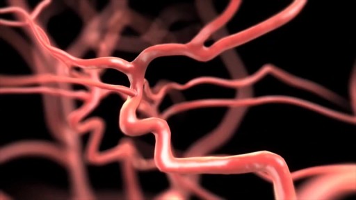

Narrated animation of stroke intervention. Video supplied by Covidien, showing the Solitaire mechanical thrombectomy device, which was the first FDA-approved device for such an indication.

Miracle baby's heart beats outside her chest

Conduct disorder (CD) is a mental disorder diagnosed in childhood or adolescence that presents itself through a repetitive and persistent pattern of behavior in which the basic rights of others or major age-appropriate norms are violated. These behaviors are often referred to as "antisocial behaviors."

G-Shot (G-Spot Amplification)
Overview HIV is a virus that affects the immune system, specifically the CD4 cells. The CD4 cells help protect the body from illness. Unlike other viruses that the immune system can fight off, HIV can’t be eliminated by the immune system. The symptoms of HIV can vary greatly from person to person. No two people with HIV will likely experience the exact same symptoms. However, HIV will generally follow this pattern: acute illness asymptomatic period advanced infection Acute illness Approximately 80 percent of people who contract HIV experience flu-like symptoms within two to four weeks. This flu-like illness is known as acute HIV infection. Acute HIV infection is the primary stage of HIV and lasts until the body has created antibodies against the virus. The most common symptoms of this stage of HIV include: body rash fever sore throat severe headaches Less common symptoms may include: fatigue swollen lymph nodes ulcers in the mouth or on the genitals muscle aches joint pain nausea and vomiting night sweats Symptoms typically last one to two weeks. Anyone who has these symptoms and thinks they may have contracted HIV should consider scheduling an appointment with their healthcare provider to get tested. Symptoms specific to men Symptoms of HIV are generally the same in women and men. One HIV symptom that is unique to men is an ulcer on the penis. HIV may lead to hypogonadism, or poor production of sex hormones, in either sex. However, hypogonadism’s effects on men are easier to observe than its effects on women. Symptoms of low testosterone, one aspect of hypogonadism, can include erectile dysfunction (ED).

TV interview with Adina Nack, Ph.D. about her own cervical HPV experiences, STD research, her new book (Damaged Goods? Women Living with Incurable Sexually Transmitted Diseases), and women's lives after genital warts, HPV and herpes infections. More info is available on STDdatings.com, which is the official STD dating & support site.

Systemic Lupus Erythematosus Animation 3d
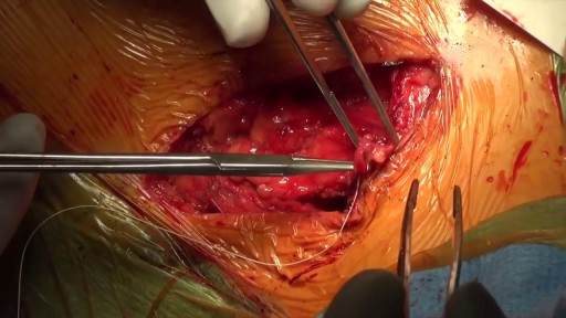
Thoracic outlet syndrome affects the space between the collarbone and first rib (thoracic outlet). Common causes include trauma, repetitive injuries, pregnancy, and anatomical defects, such as having an extra rib. Symptoms include pain in the shoulders and neck and numbness, weakness, and coldness in the fingers. Treatment involves physical therapy and pain relief. In rare cases, surgery may be needed to relieve the compression.
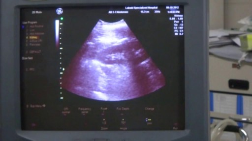
A biopsy is a diagnostic test that involves collecting small pieces of tissue, usually through a needle, for examination with a microscope. A kidney biopsy can help in forming a diagnosis and in choosing the best course of treatment.
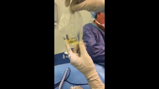
As you can see I access the left implant from the periareolar incisions which I made at the lower portion of the areola. As I entered the capsule and begin to remove the implant I noticed a lot of fluid surrounding the implant. Right away I know this is a rupture and that the mammogram was incorrect. Mammograms are very helpful in detecting cancer but often not ruptures. When implants rupture, it is important to have them replaced as soon as possible to avoid excessive scarring in the breasts. If too much scar tissue has accumulated around the deflated implant, it becomes difficult to create a normal breast shape in the future. Therefor know the signs of a ruptured implant such as, painful to touch, visible asymmetry or loss of integrity to the bag. For more information please visit: www.drlinder.com
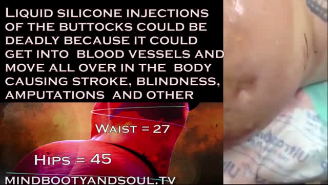
2016 marks 10 years when illegal injections started to gain momentum and become a popular alternative to butt implants. The Brazilian butt lift wasn't well know at the time but the goal of finding an unlicensed person to inject a foreign substance into the body was in high demand.

The E.coli bacteria has claimed 14 lives and infected more than 300 hundred in the country. It has now spread to other European states.
Facing an increasing number of cases, German health authorities warned people to avoid eating raw cucumber, tomatoes and lettuce.
Reinhard Burger, President of Robert Koch Institute, said, "As for the present situation there is no reason to give the all-clear yet and it is possible the original source of the infection is still active and could lead to further infections."
The first cases of the EHEC outbreak were noticed in Northern Germany, but infections are now spreading across the country.
Cases haves spiked compared to other years, and are still rising.
Daniel Bahr, German Health Minister, said, "The result is that we unfortunately still have to prepare for a rising number of cases. Exercising caution is still recommended and we ask our citizens to be particularly careful. "
But, the German government says it's working around the clock to stop the outbreak and clarify how it arose.
Ilse Aigner, German Agriculture Minister, said, "Together, we face a big challenge to piece together hundreds or thousands of mosaic pieces from Germany and abroad into an overall picture that gives us a clear answer on how this terrible infection arose. "
In the meantime, experts are advising not to eat pre-packaged or prepared salad, which may contain the bacteria.
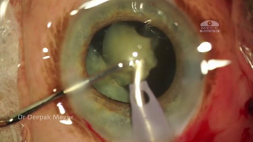
Phacolytic glaucoma usually is associated with a mature or hypermature cataract and typically occurs in elderly patients. Today, phacolytic glaucoma is rare in the United States, found primarily in areas where access to care is poor. Will the increase in the number of under- and uninsured patients lead to an increase in this condition? Evaluation and Diagnosis Signs and symptoms. Patients typically report acute-onset pain, decreased vision, tearing and photophobia. Examination will reveal injection, corneal edema, elevated IOP, anterior chamber reaction with or without pseudohypopyon, particles on the lens capsule and anterior capsule wrinkling. Patient history. The duration of symptoms should be elicited; a delayed presentation of more than five days since onset can result in glaucomatous disc damage and poorer prognosis.¹ The ocular history may reveal that the patient decided against removal of an advanced cataract. Prior intraocular surgery or trauma may have left residual lens material that could cause phacoanaphylactic glaucoma or exacerbate infectious endophthalmitis. Visual acuity and visual potential should be assessed. Exam essentials. A complete ophthalmologic examination should be done. The eye should be inflamed, and the cornea may be edematous due to the high IOP. The anterior chamber will demonstrate massive inflammation and/ or pseudohypopyon. Gonioscopy is essential; it will help rule out angle closure due to phacomorphic glaucoma or neovascularization of the angle. Assess ment of the posterior pole should be performed to rule out vitreous hemorrhage (which can result in ghost-cell glaucoma) or vitritis (which may be associated with infectious endophthalmitis or panuveitis). If the view to the fundus is obstructed, B-scan ultrasonography also should be performed. Differential diagnosis. The differential diagnosis includes infectious endophthalmitis, phacoanaphylactic glaucoma, inflammatory glaucoma, glaucoma secondary to intraocular tumor, phacomorphic glaucoma, acute-angle closure glaucoma and neovascular glaucoma. Management Medication. Medical management is used to temporarily control the glaucoma and inflammation. Initial treatment consists of hyperosmotic agents, aqueous suppressants, anti-inflammatory drugs and cycloplegics. Surgery. Definitive treatment is removal of the lens via extracapsular cataract extraction with or without an IOL. Some ophthalmologists defer placement of an IOL until after the inflammation subsides; however, there is no significant difference in final visual acuity between those patients who did receive an IOL and those who did not.¹ If the phacolytic glaucoma is of long duration (more than seven days), a combined trabeculectomy may be needed to prevent postoperative IOP spikes.² In eyes with hypermature Morgagnian cataracts, one must be especially careful, as the capsule is fragile, the zonules are weak and the view is difficult due to the white, milky cortex. Vision limited to light perception on presentation is not a contraindication to performing cataract extraction. Surgical Tips For a planned extracapsular cataract extraction with a posterior chamber IOL, fashion a superior fornix-based conjunctival flap.³ Make a partial-thickness incision along the sclerolimbal junction superiorly for 120 degrees with a No. 69 blade. Forty-five degrees away, a paracentesis should be done to decompress the eye. The anterior chamber fluid can be withdrawn for analysis, to look for macrophages and high molecular-weight proteins. Inject balanced salt solution in a cannula to wash out any residual particulate matter, then inject Healon or viscoelastic into the anterior chamber. Make an incision entering the anterior chamber at the 12 o’clock position with a keratome. A 26-gauge cystotome mounted on a syringe is then introduced through the 12 o’clock incision and used to puncture the capsular bag. The milky cortex should be aspirated as much as possible, until the nucleus is visible. Withdraw the needle through the keratome incision, then inject Healon through the 12 o’clock incision into the capsular bag. Next, enlarge the corneoscleral keratome incision with curved Westcott scissors to 120 degrees. Perform a partial V-shaped capsulotomy; this can be done either with the cystotome or with an angled Vannas scissors. Place viscoelastic under the nucleus to float the nucleus and sever any adhesions between the nucleus and the capsule. The nuclear portion of the lens can then be removed with an irrigating vectis (lens loop) with or without gentle pressure at the inferior limbus (6 o’clock). Irrigate and aspirate the residual cortex with the Simcoe cannula. Inspect the capsular bag; if it is intact, place a posterior chamber IOL into the bag. Close the incision with several interrupted 10-0 monofilament nylon sutures and reattach the conjunctival flap. Potential Sequelae and Prognosis Postoperatively, the patient should be managed with topical steroids and/or aqueous suppressants and hyperosmotics if necessary. Vitreous opacification behind the posterior capsule occurs in a small percentage of eyes. These vitreous opacities are typically absorbed by one to two weeks postoperatively. IOP usually is controlled without antiglaucoma medications after the cataract removal. A detailed glaucoma evaluation (including repeat gonioscopy to assess for peripheral anterior synechiae, visual field and optic nerve status) should be done to assess the extent of glaucomatous damage. The prognosis is dependent on the duration of elevated IOP, PAS and optic nerve damage. In one study, patients who were older than 60 and whose glaucoma was present for more than five days did significantly worse than a comparison group of younger individuals with shorter disease duration.

Brushing flossing and rinsing -the winning combination for preventing gingivitis

Brain Worm Infection
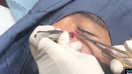
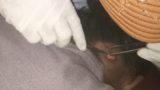
Nose Cyst Extraction
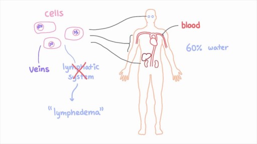
The lymphatic system is a network of specialized vessels (lymph vessels) throughout the body whose purpose is to collect excess lymph fluid with proteins, lipids, and waste products from the tissues. This fluid is then carried to the lymph nodes, which filter waste products and contain infection-fighting cells called lymphocytes. The excess fluid in the lymph vessels is eventually returned to the bloodstream. When the lymph vessels are blocked or unable to carry lymph fluid away from the tissues, localized swelling (lymphedema) is the result.

Benzodiazepines are a class of agents that work on the central nervous system, acting selectively on gamma-aminobutyric acid-A (GABA-A) receptors in the brain. GABA is a neurotransmitter that inhibits or reduces the activity of nerve cells (neurons) within the brain. Benzodiazepines open GABA-activated chloride channels, and allow chloride ions to enter the neuron. This makes the neuron negatively charged and resistant to excitation.
