- Physical Examination
- Surgical Examination
- Ophthalmology
- Clinical Skills
- Orthopedics
- Surgery Videos
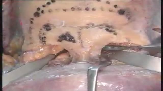
- Laparoscopy
- Pediatrics
- Funny Videos
- Cardiothoracic Surgery
- Nursing Videos
- Plastic Surgery
- Otorhinolaryngology
- Histology and Histopathology
- Neurosurgery
- Dermatology
- Pediatric Surgery
- Urology
- Dentistry
- Oncology and Cancers
- Anatomy Videos
- Health and Fitness
- Radiology
- Anaesthesia
- Physical Therapy
- Pharmacology
- Interventional Radiology
- Cardiology
- Endocrinology
- Gynecology
- Emergency Medicine
- Psychiatry and Psychology
- Childbirth Videos
- General Medical Videos
- Nephrology
- Physiology
- Diet and Food Health
- Diabetes Mellitus
- Neurology
- Women Health
- Osteoporosis
- Gastroenterology
- Pulmonology
- Hematology
- Rheumatology
- Toxicology
- Nuclear Medicine
- Infectious Diseases
- Vascular Disease
- Reproductive Health
- Burns and Wound Healing
- Other
Top videos
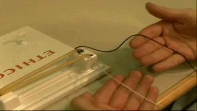
One Handed Knot Tie with Right Hand
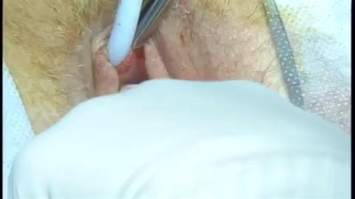
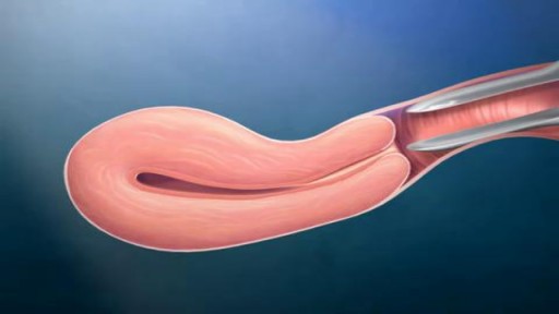
An intrauterine device (IUD), also known as intrauterine contraceptive device (IUCD or ICD) or coil, is a small, often T-shaped birth control device that is inserted into a woman's uterus to prevent pregnancy. IUDs are one form of long-acting reversible birth control (LARC).
lesions at the anterior skull base invading the paranasal area and the paracavernous area can be reached without brain retraction by the shown subfrontal approach. it enables to control the paranasal sinus, optic nerve, periorbital tissue, carotid artery and pituary gland. reconstruction is not easy... but cosmetically appealing. CSF leaks are rare with the use of fascia lata and tissucol ( fibrin glue). osseous reconstruction is done by microsrews and calciumpyrophosphate ( norian, synthes).
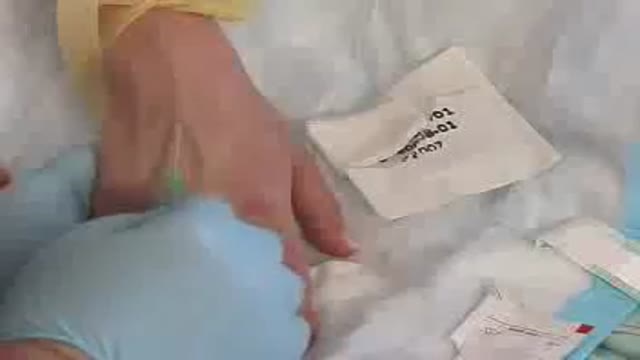
Starting an IV
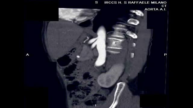
J Vasc Surg. 2009 Jul;50(1):134-9. Celiac artery compression syndrome managed by laparoscopy. Baccari P, Civilini E, Dordoni L, Melissano G, Nicoletti R, Chiesa R. Department of General Surgery, Scientific Institute San Raffaele University Hospital, Milan, Italy. paolo.baccari@hsr.it Abstr...
act OBJECTIVE: Celiac artery compression syndrome (CACS) is an unusual condition caused by abnormally low insertion of the median fibrous arcuate ligament and muscular diaphragmatic fiber resulting in luminal narrowing of the celiac trunk. Surgical treatment is the release of the extrinsic compression by division of the median arcuate ligament overlying the celiac axis and skeletonization of the aorta and celiac trunk. The laparoscopic approach has been recently reported for single cases. Percutaneous transluminal angioplasty (PTA) and stenting of the CA alone, before or after the surgical relief of external compression to the celiac axis, has also been used. We report our 7-year experience with the laparoscopic management of CACS caused by the median arcuate ligament. METHODS: Between July 2001 and May 2008, 16 patients (5 men; mean age, 52 years) were treated. Diagnosis was made by duplex ultrasound scan and angiogram (computed tomography [CT] or magnetic resonance). The mean body mass index of the patients was 21.2 kg/m(2). One patient underwent laparoscopic surgery after failure of PTA and stenting of the CA, and two patients after a stenting attempt failed. RESULTS: All procedural steps were laparoscopically completed, and the celiac trunk was skeletonized. The laparoscopic procedures lasted a mean of 90 minutes. Two cases were converted to open surgery for bleeding at the end of the operation when high energies were used. The postoperative course was uneventful. Mean postoperative hospital stay was 3 days. On follow-up, 14 patients remained asymptomatic, with postoperative CT angiogram showing no residual stenosis of the celiac trunk. One patient had restenosis and underwent aortoceliac artery bypass grafting after 3 months. Another patient had PTA and stenting 2 months after laparoscopic operation. All patients reported complete resolution of symptoms at a mean follow-up of 28.3 months. CONCLUSIONS: The laparoscopic approach to CACS appears to be feasible, safe, and successful, if performed by experienced laparoscopic surgeons. PTA and stenting resulted in a valid complementary procedure only when performed after the release of the extrinsic compression on the CA. Additional patients with longer follow-up are needed.
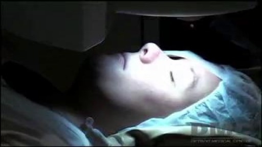
Lasik eye surgery at the Detroit Medical Center's Advanced Laser and Clear Vision Center offer patients pain-free, life-changing procedures that correct nearsightedness, farsightedness, and astigmatism. ~ Detroit Medical Center
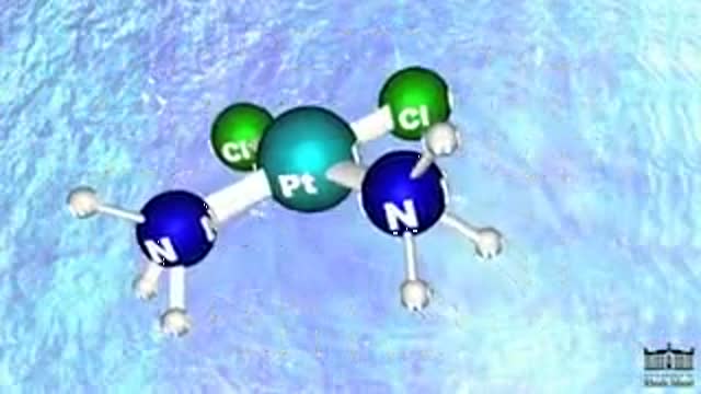
Cisplatin is in a class of drugs known as platinum-containing compounds used to treat various types of cancers including metastatic testicular and ovarian tumors. The molecule was first discovered in 1845, but did not receive FDA approval until 1978. Today it is known as the "penicillin of cancer drugs," because it is so effective for many different cancers. There are three key players involved in Cisplatin's mechanism: (1) Cisplatin, (2) DNA (3) and an HMG Protein. Most Cisplatin enters the body through active transport, but some molecules are passively defused through the cell membrane. Once in the nucleus, Cisplatin can form an adduct with two consecutive guanine bases within a strand of DNA. The molecule loses its chlorine atoms in exchange for the nitrogen atoms of the target guanines. Cisplatin can bond more tightly with nitrogen because nitrogen balances the platinum charge more effectively than chlorine. It is this adduct-induced DNA bend that allows binding of proteins which contain the high mobility group, HMG domain. Once the protein is bound to the DNA, it inserts a wedge-like phenyl group of phenylalanine 37 into the widened minor groove created by the bend. The tightly bound HMG protein causes destacking of the nucleotide bases, resulting in the DNA helix becoming kinked. In this way, Cisplatin can be thought of as a monkey wrench in the DNA repair system. With the HMG protein bound to the DNA, the modified strand is not repaired properly and so the cell dies. The success of Cisplatin depends on its ratio of efficacy between cancerous and healthy cells.
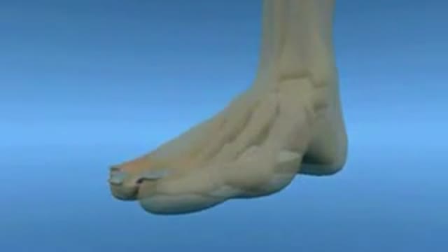
A "Hallux Valgus" or "Hallux Abducto-Valgus" deformity, is commonly referred to as a "Bunion." This describes a pathological condition involving the position of the "hallux" in relation to the first metatarsal.
A bunion deformity can clinically present with a variety of characteristics. The foot itself may present with a wide splaying of the forefoot and a painful bump on the medial aspect of the first metatarsal phalangeal joint. In addition, the hallux may be abducted from the midline of the body, with a valgus rotation in the frontal plane.
A radiographic analysis of a bunion deformity in the Anterior/Posterior or Dorsal/Plantar view will reveal a variety of pathological components. Most notably so, is the exaggerated inter-metatarsal angle between the first and second metatarsal. This may be accompanied by a displacement of the first metatarsal from its position over the sesamoids, such that the metatarsal demonstrates a medial alignment away from the sesamoids which lie to the lateral side.
In some cases, the proximal articular set angle at the head of the first metatarsal may be off-set. This "PASA" is one of the factors which determines the position of the proximal phalanx on the metatarsal during movement as well as at rest.
Although conservative care may involve shoe modifications, padding, strapping, and custom orthosis; surgical reconstruction may be required to alleviate painful and immobilizing bunion conditions.
Soft tissue components of the bunion deformity are primarily addressed by means of a capsular modification, as well as a tenotomy of the adductor tendon at its insertion on the base of the proximal phalanx. The fibular sesamoid may be repositioned by a release of the surrounding ligaments.
Surgical management of the bone or osseous components of a bunion deformity will commonly include an osteotomy and correction to re-establish a more functional position of the first metatarsal within the forefoot. This capital fragment of bone is held in place with hardware fixation in order to secure a proper alignment during the healing phase, thus allowing the hallux to return to a more functionally useful position in the sagittal plane.

Endoscopic Brain Surgery, third Ventriculostomy

A new procedure, laparoscopic hysterectomy, means there's no reason for a woman to undergo an invasive abdominal hysterectomy unless she has a severe medical problem. ~ Detroit Medical Center
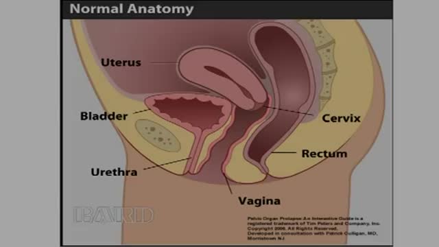
-Rectocele is a relatively common condition in older women and is characterized by the displacement of the rectum through posterior vaginal wall defect(s). The condition is typically caused by damage to the rectovaginal septum incurred during vaginal childbirth and is exacerbated by periodic increases in intraabdominal pressure (e.g., when laughing or coughing) and the effects of gravity. Women with symptomatic rectoceles who are poor surgical candidates may be treated with pessaries, which are structures designed to support the vaginal wall. Pessaries should only be used in conjunction with vaginal
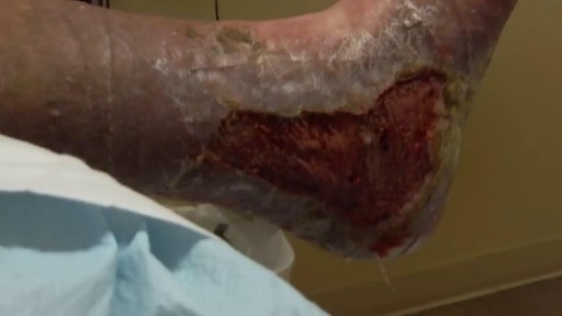
Debridement is the removal of necrotic tissue, foreign debris, bacterial growth, callus, wound edge, and wound bed tissue from chronic wounds in order to stimulate the wound healing process. Stimulation of wound healing mediated by debridement is thought to occur by the conversion of a chronic non-healing wound environment to an acute healing environment through the removal of cells that are not responsive to endogenous healing stimuli. Debridement is used commonly in standard wound treatment of diabetic foot ulcers (DFUs). Methods of debridement include surgery (sharp debridement), chemical debridement (antiseptics, polysaccharide beads, pastes), autolytic (hydrogels, hydrocolloids and transparent films), biosurgery (maggots), mechanical (hydrodebridement), and biochemical debridement (enzyme preparations). Callus is a buildup of keratinized skin formed under conditions of repeated pressure or friction and may contribute to ulcer formation by creating focal areas of high plantar pressure. The debridement of callus has been proposed to be relevant for both treatment and prevention of DFU. The purpose of this report is to retrieve and review existing evidence of comparative clinical effectiveness of different methods of debridement for the treatment of DFUs. Additionally examined in this report is the clinical effectiveness for treatment and prevention of DFU using callus debridement. Cost-effectiveness, and existing debridement guidelines for the treatment of DFUs will also be reviewed.

Life Before Birth
Needle Insertion Transversus Abdominus Block

Inspection of the mouth

After the diagnosis of primary melanoma of pectoral region had been established, the patient was referred to lymphoscintigraphy with gamma camera (techencium; nanno colloid). Two hours after the administration of the contrast medium, the operation commenced. During the operation the primary tumor wa...s excised, and the sentinel node was detected with the use of gamma probe and also excised.
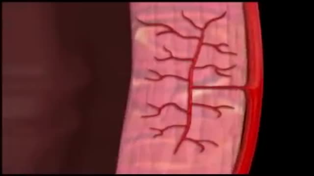
Coronary circulation is the circulation of blood in the blood vessels of the heart muscle (myocardium). The vessels that deliver oxygen-rich blood to the myocardium are known as coronary arteries. The vessels that remove the deoxygenated blood from the heart muscle are known as cardiac veins.
A Medical Video showing an overview of the endocrine and gland system of the human body
Urological surgeons have become proficient at performing complex pelvic urological procedures, such as radical prostatectomy, using the laparoscopic approach. Declan Murphy and Daniel Moon share their experience of four less common procedures they have performed recently using laparoscopic techniques. These include: excision of a urachal cyst; partial cystectomy for endometriosis (combined endoscopic-laparoscopic approach); repair of an intra-peritoneal bladder rupture; and repair of a ureteric injury (combined endoscopic-laparoscopic approach).
