- Physical Examination
- Surgical Examination
- Ophthalmology
- Clinical Skills
- Orthopedics
- Surgery Videos
- Laparoscopy
- Pediatrics
- Funny Videos
- Cardiothoracic Surgery
- Nursing Videos
- Plastic Surgery
- Otorhinolaryngology
- Histology and Histopathology
- Neurosurgery
- Dermatology
- Pediatric Surgery
- Urology
- Dentistry
- Oncology and Cancers
- Anatomy Videos
- Health and Fitness
- Radiology
- Anaesthesia
- Physical Therapy
- Pharmacology
- Interventional Radiology
- Cardiology
- Endocrinology
- Gynecology
- Emergency Medicine
- Psychiatry and Psychology
- Childbirth Videos
- General Medical Videos
- Nephrology
- Physiology
- Diet and Food Health
- Diabetes Mellitus
- Neurology
- Women Health
- Osteoporosis
- Gastroenterology
- Pulmonology
- Hematology
- Rheumatology
- Toxicology
- Nuclear Medicine
- Infectious Diseases
- Vascular Disease
- Reproductive Health
- Burns and Wound Healing
- Other
Top videos
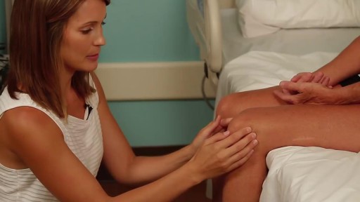
Detailed examination of the joints is usually not included in the routine medical examination. However, joint related complaints are rather common, and understanding anatomy and physiology of both normal function and pathologic conditions is critically important when evaluating the symptomatic patient. By gaining an appreciation for the basic structures and functioning of the joint, you'll be able to "logic" your way thru the exam, even if you can't remember the eponym attached to each specific test!
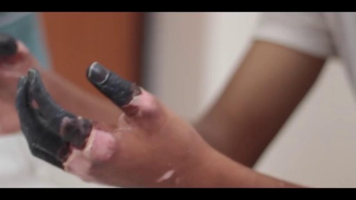
Frostbite is an injury caused by freezing of the skin and underlying tissues. First your skin becomes very cold and red, then numb, hard and pale. Frostbite is most common on the fingers, toes, nose, ears, cheeks and chin. Exposed skin in cold, windy weather is most vulnerable to frostbite. But frostbite can occur on skin covered by gloves or other clothing. Frostnip, the first stage of frostbite, doesn't cause permanent skin damage. You can treat very mild frostbite with first-aid measures, including rewarming your skin. All other frostbite requires medical attention because it can damage skin, tissues, muscle and bones. Possible complications of severe frostbite include infection and nerve damage.
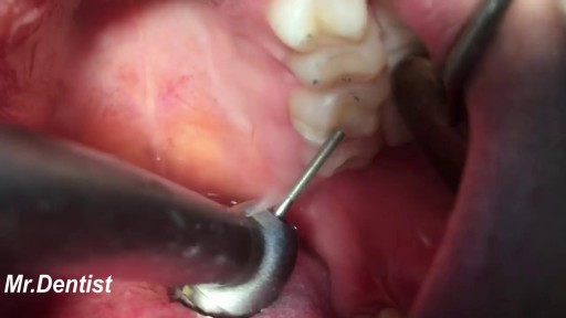
Fillings are a way for dentists to restore a partially decayed tooth. While many people fear the dentist, this procedure is typically quick, effective, and inexpensive. Without fillings, cavities can rapidly worsen. Seeing a dentist regularly can help you to monitor the condition of your teeth and plan for corrective procedures. According to the National Institute of Dental and Craniofacial Research, nearly 93 percent of adults between the ages of 20 and 64 have cavities, and at least 29 percent have decay that is untreated. Dentists can quickly identify tooth decay and then come up with a plan of action that involves filling teeth and restoring adverse conditions. You can do your part by sticking to a solid at-home oral hygiene routine. By simply brushing twice a day with a fluoride-treated toothpaste and flossing regularly, you can prevent the build up of bacteria-rich plaque and eliminate cavity-causing conditions.
Sex reassignment surgery for male-to-female involves reshaping the male genitals into a form with the appearance of, and, as far as possible, the function of female genitalia. Prior to any surgeries, patients usually undergo hormone replacement therapy (HRT), and, depending on the age at which HRT begins, facial hair removal. There are associated surgeries patients may elect to, including facial feminization surgery, breast augmentation, and various other procedures.
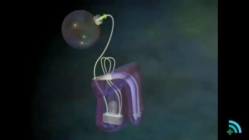
he inflatable penile prosthesis consists of two attached cylinders -- a reservoir and a pump -- which are placed surgically in the body. The two cylinders are inserted in the penis and connected by tubing to a separate reservoir of saline. The reservoir is implanted under the rectus muscles in the lower abdomen.
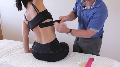
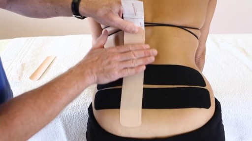
this video he is demonstrating how to apply Kinesiology Tape for a patient that presents with rib or intercostal pain
Dysfunction in the sacroiliac joint, also called the SI joint, can sometimes cause lower back and/or leg pain. Leg pain from sacroiliac joint dysfunction can be particularly difficult to differentiate from radiating leg pain caused by a lumbar disc herniation (sciatica) as they can feel quite similar.
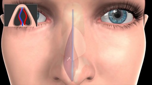
Septoplasty (SEP-toe-plas-tee) is a surgical procedure to correct a deviated septum — a displacement of the bone and cartilage that divides your two nostrils. During septoplasty, your nasal septum is straightened and repositioned in the middle of your nose.
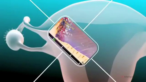
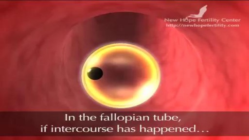
The menstrual cycle is the regular natural change that occurs in the female reproductive system that makes pregnancy possible. The cycle is required for the production of oocytes, and for the preparation of the uterus for pregnancy.
Before ovulation occurs, the average diameter of the dominant follicle is 22 to 24 mm (range 18-36 mm). It is the only marker that can predict ovulation with ease. * In stimulated cycle (hormonal treatment), generally, all or most of the antral follicles grow. The growth rate will be different for each of them.
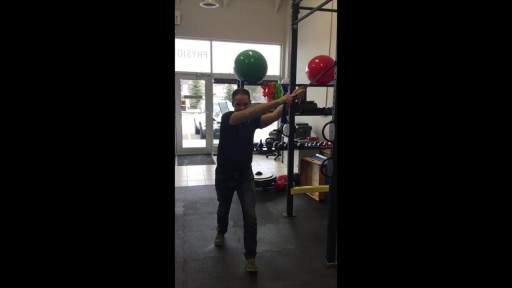
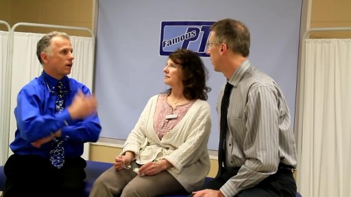
Curious about physiotherapy or wanting to know how to properly perform an exercise? Check us out on Social Media! Facebook: https://www.facebook.com/striveptandperformance/ Instagram: https://www.instagram.com/striveptandperf/ Twitter: https://twitter.com/StrivePTandPerf Blog: http://www.strivept.ca/blog
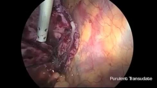
Thoracoscopic Management of Lung Abscess Before Empyema
A computed tomography (CT) scan uses a special X-ray machine to take detailed pictures of the body’s organs and tissues. In a biopsy, a small piece of tissue is removed from your body. This tissue sample is then examined in the lab. A needle biopsy is the safest and easiest way to remove this tissue safely from the body. To do a needle biopsy, the radiologist will insert a needle through your skin and into your tissue. A syringe or an automated needle may be used to take the tissue sample.
There can be a number of psychological triggers that cause nightmares in adults. For example, anxiety and depression can cause adult nightmares. Post-traumatic stress disorder (PTSD) also commonly causes people to experience chronic, recurrent nightmares. Nightmares in adults can be caused by certain sleep disorders
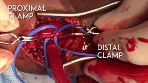
A proper embolectomy should have a good proximal and distal flow to the arteriotomy :)
EKG/ECG Interpretation Explained Clearly

Mammogram are great technologies, however, sometimes it cannot detect many things under our bodies. In this video, Dr. Linder is performing a breast implant removal and revision on a patient who has a rupture breast implants. Dr. Stuart Linder is a Beverly Hills board certified plastic surgeon, specializing in body sculpting and reconstructive procedures including breast augmentation, reduction, lift, liposuction and tummy tuck. He is board-certified by the American Board of Plastic Surgery and is affiliated with the American College of Surgeons, the American Society of Plastic and Reconstructive Surgeons and the American Medical Association.
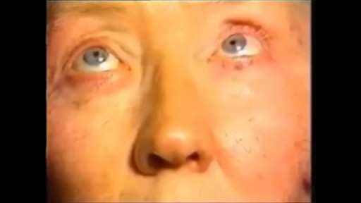
Nystagmus is a vision condition in which the eyes make repetitive, uncontrolled movements. These movements often result in reduced vision and depth perception and can affect balance and coordination. These involuntary eye movements can occur from side to side, up and down, or in a circular pattern.
The Epley maneuver or repositioning maneuver is a maneuver used to treat benign paroxysmal positional vertigo of the posterior or anterior canals
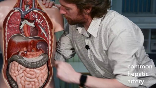
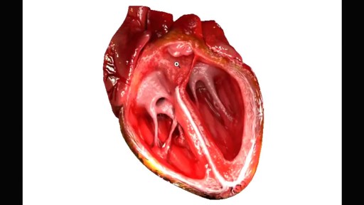
The primary functions of the liver are: Bile production and excretion. Excretion of bilirubin, cholesterol, hormones, and drugs. Metabolism of fats, proteins, and carbohydrates. Enzyme activation. Storage of glycogen, vitamins, and minerals. Synthesis of plasma proteins, such as albumin, and clotting factors.
