- Physical Examination
- Surgical Examination
- Ophthalmology
- Clinical Skills
- Orthopedics
- Surgery Videos
- Laparoscopy
- Pediatrics
- Funny Videos
- Cardiothoracic Surgery
- Nursing Videos
- Plastic Surgery
- Otorhinolaryngology
- Histology and Histopathology
- Neurosurgery
- Dermatology
- Pediatric Surgery
- Urology
- Dentistry
- Oncology and Cancers
- Anatomy Videos
- Health and Fitness
- Radiology
- Anaesthesia
- Physical Therapy
- Pharmacology
- Interventional Radiology
- Cardiology
- Endocrinology
- Gynecology
- Emergency Medicine
- Psychiatry and Psychology
- Childbirth Videos
- General Medical Videos
- Nephrology
- Physiology
- Diet and Food Health
- Diabetes Mellitus
- Neurology
- Women Health
- Osteoporosis
- Gastroenterology
- Pulmonology
- Hematology
- Rheumatology
- Toxicology
- Nuclear Medicine
- Infectious Diseases
- Vascular Disease
- Reproductive Health
- Burns and Wound Healing
- Other
Top videos

Ear Infection Drainage Time Lapse Video
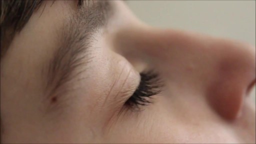
Retinitis pigmentosa is a rare, inherited degenerative eye disease that causes severe vision impairment. Symptoms often begin in childhood. They include decreased vision at night or in low light and loss of side vision (tunnel vision).

Nasal polyps are soft, painless, noncancerous growths on the lining of your nasal passages or sinuses. They hang down like teardrops or grapes. They result from chronic inflammation due to asthma, recurring infection, allergies, drug sensitivity or certain immune disorders. Small nasal polyps may not cause symptoms. Larger growths or groups of nasal polyps can block your nasal passages or lead to breathing problems, a lost sense of smell and frequent infections. Nasal polyps can affect anyone, but they're more common in adults. Medications can often shrink or eliminate nasal polyps, but surgery is sometimes needed to remove them. Even after successful treatment, nasal polyps often return.
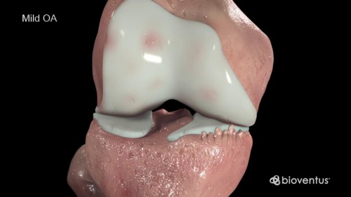
Cartilage is a slippery tissue that provides a smooth surface for joint motion and acts as a cushion between the bones. Synovium is soft, and it lines the joints. It produces fluid, called synovial fluid, for lubrication, and it supplies nutrients and oxygen to the cartilage. As these functions break down, they no longer protect the bones of the knee joint, and bone damage occurs. OA of the knee can cause pain and stiffness. The symptoms worsen over time

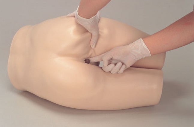
Site enhancement oil, often called "santol" or "synthol" (no relation to the Synthol mouthwash brand), refers to oils injected into muscles to increase the size or change the shape. Some bodybuilders, particularly at the professional level, inject their muscles with such mixtures to mimic the appearance of developed muscle where it may otherwise be disproportionate or lagging. This is known as "fluffing".Synthol is 85% oil, 7.5% lidocaine, and 7.5% alcohol. It is not restricted, and many brands are available on the Internet. The use of injected oil to enhance muscle appearance is common among bodybuilders, despite the fact that synthol can cause pulmonary embolisms, nerve damage, infections, sclerosing lipogranuloma,[60] stroke,[55] and the formation of oil-filled granulomas, cysts or ulcers in the muscle. Rare cases might require surgical intervention to avoid further damage to the muscle and/or to prevent loss of life. Sesame oil is often used in such mixtures, which can cause allergic reactions such as vasculitis.
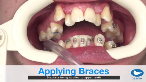
How teeth braces are put

Synthol, otherwise known as site enhancement oil is used by some people (including bodybuilders) to increase the apparent size of their muscles by directly injecting the oil into their muscle tissue. Users treat it as a short cut of looking like a body builder, without the actual hard work of bodybuilding training. With repeated injections, a larger volume of synthol builds up inside the muscle, expanding its size like a balloon filling up with air. Side effects of synthol can cause nerve damage, stroke, ulcers, pulmonary embolisms, and much more. Injecting synthol is very dangerous and if that doesn’t deter potential users, there is also a problem from an aesthetic standpoint; synthol use makes ones body look deformed (just see for yourself in the pictures below).

Watch that video to know the Premature Ejaculation Causes and Cures
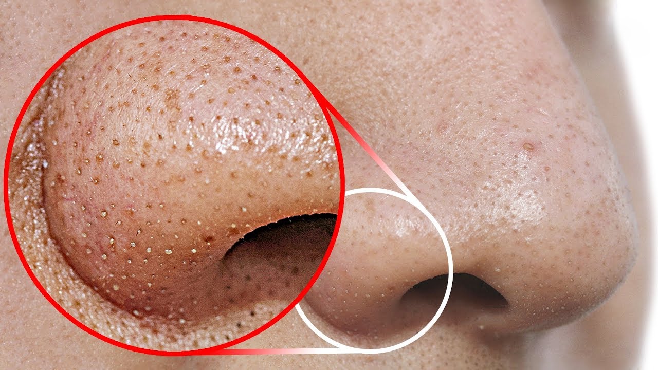
Watch that video to know How to Get Rid of Blackheads From Your Nose
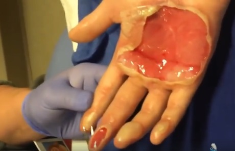
watch that video of Popping a Huge Hand Burn Blister
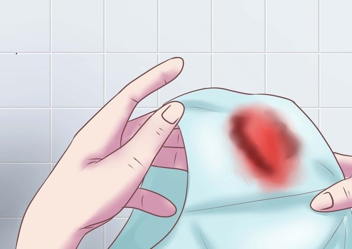
Watch that video to know the Abnormal Female Genital Bleeding Causes


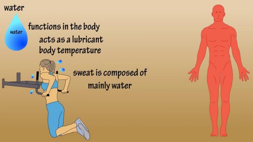
Water is an essential nutrient for the body, as the body loses water through perspiration, breathing, bowel movements, and in urine. Water must be consumed regularly to maintain a sufficient level. Water has many vital functions in the body, including… Serving as a lubricant. Water is a main component of saliva, which helps moisten food making it easier to swallow. Water also helps lubricate joints, reducing friction and inflammation. Water is important in body temperature regulation. When body heat rises, such as during strenuous activities, the body starts to sweat to cool itself. And sweat is made up almost entirely of water.

Watch that video of a Model's Leg and Butt Cosmetic Implants Exploded Inside Her

Mothers can do everything for her baby
watch that Enema Insertion Medical Procedure
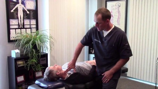
Wisdom teeth extractions can rear their ugly head later in life. This is a video of a patient with neck pain and neck weakness. When we stimulated the nerve fibers in the area of the extracted teeth there was an immediate improvement in her ability to control her neck muscles.

symptoms of kidney dysfunction. I find kidney dysfunction in my patients very frequently. Lower back pain is a common indicator that the kidneys are starting to become irritated. Yes, lower back pain can come from many different areas, but one of the areas I always rule out is kidney congestion.
