- Physical Examination
- Surgical Examination
- Ophthalmology
- Clinical Skills
- Orthopedics
- Surgery Videos
- Laparoscopy
- Pediatrics
- Funny Videos
- Cardiothoracic Surgery
- Nursing Videos
- Plastic Surgery
- Otorhinolaryngology
- Histology and Histopathology
- Neurosurgery
- Dermatology
- Pediatric Surgery
- Urology
- Dentistry
- Oncology and Cancers
- Anatomy Videos
- Health and Fitness
- Radiology
- Anaesthesia
- Physical Therapy
- Pharmacology
- Interventional Radiology
- Cardiology
- Endocrinology
- Gynecology
- Emergency Medicine
- Psychiatry and Psychology
- Childbirth Videos
- General Medical Videos
- Nephrology
- Physiology
- Diet and Food Health
- Diabetes Mellitus
- Neurology
- Women Health
- Osteoporosis
- Gastroenterology
- Pulmonology
- Hematology
- Rheumatology
- Toxicology
- Nuclear Medicine
- Infectious Diseases
- Vascular Disease
- Reproductive Health
- Burns and Wound Healing
- Other
Top videos

breastfeeding tiny infant
samer kareem
3,997 Views • 2 years ago

Breastfeeding Position and Latch
samer kareem
3,560 Views • 2 years ago
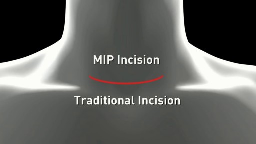
Minimally Invasive Parathyroid Surgery
samer kareem
8,628 Views • 2 years ago

Symptoms of male hypogonadism or low testosterone
samer kareem
5,769 Views • 2 years ago

The Cause of Early Puberty
samer kareem
5,155 Views • 2 years ago

Aphthous ulcers
samer kareem
2,213 Views • 2 years ago
What is Diabetic Neuropathy? Symptoms, Treatments
samer kareem
1,951 Views • 2 years ago
Meningitis Examination
samer kareem
2,280 Views • 2 years ago

Elbow examination
samer kareem
2,716 Views • 2 years ago

Tongue Piercing
Scott
6,713 Views • 2 years ago
This video demonstrates tongue piercing procedure done OUTSIDE a clinical setting
Eye Jewelry Implant
Mohamed Ibrahim
4,432 Views • 2 years ago
Procedure showing how to implant jewelry in the eye

Popping Cyst in the Ear Lobe
Scott
52,103 Views • 2 years ago
Popping Cyst in the Ear Lobe

Ingrown hair removal on thigh
Scott
17,146 Views • 2 years ago
Ingrown hair removal on thigh #18
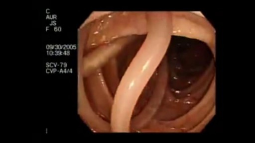
Parasites Accidentally Seen During Colonoscopy
Scott
49,002 Views • 2 years ago
Parasites Accidentally Seen During Colonoscopy

What Unborn Babies Can Do in the Womb
samer kareem
3,141 Views • 2 years ago

Surprising Facts About High Blood PressureMust #W #A #T #C #H
samer kareem
2,137 Views • 2 years ago
Surprising Facts About High Blood Pressure

How does blood pressure change DURING exercise?
samer kareem
2,339 Views • 2 years ago
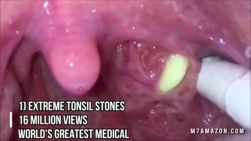
Tonsil Stone Removal Techniques
Scott
96,775 Views • 2 years ago
Tonsil Stone Removal Techniques

Doctor distracts baby from her shots with goofy tune
samer kareem
2,874 Views • 2 years ago
Multiple Lipoma removal surgery
samer kareem
10,082 Views • 2 years ago
Showing 91 out of 103
