- Physical Examination
- Surgical Examination
- Ophthalmology
- Clinical Skills
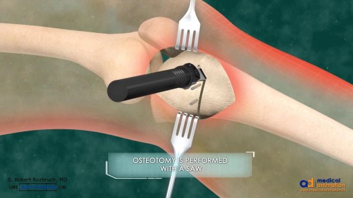
- Orthopedics
- Surgery Videos
- Laparoscopy
- Pediatrics
- Funny Videos
- Cardiothoracic Surgery
- Nursing Videos
- Plastic Surgery
- Otorhinolaryngology
- Histology and Histopathology
- Neurosurgery
- Dermatology
- Pediatric Surgery
- Urology
- Dentistry
- Oncology and Cancers
- Anatomy Videos
- Health and Fitness
- Radiology
- Anaesthesia
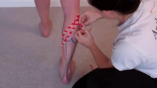
- Physical Therapy
- Pharmacology
- Interventional Radiology
- Cardiology
- Endocrinology
- Gynecology
- Emergency Medicine
- Psychiatry and Psychology
- Childbirth Videos
- General Medical Videos
- Nephrology
- Physiology
- Diet and Food Health
- Diabetes Mellitus
- Neurology
- Women Health
- Osteoporosis
- Gastroenterology
- Pulmonology
- Hematology
- Rheumatology
- Toxicology
- Nuclear Medicine
- Infectious Diseases
- Vascular Disease
- Reproductive Health
- Burns and Wound Healing
- Other
Top videos
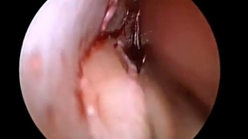
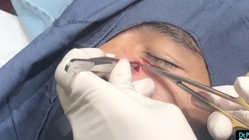
Repair Deviated Nasal Septum, Endoscopic Septoplasty, endoscopic surgery, Stapler repair of nasal septum, Dr B. Todd Schaeffer.
Most people develop several moles (nevi) throughout adulthood. Moles can be found anywhere on the body, usually in sun-exposed areas, and are usually brown, smooth, and slightly raised. In most cases, a nevus is benign and doesn't require treatment. Rarely, they turn into melanoma or other skin cancers. A nevus that changes shape, grows bigger, or darkens should be evaluated for removal.

Menorrhagia is the medical term for menstrual periods with abnormally heavy or prolonged bleeding. Although heavy menstrual bleeding is a common concern, most women don't experience blood loss severe enough to be defined as menorrhagia. With menorrhagia, you can't maintain your usual activities when you have your period because you have so much blood loss and cramping. If you dread your period because you have such heavy menstrual bleeding, talk with your doctor. There are many effective treatments for menorrhagia. Symptoms Signs and symptoms of menorrhagia may include: Soaking through one or more sanitary pads or tampons every hour for several consecutive hours Needing to use double sanitary protection to control your menstrual flow Needing to wake up to change sanitary protection during the night Bleeding for longer than a week Passing blood clots larger than a quarter Restricting daily activities due to heavy menstrual flow Symptoms of anemia, such as tiredness, fatigue or shortness of breath

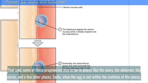
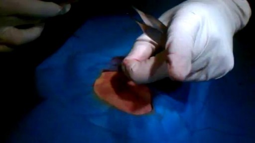
When Does Implantation Occur in Pregnancy? || Common gynaecological problems in women There are a lot of things going on in early pregnancy. The first thing that you need to understand is the menstrual cycle. A good understanding of this can help you understand how the other parts play into the process. A huge part of the menstrual cycle that is the basis of pregnancy is ovulation. Ovulation typically occurs fourteen days prior to the beginning of menstruation, the point when the uterine lining is sloughed off if no pregnancy has occurred.
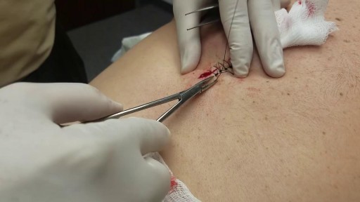
Draining HUGE back abscess
Dental Abscess Drainage and Extraction
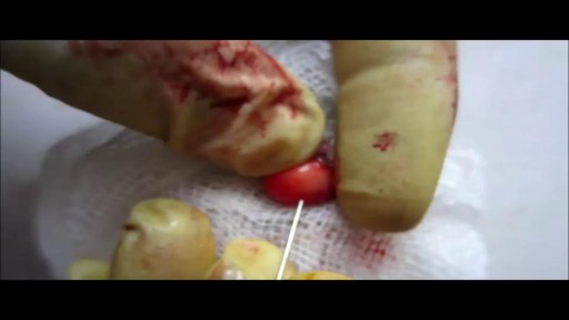
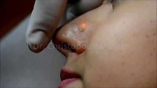
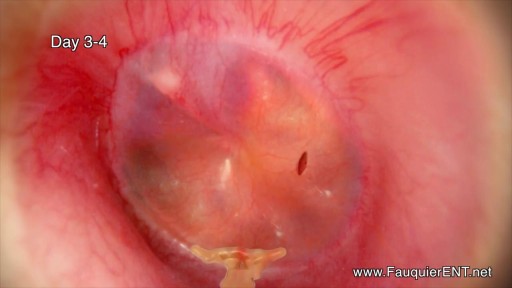
Nose Cyst Extraction
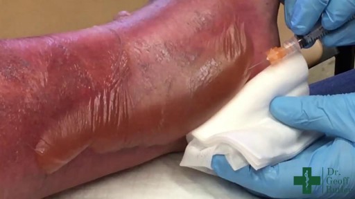
his patient had spilled boiling water on his lower leg a couple days before. This isn't complicated but the teaching points should focus on draining the large blistered areas and attempting to maintain moisture as long as we can so the skin doesn't contract down on itself.
Ear Infection Drainage Time Lapse Video
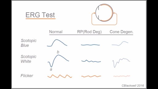
Retinitis pigmentosa is a rare, inherited degenerative eye disease that causes severe vision impairment. Symptoms often begin in childhood. They include decreased vision at night or in low light and loss of side vision (tunnel vision). There's no effective treatment for this condition. Wearing sunglasses may help protect remaining vision.
Retinitis pigmentosa is a rare, inherited degenerative eye disease that causes severe vision impairment. Symptoms often begin in childhood. They include decreased vision at night or in low light and loss of side vision (tunnel vision).