
- Physical Examination
- Surgical Examination
- Ophthalmology
- Clinical Skills
- Orthopedics
- Surgery Videos
- Laparoscopy
- Pediatrics
- Funny Videos
- Cardiothoracic Surgery
- Nursing Videos
- Plastic Surgery
- Otorhinolaryngology
- Histology and Histopathology
- Neurosurgery
- Dermatology
- Pediatric Surgery
- Urology
- Dentistry
- Oncology and Cancers
- Anatomy Videos
- Health and Fitness
- Radiology
- Anaesthesia
- Physical Therapy
- Pharmacology
- Interventional Radiology
- Cardiology
- Endocrinology
- Gynecology
- Emergency Medicine
- Psychiatry and Psychology
- Childbirth Videos
- General Medical Videos
- Nephrology
- Physiology
- Diet and Food Health
- Diabetes Mellitus
- Neurology
- Women Health
- Osteoporosis
- Gastroenterology
- Pulmonology
- Hematology
- Rheumatology
- Toxicology
- Nuclear Medicine
- Infectious Diseases
- Vascular Disease
- Reproductive Health
- Burns and Wound Healing
- Kidney Zone Podcast
- Other



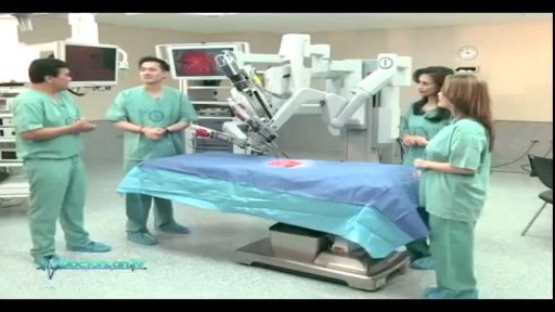
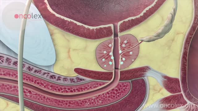

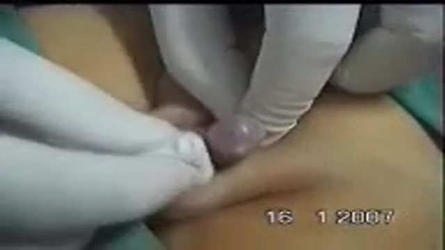
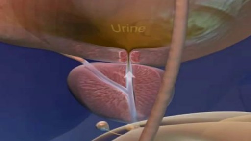
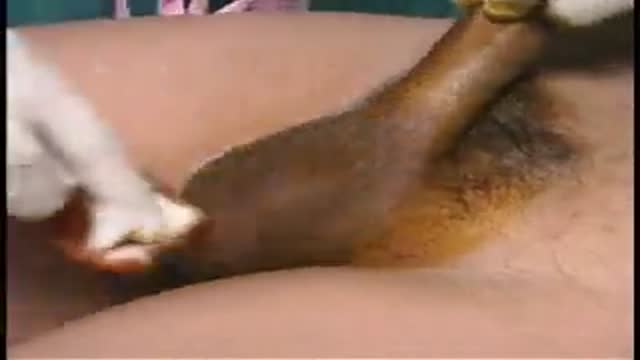

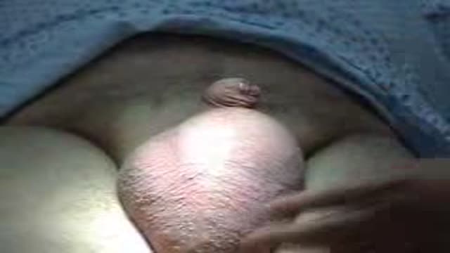
Penile Cancer Explained

Educational only: videos on MedicalVideos.com are for general information and are not medical advice. Always consult your own doctor about your health.
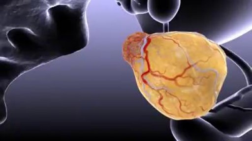
Urogenital neoplasms spreading to the inguinal lymph nodes are penile carcinoma (the most frequent), urethral and scrotum cancers, tumors of the testis with scrotal violation. Penile carcinoma is an uncommon malignant disease and accounts for as many 0.4-0.6% of male cancers. Most patients are elder...ly. It rarely occurs in men under age 60 and its incidence increases progressively until it reaches a peak in the eighth decade 1. The risk of a lymph node invasion is greater with high grade and high stage tumors 2. Some investigators have reported the inaccuracy of the sentinel node biopsy 3, 4, described by Cabanas 5. Patients with metastatic lymph node penis cancer have a very poor prognosis if penectomy only is performed. Ilioinguinal lymphadenectomy is basically carried out as a treatment modality and not only as a staging act. Patients with lymph node invasion have a 30-40% cure rate. Ilioinguinal lymphadenectomy should be also performed in patients with disseminated neoplasms for the local control of the disease. The 5 years survival rate of patients with clinically negative lymph nodes treated with a modified inguinal lymphadenectomy is 88% versus 38% in patients not initially treated with lymphadenectomy 6. This video-tape clearly shows a therapeutic algorithm, the anatomy of the inguinal lymph nodes, according to Rouviere 7 and Daseler 8, the radical ilioinguinal node dissection with transposition of the sartorius muscle and the modified inguinal lymphadenectomy proposed by Catalona 9. References: 1. Lynch D.F. and Schellhammer P: Tumors of the penis. In Campbell’s Urology Seventh Edition, edited by Walsh P.C., Retik A.B., Darracott Vaughan E. and Wein A.J. W.B. Saunders Company, Vol. 3, chapt. 79, p. 2458, 1998. 2. Pizzocaro G., Piva L., Bandieramonte G., Tana S. Up-to-date management of carcinoma of the penis. Eur. Urol. 32: 5-15, 1997 3. Perinetti E., Crane D.B. and Catalona W.J. Unreliability of sentinel lymph node biopsy for staging penile carcinoma. J. Urol. 124: 734, 1980 4. Fowler J.E. Jr. Sentinel lymph node biopsy for staging penile cancer. Urology 23: 352, 1984 5. Cabanas R.M. An approach for the treatment of penile carcinoma. Cancer 39: 456, 1977 6. Russo P. and Gaudin P. Management strategies for carcinoma of the penis. Contemporary Urology;5:48-66, 2000 7. Rouviere H. Anatomy of the human lymphatic system. Edwards Brothers, p. 218, 1938 8. Daseler E.H., Anson B.J., Reimann A.F. Radical excision of the inguinal and iliac lymph glands: a study based on 450 anatomical dissections and upon supportive clinical observations. Surg. Gynecol. Obstet. 87: 679, 1948 9. Catalona W.J. Modified inguinal lymphadenectomy for carcinoma of the penis with preservation of saphenous veins: technique and preliminary results. J. Urol. 140: 306-310, 1988
This video in 30 seconds
A narrated surgical-atlas video of ilioinguinal lymphadenectomy for penile cancer. The commentary reviews when node dissection is offered, then follows two dissection approaches in detail, covering superficial and deep inguinal node removal within defined anatomical boundaries and muscle transposition to cover the femoral vessels.
Key points
- Inguinal lymph node groups and their anatomy are reviewed
- Indications for lymphadenectomy are discussed in the commentary
- Superficial and deep inguinal node dissection are shown within defined boundaries
- A second dissection approach with different boundaries is followed
- Muscle transposition to cover the femoral vessels is described
Chapters
- 0:29 — Inguinal node groups and anatomy
- 1:31 — Indications for lymphadenectomy
- 2:22 — First dissection boundaries and technique
- 4:47 — Deep node dissection
- 5:35 — Sartorius transposition and closure
- 6:32 — Second dissection approach
Video Transcript
A narrated surgical-atlas video of ilioinguinal lymphadenectomy for penile cancer. The commentary reviews when node dissection is offered, then follows two dissection approaches in detail — superficial and deep inguinal node removal within defined anatomical boundaries, and sartorius muscle transposition to cover the femoral vessels. The metadata language tag says Latin; the narration is in fact English.
Read the full transcript
The inguinal lymph nodes may be divided into two groups, deep and superficial, separated by the fascia lata. The superficial inguinal lymph nodes lie underneath the camper's fascia, and may be divided into five groups, superolateral, superomedial, infeoromedial, infeorolateral, central. The deep ileoinguinal glands are placed under the fascia lata, on the medial side of the femoral vein. The highest is the gland of cloket, or rosemular. The profilactic lymphadenectomy impatient with penicarsinoma is controversial for a significant incidence of morbidity. It is usually performed impatient with lymph node invasion, but without distant metastases.
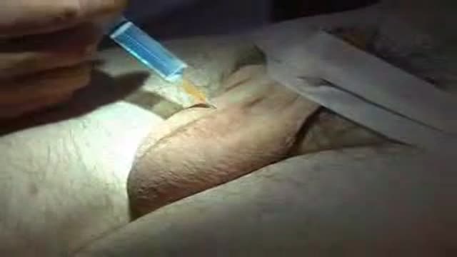
Eleoinguinal lymphadenectomy should be performed impatient with clinically positive lymph nodes, and impatient with stage PT3, PT4- penicarsinoma. Lymphadenectomy can be performed in patient without evident clinical lymphenode invasion, with the stage Pg2 or high grade Pg1 tumors. Patient with metastatic inguinal disease should be treated with pelvic nod dissection. The boundaries of dissection described by DASLAV are superior to the inguinal ligament, laterally to the sartolius muscle, medially to the adducto-longus muscle. Inferiorly to the base of femora triangle, the dissection begins with a 12 cm elliptic incision, 1 cm below and parallel to the inguinal ligament.
Kampers fascia must be located because it represents the superficial boundary of the lymphadenectomy to preserve skin's blood supply. Superficial epigastric vein is now ligated and dissected in order to gain access to the deeper vascular structures. After the dissection of the external pudendal vein, the spermatic cord is located. The superior superficial lymphenotes are dissected up to the circumflex superficial iliac vessels that are previously ligated. The superior superficial lymphenotes are dissected first from the pubic bone, then from the iliac bone and finally from the inguinal ligament so that femoral vessels are revealed.
The dissection of the sub-inguinal lymphenotes proceeds superficially following the campers fascia, reaching the epics of the scarpa's triangle where the major saphenous vein and the lateral accessory saphenous vein are ligated. The major saphenous vein is dissected at the epics of the scarpa's triangle and the lymphenotes are removed from the fascia lata up to the ovalis posa. Superficial lymphadenectomy is completed. Underneath the fascia lata and medially to the femoral vein, deep ilioinquinal nodes are found. Removal of these lymphenotes, including the cloketz lymphenote, is shown here. The fascia lata covering the scarpa's triangle is removed.
The saphenous femoral nerve is located and preserved. As soon as the lymphadenectomy is completed, the sartallius muscle is dissected for a 10-12 cm length starting from the proximal end and sectioned from the iliac bone. The sartallius muscle is transposed over the femoral vessels and fixed to the inguinal ligament with an interuptured suture. A closed suction drain is placed. The skin incision is closed. Contralateral ilioinquinal lymphadenectomy and bilateral pelvic lymphadenectomy are performed if metastatic lymph node disease is found. The pelvic lymphadenectomy may be done laparoscopically. The boundaries of dissection, described by Catalona,
are superior to the inguinal ligament with a shorter skin incision, laterally to the femoral artery, inferiorly to the base of obalis fossa, and medially to the adductor lungus muscle. The dissection begins with a 10 cm elliptic incision approximately 2 cm below the groin crease. Campers fascia must be located because it represents the superficial boundary of the lymphadenectomy to preserve the skin's blood supply. The spermatic core is identified and reflected medially. Superior and inferior skin flaps are developed to the level of the external oblique aponeurosis and of the adductor lungus muscle respectively.
The greater saphenous vein and the lateral accessory saphenous vein are carefully dissected and preserved. Superficial epigastic vein and the superficial circumflex vein are ligated and transacted at the level of the superior boundaries of dissection. The superficial lymphatic tissue is superiorly removed. Lymphadenectomy is continued developing the plane from the fascialata up to the obalis fossa. The saphenous femoral junction is reached and the superficial circumflex vein is ligated and transacted. The greater saphenous vein is carefully dissected and preserved. The external pudendal vein and the superficial epigastic vein are clipped and transacted.
The superior and inferior groups of superficial lymphenose are easily removed. The deep inguinal nodes which are located below the fascialata and medially to the femoral vein are dissected from the inguinal ligaments superiorly and just distally to the fossa ovalis inferiorly. The specimen is clipped and transacted at the level of the femoral canal where the cloked lymph node is located. Boundaries of dissection are clearly seen. The femoral canal, the inguinal ligament, the spermatic cord, the abductor-langus muscle, the pectineus muscle and the inferior part of the fasal ovalis
with the preserved veins. The transposition of the sartovius muscle is unnecessary and the skin incision is closed after placing a closed suction drain.
The superior and inferior groups of superficial lymphenose are easily removed from the fasal ovalis inferiorly.
Transcript auto-generated from the video’s audio for accessibility and study. Educational content only — not medical advice.
Video Overview
From our Urology collection: Prostate surgery (TURP), cystoscopy, kidney stone treatment, vasectomy and male health procedures explained step by step. Browse all Urology videos →
Medical Videos
Free medical and surgery videos for patients, students and professionals — trusted since 2007.
Founded by Dr. Mohamed Ibrahim, MD, PhD — physician-scientist.
Follow us on Facebook · Instagram
SORT BY-
Top Comments
-
Latest comments