
- Physical Examination
- Surgical Examination
- Ophthalmology
- Clinical Skills
- Orthopedics
- Surgery Videos
- Laparoscopy
- Pediatrics
- Funny Videos
- Cardiothoracic Surgery
- Nursing Videos
- Plastic Surgery
- Otorhinolaryngology
- Histology and Histopathology
- Neurosurgery
- Dermatology
- Pediatric Surgery
- Urology
- Dentistry
- Oncology and Cancers
- Anatomy Videos
- Health and Fitness
- Radiology
- Anaesthesia
- Physical Therapy
- Pharmacology
- Interventional Radiology
- Cardiology
- Endocrinology
- Gynecology
- Emergency Medicine
- Psychiatry and Psychology
- Childbirth Videos
- General Medical Videos
- Nephrology
- Physiology
- Diet and Food Health
- Diabetes Mellitus
- Neurology
- Women Health
- Osteoporosis
- Gastroenterology
- Pulmonology
- Hematology
- Rheumatology
- Toxicology
- Nuclear Medicine
- Infectious Diseases
- Vascular Disease
- Reproductive Health
- Burns and Wound Healing
- Kidney Zone Podcast
- Other
Ophthalmology
Ophthalmology videos: eye examinations, cataract and LASIK surgery, retina procedures and common eye conditions explained by specialists. Free to watch.
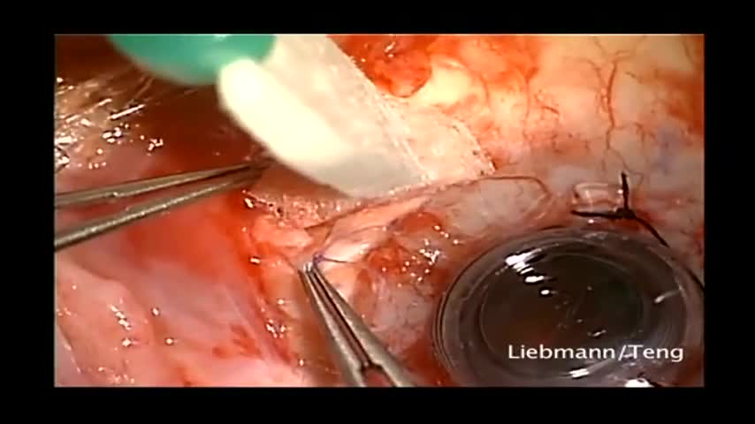

This patient had a history of herpes and had a previous corneal transplant that subsequently became opacified. There was also a previous Baerveldt implant placed into the anterior chamber. This surgery consists of Baerveldt tube being moved from the anterior chamber to posterior chamber, removal of failed graft, placement of temporary keratoprosthesis, pars plana vitectomy and corneal transplant. Jeffrey Liebmann, MD. Mark Speaker, MD. Uri Shabto, MD. Christopher Teng, MD.
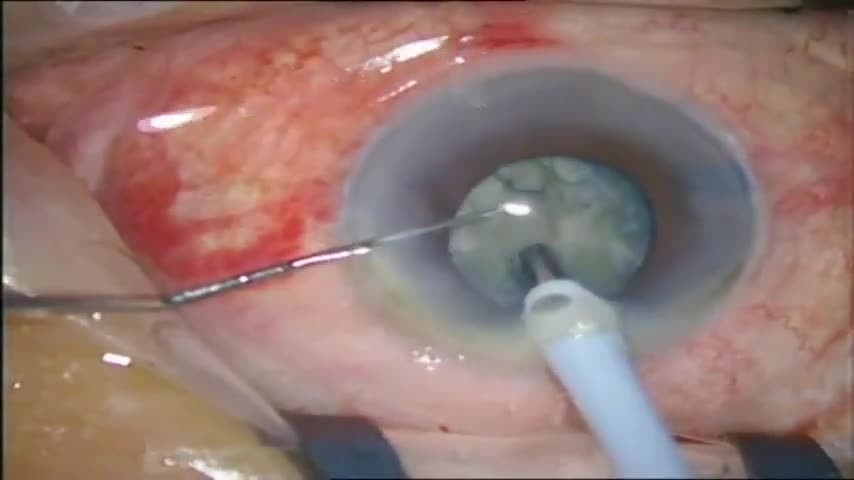
Stop and chop surgery on a hard cataract on a small pupil.
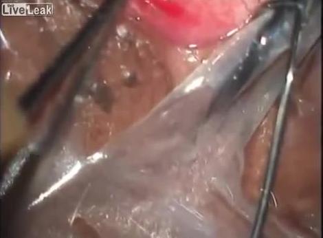
Loa loa filariasis (also known as loiasis, loaiasis, Calabar swellings, Fugitive swelling, Tropical swelling and African eyeworm) is a skin and eye disease caused by the nematode worm, loa loa. Humans contract this disease through the bite of a Deer fly or Mango fly (Chrysops spp), the vectors for Loa loa. The adult Loa loa filarial worm migrates throughout the subcutaneous tissues of humans, occasionally crossing into subconjunctival tissues of the eye where it can be easily observed. Loa loa does not normally affect one's vision but can be painful when moving about the eyeball or across the bridge of the nose.The disease can cause red itchy swellings below the skin called "Calabar swellings". The disease is treated with the drug diethylcarbamazine (DEC), and when appropriate, surgical methods may be employed to remove adult worms from the conjunctiva.

Nearly 300 million people experience the world without certain colors every day. ‘Color For the Colorblind’ is a short documentary about what happened when we partnered with EnChroma, maker of color blindness-correcting glasses, to help people experience colors for the first time.
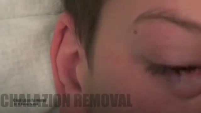
Chalazions are extremely common, and having a sound surgical technique to drain a chalazion is a fundamental in general ophthalmology and oculoplastic surgery. I believe one of the biggest downfalls in treating chalazions is inadequate local anesthetic. Please that both the outer and inner surface to the eyelid need to receive local anesthesia to make the patient totally comfortable. It is important to be careful in delivering the local anesthetic and making sure you have control of the head position, and the position of your needle is bent to minimize any possibility of contact with the globe.
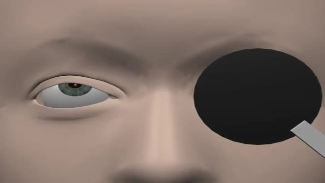
Eye Movement Terminology
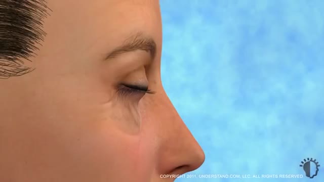
Eyelid surgery, or blepharoplasty, is a surgical procedure to improve the appearance of the eyelids. Surgery can be performed on either the upper and lower lids, or both. Whether you want to improve your appearance or are experiencing functional problems with your eyelids, eyelid surgery can rejuvenate the area surrounding your eyes.
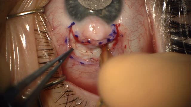
Limbal dermoid is a congenital growth on the eye that forms from germline cells that get trapped in this region during embryogenesis. The dermoid often has tissue from multiple germ layers including hair and fat. Dermoids on the eye can cause astigmatism and be unsightly.
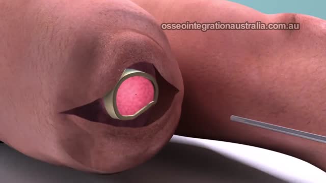
Integral Leg Prosthesis System surgical procedure performed
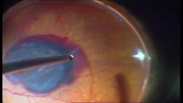
The fovea was moved 425 to 1,700 microm (965+/-262 microm) superiorly or inferiorly. Follow-up time was 2 to 12 months (median 8 months). Complications included macular pucker (3 eyes), subfoveal hemorrhage (2 eyes), macular hole (1 eye), and progression of cataract in phakic eyes (3 eyes). Thirteen of 20 eyes showed various degrees of proliferative vitreoretinopathy with epiretinal membrane formation over the inferior peripheral retina with the inferior retinal detachment stabilized by the silicone oil. One eye progressed to phthisis bulbi. Initial visual acuity ranged from 20/80 to 20/800 (median 20/150) and final visual acuity ranged from light perception to 20/200 (median 20/1000).
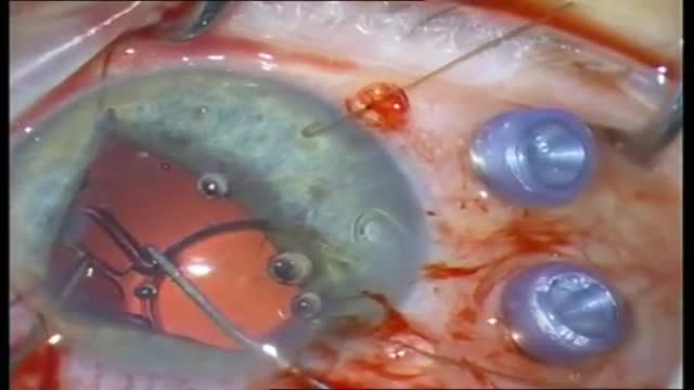
Patients with candida endophthalmitis who have chorioretinitis with vitreal involvement should be treated with vitrectomy and systemic antifungal therapy with amphotericin B (Choice B) and/or fluconazole. An early vitrectomy improves the likelihood of a positive outcome, and intravitreal injection of amphotericin B may be of help. Prompt diagnosis and treatment of candida endophthalmitis is essential, as the condition can worsen quickly.

Diabetic retinopathy is classified into two types: Non-proliferative diabetic retinopathy (NPDR) is the early stage of the disease in which symptoms will be mild or nonexistent. In NPDR, the blood vessels in the retina are weakened. ... Proliferative diabetic retinopathy (PDR) is the more advanced form of the disease.

Diabetic retinopathy involves changes to retinal blood vessels that can cause them to bleed or leak fluid, distorting vision. Diabetic retinopathy is the most common cause of vision loss among people with diabetes and a leading cause of blindness among working-age adults.
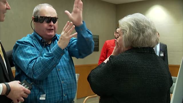
A visual prosthesis, often referred to as a bionic eye, is an experimental visual device intended to restore functional vision in those suffering from partial or total blindness. In 1983 Joao Lobo Antunes, a Portuguese doctor, implanted a bionic eye in a person born blind.
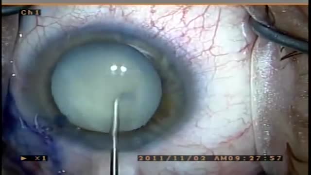
A cataract is a clouding of the lens in the eye that affects vision. Most cataracts are related to aging. Cataracts are very common in older people. By age 80, more than half of all Americans either have a cataract or have had cataract surgery. A cataract can occur in either or both eyes. It cannot spread from one eye to the other.

Rheum is made up of mucus, skin cells, oils and dust. The rheum that comes from the eyes and forms eye boogers is called gound, which you may know as eye sand, eye gunk, sleep dust, sleep sand, sleep in your eyes, or eye shnooters. When you're awake, gound doesn't cause any problems.
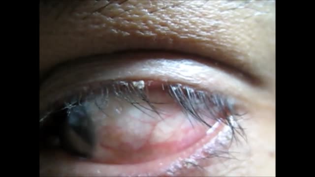
Blepharitis is an inflammation of the eyelids in which they become red, irritated and itchy and dandruff-like scales form on the eyelashes. It is a common eye disorder caused by either bacteria or a skin condition, such as dandruff of the scalp or acne rosacea. It affects people of all ages. Although uncomfortable, blepharitis is not contagious and generally does not cause any permanent damage to eyesight.
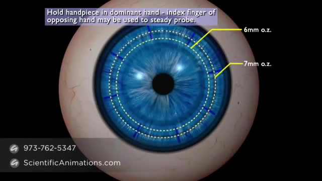
Keratoplasty is the procedure whereby abnormal corneal tissue is replaced by a healthy donor cornea.

The eyes A close up of a young person's eyes. The eyes are responsible for four-fifths of all the information our brain receives. Here you can find out a bit more about how they work, common problems that affect vision and the work Sightsavers does to treat and prevent avoidable blindness. You can also find out more about the people whose lives have been changed thanks to donations from people like you. How do eyes work? (click image to see enlarged version or click here for text alternative) Graphic of an eye with information about its different parts The images we see are made up of light reflected from the objects we look at. This light enters the eye through the cornea. Because this part of the eye is curved, it bends the light, creating an upside down image on the retina (this is eventually put the right way up by the brain). The retina is a complex part of the eye, but only the very back of it is light sensitive. This part of the retina has roughly the area of a 10p coin, and is packed with photosensitive cells called rods and cones. Cones are the cells responsible for daylight vision. There are three kinds – each responding to a different wavelength of light: red, green and blue. The cones allow us to see images in colour and detail. Rods are responsible for night vision. They are sensitive to light but not to colour. In darkness, the cones do not function at all. How do we see an image? The lens focuses the image. It can do this because it is adjustable – using muscles to change shape and help us focus on objects at different distances. The automatic focusing of the lens is a reflex response and is not controlled by the brain. Once the image is clearly focused on the sensitive part of the retina, energy in the light that makes up that image creates an electrical signal. Nerve impulses can then carry information about that image to the brain through the optic nerve.
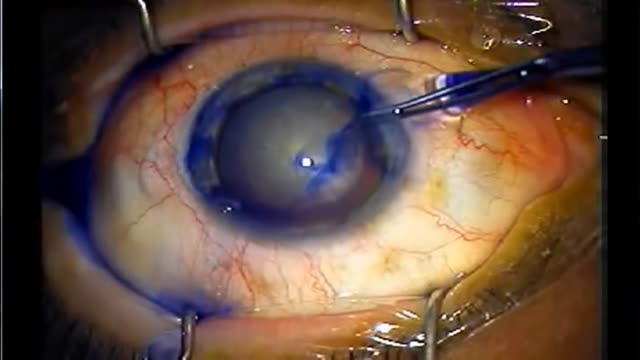
In cataract surgery, the lens inside your eye that has become cloudy is removed and replaced with an artificial lens (called an intraocular lens, or IOL) to restore clear vision. The procedure typically is performed on an outpatient basis and does not require an overnight stay in a hospital or other care facility.
Medical Videos
Free medical and surgery videos for patients, students and professionals — trusted since 2007.
Founded by Dr. Mohamed Ibrahim, MD, PhD — physician-scientist.
Follow us on Facebook · Instagram