
- Physical Examination
- Surgical Examination
- Ophthalmology
- Clinical Skills
- Orthopedics
- Surgery Videos
- Laparoscopy
- Pediatrics
- Funny Videos
- Cardiothoracic Surgery
- Nursing Videos
- Plastic Surgery
- Otorhinolaryngology
- Histology and Histopathology
- Neurosurgery
- Dermatology
- Pediatric Surgery
- Urology
- Dentistry
- Oncology and Cancers
- Anatomy Videos
- Health and Fitness
- Radiology
- Anaesthesia
- Physical Therapy
- Pharmacology
- Interventional Radiology
- Cardiology
- Endocrinology
- Gynecology
- Emergency Medicine
- Psychiatry and Psychology
- Childbirth Videos
- General Medical Videos
- Nephrology
- Physiology
- Diet and Food Health
- Diabetes Mellitus
- Neurology
- Women Health
- Osteoporosis
- Gastroenterology
- Pulmonology
- Hematology
- Rheumatology
- Toxicology
- Nuclear Medicine
- Infectious Diseases
- Vascular Disease
- Reproductive Health
- Burns and Wound Healing
- Kidney Zone Podcast
- Other
Ophthalmology
Ophthalmology videos: eye examinations, cataract and LASIK surgery, retina procedures and common eye conditions explained by specialists. Free to watch.


When foreign organisms such as bacteria enter the body, the immune system sends white blood cells to fight the infection. This causes swelling (inflammation) at the site of infection and the death of nearby tissue, creating a hole called a cavity, which fills with pus to form an abscess.
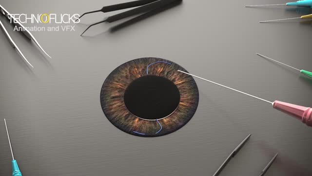
Cosmetic iris implants have not been evaluated by any U.S. regulatory agency or tested for safety in clinical trials. While the implants are not approved by the U.S. Food and Drug Administration, it has been reported in the media this month that the surgery is being performed overseas. During iris implant surgery, an artificial iris made of silicone is folded and inserted into a slit that has been cut into the cornea. Then the iris is unfolded and adjusted to cover the natural iris. Local anesthesia is used.
The cornea occupies the front center part of the outer wall of the eye. It is made of collagen fibers in a very special arrangement so that the cornea is clear. One looks through the cornea to see the iris and pupil. The cornea bends light coming into the eye so that it is focused on the retina.
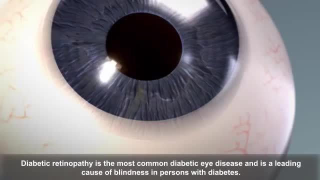
Diabetic retinopathy (die-uh-BET-ik ret-ih-NOP-uh-thee) is a diabetes complication that affects eyes. It's caused by damage to the blood vessels of the light-sensitive tissue at the back of the eye (retina). At first, diabetic retinopathy may cause no symptoms or only mild vision problems. Eventually, it can cause blindness. The condition can develop in anyone who has type 1 or type 2 diabetes. The longer you have diabetes and the less controlled your blood sugar is, the more likely you are to develop this eye complication.
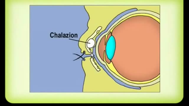
A chalazion is a lump of the lid that is caused by obstruction of the drainage duct of an oil gland within the upper or lower eyelid. This lump may increase in size over days to weeks and may occasionally become red, warm, or painful.

contact lens for beginners

How To Apply Contact Lenses
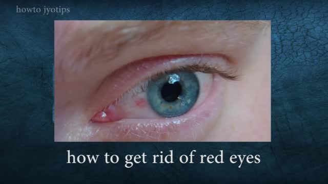
Red eyes usually are caused by allergy, eye fatigue, over-wearing contact lenses or common eye infections such as pink eye (conjunctivitis). However, redness of the eye sometimes can signal a more serious eye condition or disease, such as uveitis or glaucoma.

Keratitis is an inflammation of the cornea — the clear, dome-shaped tissue on the front of your eye that covers the pupil and iris. Keratitis is sometimes caused by an infection involving bacteria, viruses, fungi or parasites. Noninfectious keratitis can be caused by a minor injury, wearing your contact lenses too long or other noninfectious diseases. If you have eye redness or other symptoms of keratitis, make an appointment to see your doctor. With prompt attention, mild to moderate cases of keratitis can usually be effectively treated without loss of vision. If left untreated, or if an infection is severe, keratitis can lead to serious complications that may permanently damage your vision.

Pink eye (conjunctivitis) is an inflammation or infection of the transparent membrane (conjunctiva) that lines your eyelid and covers the white part of your eyeball. When small blood vessels in the conjunctiva become inflamed, they're more visible. This is what causes the whites of your eyes to appear reddish or pink. Pink eye is commonly caused by a bacterial or viral infection or an allergic reaction. It may affect one or both eyes. Pink eye can be irritating, but it rarely affects your vision. Treatments can help ease the discomfort of pink eye. Because pink eye can be contagious, early diagnosis and treatment can help limit its spread.
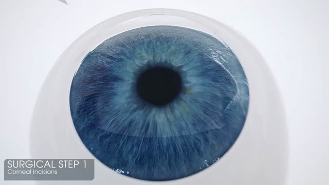
Less than a decade ago, corneal transplantation took a big leap forward with the introduction of Descemet’s stripping endothelial keratoplasty (DSEK), which removes only Descemet’s membrane and the diseased endothelium and replaces them with a thin, tripartite donor graft of posterior corneal stroma, Descemet’s membrane, and healthy endothelium. Then came DSAEK, in which the donor graft is prepared with an automated microkeratome, allowing for easier donor preparation and reproducible results by surgeons and eye bank technicians.1 DSAEK has proved to have many advantages over penetrating keratoplasty (PK) and its endothelial predecessors (see “A Brief History of Endothelial Keratoplasty”). Now DSAEK is being compared with a newer technique, Descemet’s membrane endothelial keratoplasty (DMEK), which has emerged as a promising alternative—grafting only Descemet’s membrane and endothelium, allowing for a pure anatomical replacement of only what was removed and the possibility of even better vision with quicker healing.1 Although indications for these procedures are similar, each has unique benefits and drawbacks. Five cornea surgeons offer their perspectives on the procedures and their thoughts on whether it may be time to move to the newer surgery.
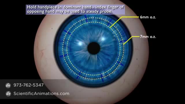
A cornea transplant, also called keratoplasty, is a surgical procedure to replace part of your cornea with corneal tissue from a donor. Your cornea is the transparent, dome-shaped surface of your eye that accounts for a large part of your eye's focusing power. A cornea transplant can restore vision, reduce pain and improve the appearance of a damaged or diseased cornea. Most cornea transplant procedures are successful. But cornea transplant carries a small risk of complications, such as rejection of the donor cornea.
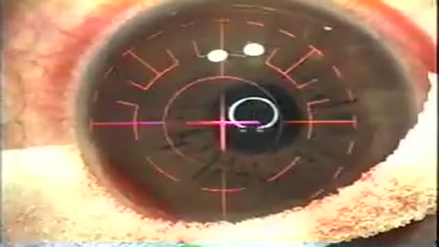
LASIK, which stands for laser in-situ keratomileusis, is a popular surgery used to correct vision in people who are nearsighted, farsighted, or have astigmatism. All laser vision correction surgeries work by reshaping the cornea, the clear front part of the eye, so that light traveling through it is properly focused onto the retina located in the back of the eye. LASIK is one of a number of different surgical techniques used to reshape the cornea.

Get the facts and fight back against Dry Eye disease.

Take a trip around the world while ensuring healthy eye habits and optimal vision

How amblyopia develops in children. Basically, if one eye doesn't see well from an early age, the wiring never forms correctly back to the occipital cortex.
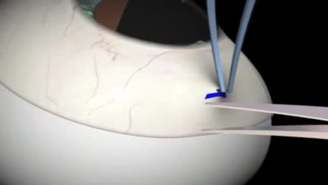
Glaucoma Surgery 3D Animation
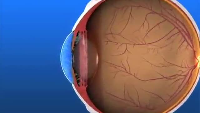
Glaucoma Pathogenesis Simplified
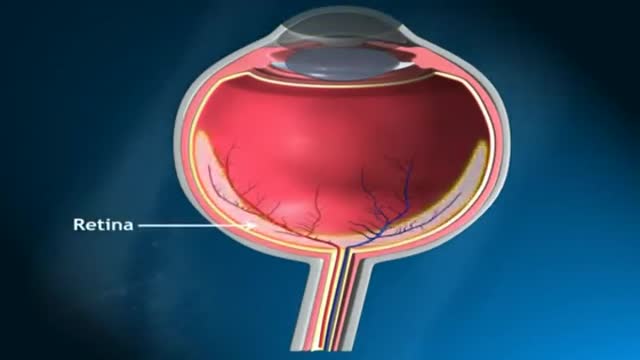
Glaucoma is called the silent thief of sight. It does not have symptoms during the early stages of the diseases and can make a patient blind over several years
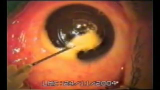
Big Bubble Technique
Medical Videos
Free medical and surgery videos for patients, students and professionals — trusted since 2007.
Founded by Dr. Mohamed Ibrahim, MD, PhD — physician-scientist.
Follow us on Facebook · Instagram