- Physical Examination
- Surgical Examination
- Ophthalmology
- Clinical Skills
- Orthopedics
- Surgery Videos
- Laparoscopy
- Pediatrics
- Funny Videos
- Cardiothoracic Surgery
- Nursing Videos
- Plastic Surgery
- Otorhinolaryngology
- Histology and Histopathology
- Neurosurgery
- Dermatology
- Pediatric Surgery
- Urology
- Dentistry
- Oncology and Cancers
- Anatomy Videos
- Health and Fitness
- Radiology
- Anaesthesia
- Physical Therapy
- Pharmacology
- Interventional Radiology
- Cardiology
- Endocrinology
- Gynecology
- Emergency Medicine
- Psychiatry and Psychology
- Childbirth Videos
- General Medical Videos
- Nephrology
- Physiology
- Diet and Food Health
- Diabetes Mellitus
- Neurology
- Women Health
- Osteoporosis
- Gastroenterology
- Pulmonology
- Hematology
- Rheumatology
- Toxicology
- Nuclear Medicine
- Infectious Diseases
- Vascular Disease
- Reproductive Health
- Burns and Wound Healing
- Other
Top videos
Learn what's working for other Nursing Students! Check out our Top 10 Most Popular Lessons Here: https://bit.ly/3nda5u3
Get the full lesson here: https://nursing.com/lesson/ski....lls-03-04-trach-care
Welcome to the NURSING Family, we call it the most supportive nursing cohort on the planet.
At NURSING.com, we want to help you remove the stress and overwhelm of nursing school so that you can focus on becoming an amazing nurse.
Check out our freebies and learn more at: (http://www.nursing.com)
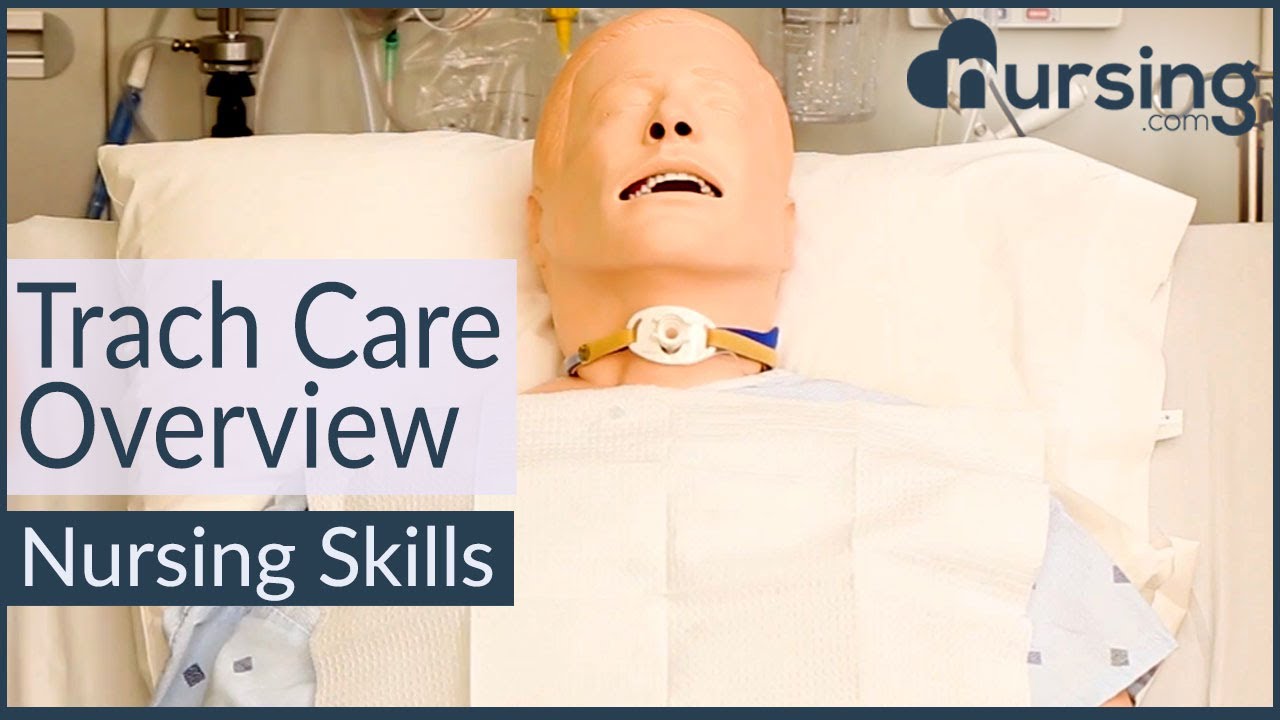
Trach Care Overview (Nursing Skills):
In this video we’re going to look at trach care. Remember you should always suction the patient before trach care, so if you haven’t watched that skill video yet, make sure you watch it!
Click here: https://nursing.com/lesson/ski....lls-03-03-trach-suct
And remember as you’re doing this, you want to be assessing the stoma for signs of infection or skin breakdown.
Bookmarks:
0:00 Introduction
0:30 Set up sterile field
1:00 Apply gloves
1:12 Remove inner canula and dressing
1:30 Apply sterile gloves
2:05 Clean secretions
2:56 Clean stoma
3:48 Replace inner canula
4:14 Change trach ties
5:50 Apply dressing
Visit us at https://nursing.com/medical-disclaimer/ for disclaimer information.
NCLEX®, NCLEX-RN® are registered trademarks of the National Council of State Boards of Nursing, INC. and hold no affiliation with NURSING.com.

Nursing skills lab procedure for IV insertion.

The head-to-toe assessment in nursing is an important physical health assessment that you'll be performing as a nursing student and nurse.
Head-to-toe assessments allow nurses to assess the health status of patients by following a checklist of criteria.
On the job, your head-to-toe nursing assessment will be performed much faster, and it may be different or more specialized to accommodate the patients' needs within your nursing specialty.
This assessment represents a general assessment checklist (or cheat sheet) that you might encounter in nursing school. (Note: Always follow your instructor's requirements or your employer's assessment protocols).
This nursing head-to-toe examination video guide will focus on the following areas/skills:
-Vital Signs (pulse rate, respiration rate, temperature, oxygen saturation, blood pressure, pain assessment)
https://www.youtube.com/watch?v=gUWJ-6nL5-8
-Cranial Nerve examination
-Head assessment (hair, cranium, eyes, nose, mouth, ears, sinuses)
-Neck assessment (jugular vein, thyroid, trachea, carotid)
-Heart sounds assessment: https://www.youtube.com/watch?v=H48WsyIjFs0&t=73s
-Lung sounds assessment: https://www.youtube.com/watch?v=KNrcG077brQ
-Abdominal assessment
-Assessing extremities (arms, hands, legs, feet)
-Back assessment
-and more
While performing your comprehensive head-to-toe assessment, you'll want to record your findings in the documentation.
Nursing Gear: https://teespring.com/stores/registerednursern
Subscribe: http://www.youtube.com/subscri....ption_center?add_use
Notes: http://www.registerednursern.c....om/head-toe-assessme
Nursing School Supplies: http://www.registerednursern.c....om/the-ultimate-list
Visit our website RegisteredNurseRN.com for free quizzes, nursing care plans, salary information, job search, and much more: http://www.registerednursern.com
Check out other Videos: https://www.youtube.com/user/R....egisteredNurseRN/vid
All of our videos in a playlist: https://www.youtube.com/watch?v=pAhHxt663pU&list=PLQrdx7rRsKfXMveRcN4df0bad3ugEaQnk
Popular Playlists:
NCLEX Reviews: https://www.youtube.com/playli....st?list=PLQrdx7rRsKf
Fluid & Electrolytes: https://www.youtube.com/playli....st?list=PLQrdx7rRsKf
Nursing Skills: https://www.youtube.com/playli....st?list=PLQrdx7rRsKf
Nursing School Study Tips: https://www.youtube.com/playli....st?list=PLQrdx7rRsKf
Nursing School Tips & Questions" https://www.youtube.com/playli....st?list=PLQrdx7rRsKf
Teaching Tutorials: https://www.youtube.com/playli....st?list=PLQrdx7rRsKf
Types of Nursing Specialties: https://www.youtube.com/playli....st?list=PLQrdx7rRsKf
Healthcare Salary Information: https://www.youtube.com/playli....st?list=PLQrdx7rRsKf
New Nurse Tips: https://www.youtube.com/playli....st?list=PLQrdx7rRsKf
Nursing Career Help: https://www.youtube.com/playli....st?list=PLQrdx7rRsKf
EKG Teaching Tutorials: https://www.youtube.com/playli....st?list=PLQrdx7rRsKf
Dosage & Calculations for Nurses: https://www.youtube.com/playli....st?list=PLQrdx7rRsKf
Diabetes Health Managment: https://www.youtube.com/playli....st?list=PLQrdx7rRsKf

Thank you so much for watching❤
If you enjoyed this video ▶Please leave a LIKE👍 ▶SHARE this video ▶【SUBSCRIBE】my channel for more new videos And click the BELL 🔔so you don't miss any of my videos HERE
https://www.youtube.com/c/nurs....eminder?sub_confirma
You can support my work by purchasing your NurseMinder Merch https://teespring.com/stores/nurseminder-nation (or click on merch pics under the video)
Or simply do your Amazon shopping after clicking on one of the links below
-------------------------------------------------------------------------
Thank you so much! I appreciate you!♥♥
------------------------------------------------------------------------
Nurses often prime IV lines with the hopes that there are no air bubbles. In this video, I will share a couple of tips to help reduce the risk or frequency of air bubbles during line priming. I will also talk about how to troubleshoot the air bubbles when they appear during an infusion
Providing patient care and influencing safe patient outcomes requires that registered nurses and licensed practice nurses maintain air free IV lines. Learn the strategies and tips to decrease the risk of air bubbles appearing in your primary or secondary medication line as well as troubleshooting tips to remove those alarming bubbles. Your patients will thank you!
Whether you are providing normal saline, a medication, or a combination, ensure that all fluids are compatible.
Supplies used in this video include the Alaris Primary Infusion line, alcohol swabs and a sterile 10 cc syringe ... and a nail in the wall :)
------------------------------------------------------------------------
❤️ ~ You may also be interested in watching ~ ❤️
PICC line assessment https://youtu.be/tnKClpU-J1g
How To Access a PICC line https://youtu.be/SCF6bmk8KWc
Putting on Sterile Gloves https://youtu.be/xNwkKLqDJn4
Organizational Plans for Nursing https://youtu.be/_NATxwPwHzc
Medication Conversions https://youtu.be/TCPBXg2TYCs
------------------------------------------------------------------------
💻COMMENT in the description box below and share your ideas
👍 LIKE the video
🗣 SHARE with your friends
📥 SUBSCRIBE ... hit the BELL 🔔
Subscribe to NurseMinder https://www.youtube.com/c/nurs....eminder?sub_confirma
------------------------------------------------------------------------
Amazon Affiliate Links
------------------------------------------------------------------------
Want to support me in another way? Enter Amazon through my links and continue to do your shopping. Simple and Easy Way to support the work I do.
The following list is the equipment I use (or if my version is no longer sold, a close replica).
📱 Phone 11 Cell Phone https://amzn.to/2WpOJfz
💻 MacBook Pro https://amzn.to/2YyxQC1
👉 Final Cut Video Editing software https://amzn.to/3fqlAd9
🎙️ Rode NT USB microphone (Audio Recording) for post-production voiceover https://amzn.to/2W2RJj1
👉 Neewer Professional Recording Stand – mount microphone and adjust positioning to keep it close but out of the camera’s view: https://amzn.to/3fjB4zs
👉 Manfrotto Tripod (hold cell phone) https://amzn.to/2YKGYUz
💡 Neewer Ring Light to reduce shadows and improve lighting. https://amzn.to/3dk5OP5
Disclaimer: I recommend only products that I know and trust to be of high quality. Links are provided for quick access. Some of the links contained in this checklist are affiliate links and I may receive a commission if make a purchase from the affiliate. This helps me to keep creating and offering free content.

Vial medication administration nursing skill. Learn techniques to withdraw medication from a vial using a syringe with a needle.
Medications can come in different forms, such as ampules, vials, tablets, capsules, and so forth. When withdrawing medication from a vial, there are a few things you'll want to know as a nursing student or nurse.
First, there are different needles that can be attached to the syringe. You can use a traditional needle with a beveled tip; you can use a blunt-tip needle to reduce the risk of needle sticks; or you can use a filter needle, which is sometimes required or recommended when drawing medication from a vial, particularly in cases of reconstituted medication.
When withdrawing from a vial, you'll want to do these things (assuming they fit with the protocols and manufacturer's instructions):
NOTE: Some medications or vaccines may require a different technique, so always consult with the manufacturer's instructions.
-gather your supplies
-perform hand hygiene
-clean the vial's top with alcohol prep
-attach the appropriate needle
-stick the needle using a technique to prevent coring of the rubber on the vial (start with 45 degree angle, and as you puncture the vial, rotate the needle to a 90 degree angle in one smooth motion).
-push air into the vial equal to the amount of medication you plan to draw
-invert the vial to withdraw medication
-remove air bubbles
-and much more
See more Nursing Skills: https://www.youtube.com/playli....st?list=PLQrdx7rRsKf
Notes: https://www.registerednursern.....com/how-to-withdraw-
Website: https://www.registerednursern.com/
More Videos: https://www.youtube.com/watch?v=R2XMro13dD0&list=UUPyMN8DzkFl2__xnTEiGZ1w
Nursing Gear: https://teespring.com/stores/registerednursern
Instagram: https://www.instagram.com/registerednursern_com/
Facebook: https://www.facebook.com/RegisteredNurseRNs
Twitter: https://twitter.com/NursesRN
Popular Playlists:
NCLEX Reviews: https://www.youtube.com/playli....st?list=PLQrdx7rRsKf
Fluid & Electrolytes: https://www.youtube.com/playli....st?list=PLQrdx7rRsKf
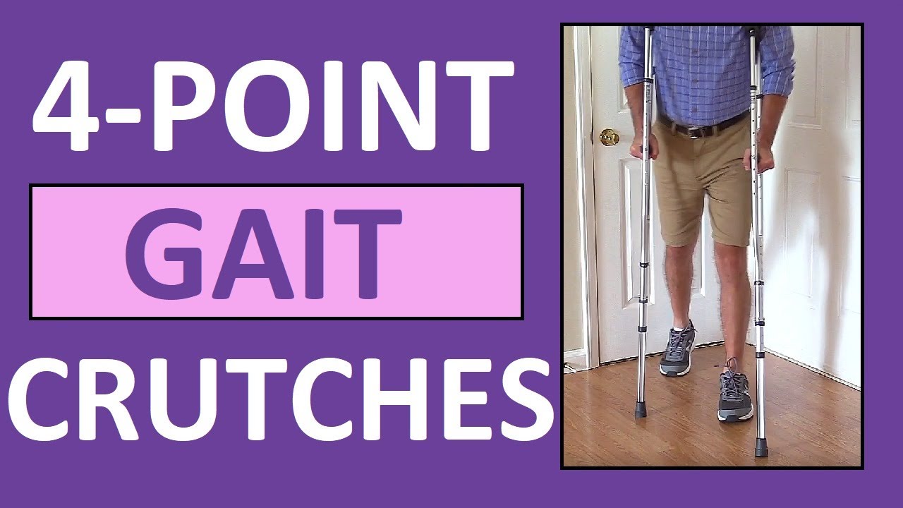
Four-point gait crutches walking pattern demonstration review for
NCLEX assistive devices and nurses.
One of the gaits that you'll have to learn for crutches is the 4-point gait. An example of a four point gait crutch pattern would be the patient moving the right crutch first (on the injured side), followed by the left foot, then the left crutch, and then the right foot. Then, you'll repeat this pattern.
In addition to this video, we have an entire compilation that features the various crutch gait patterns, as well as walkers and canes:
https://www.youtube.com/watch?v=k2-w3LZlCVk
#crutches
#nclex
#nursing
#nurse
Website: https://www.registerednursern.com/
More Videos: https://www.youtube.com/watch?v=R2XMro13dD0&list=UUPyMN8DzkFl2__xnTEiGZ1w
Nursing Gear: https://teespring.com/stores/registerednursern
Instagram: https://www.instagram.com/registerednursern_com/
Facebook: https://www.facebook.com/RegisteredNurseRNs
Twitter: https://twitter.com/NursesRN
Popular Playlists:
NCLEX Reviews: https://www.youtube.com/playli....st?list=PLQrdx7rRsKf
Fluid & Electrolytes: https://www.youtube.com/playli....st?list=PLQrdx7rRsKf
Nursing Skills: https://www.youtube.com/playli....st?list=PLQrdx7rRsKf

I talk about 5 Essential Skills you need as a nurse. These skills are timeless in the fat that you will always need to use them at some level. Of course specific skills are good to have as well but these skills are universal and can help you in other areas of life as well.
NURSING SCHOOL STUDY RESOURCES: https://sellfy.com/nursingschoolstudyNURSING
PHARMACOLOGY: https://sellfy.com/p/fnoy/
INSTAGRAM:https://www.instagram.com/your_mentor_rn/?hl=en
PERSONAL INSTAGRAM: https://www.instagram.com/crosby_steen/
MEDIUM ARTICLES: https://medium.com/@rnacademy1..../7-tips-for-nursing-
AMAZON PRIME STUDENT DISCOUNT: https://amzn.to/2OIleAe
VIDEO GEAR
Camera: G7X Markii - https://amzn.to/2na3OR8
Phone: Galaxy Note 8- https://amzn.to/2nboHM3
Audio: Zoom H4NPro Audio Recorder- https://amzn.to/2vktlf8
Computer: 13 inch Macbook Pro- https://amzn.to/2ndhISw
INSTAGRAM TV https://www.instagram.com/crosby_steen/
Hi Guys! My name is Crosby Steen. I am a Nursing Educator, and ER Travel Nurse. I do videos on daily science based news and travel, with the goal of providing value for you in science based education and travel nursing. Any questions hit me up in the comments or Email below.....
PRIVATE TUTORING OR VIDEO REQUESTS CONTACT:
crosby.steen@gmail.com
MUSIC BY: https://andrewapplepie.com/ and copyrighted by Epidemic Sound
Music by Joakim Karud http://youtube.com/joakimkarud
Music by DJ Quads


Ellis demonstrates how to connect an NG tube to suction.
#NCLEX #ClinicalSkills #HESI #Kaplan #ATI #NursingSchool #NursingStudent #Nurse #RN #PN #Education #LVN #LPN #NurseEducator
🚨 Reminder: shipping deadlines are looming 👀
🎁 Regular Shipping: Order by Friday, December 15
🚀 Expedited Shipping: Order by Monday, December 18
🔍 Still searching for last-minute gifts? Consider a Level Up RN Gift Card! 💌 It’s not only a thoughtful present but also the perfect way to share treasures like Pharmacology Flashcards OR digital treasures like Flashables Digital Nursing Flashcards & the Level Up RN membership. Give the gift of knowledge this holiday season! 🧠⚡️💖 bit.ly/LevelUpRNGC
🚪 Access our Cram Courses, Quizzes and Videos all in one ad free space with Level Up RN Membership https://bit.ly/LevelUpRNMembership
Want more ways to MASTER Clinical Skills? Check out our flashcards & videos!
👇👇👇👇👇👇👇👇👇👇
👉 https://bit.ly/clinicalnursingskills 👈
☝️👆☝️👆☝️👆☝️👆☝️👆
This is your one-stop-shop for materials to help you LEARN & REVIEW so you can PASS Nursing School.
🤔🤔🤔 DO YOU WANT TO PASS your classes, proctored exams and the NCLEX? 🤔🤔🤔 Our resources are the best you can buy. They are built with a single goal: help you pass with no fluff. Everything you need, and nothing you don’t. Don’t take our word for it, though! Check out our hundreds of ⭐️⭐️⭐️⭐️⭐️ reviews from nurses who passed their exams and the NCLEX with Level Up RN.
🗂️ Our Ultimate Nursing School Survival kit is your number 1 resource to get through nursing school and to pass the NCLEX. Whether you're just starting school or you’re already prepping for the NCLEX, this bundle of flashcards is the best you can buy. It covers all the information you need to know to pass all your exams and it has FREE shipping!
➡️ https://bit.ly/TUNSSK ⬅️
L👀king for EVEN MORE resources to survive Nursing School? Make your Nursing School experience your own! Life’s difficult enough—learning shouldn’t be.
🪅 Games https://nursesquad.com
💻 Digital resources https://bit.ly/NursingStudyCourses
📅 Organizational tools https://bit.ly/OrganizingSchool
✨Want perks? Join our channel!
https://youtube.com/leveluprn/join
🏷 Head to https://leveluprn.com/specials for all our latest deals!🥳️
📧 LOOKING FOR FREE RESOURCES TO HELP WITH YOUR EXAMS? Get exclusive tips, latest video releases and more delivered to your email!
➡️ https://leveluprn.com/signup ⬅️
⚕ 👩 LEVEL UP NURSE SQUAD 👩⚕️
All of the nurses at Level Up RN are here to help! Cathy Parkes started helping her fellow classmates back when she was in nursing school, tutoring so they could pass their exams and graduate. After she got her BSN and started working as an RN at Scripps Encinitas Hospital, she started this YouTube channel to help nursing students around the world. Since then she has built a team of top-notch dedicated nurses and nurse educators who are focused on improving nursing education and supporting career advancement for nurses everywhere. With flashcards, videos, courses, organizational tools and more, we are singularly focused on helping students and nurses Level Up on their exams and nursing careers.

Learn what's working for other Nursing Students! Check out our Top 10 Most Popular Lessons Here: https://bit.ly/3nda5u3
FREE Nursing School Cheat Sheets at: http://www.NURSING.com
Get the full lesson here: https://nursing.com/lesson/ski....lls-04-02-ng-tube-ma
Welcome to the NURSING Family, we call it the most supportive nursing cohort on the planet.
At NURSING.com, we want to help you remove the stress and overwhelm of nursing school so that you can focus on becoming an amazing nurse.
Check out our freebies and learn more at: (http://www.nursing.com)
NG Tube Management (Nursing Skills)
In this video lesson, we will look at some of the things that you need to do when you are managing a patient that already has an NG tube or (nasogastric tube). Level up your nursing skills game with these helpful nursing tips. See video bookmarks below:
Bookmarks:
0:00 Intro
0:19 Measure tube length
0:58 Flush tube
1:52 Measure residuals
3:07 Return residuals
4:09 Clamp tube
4:20 Provide oral and nasal care
Visit us at https://nursing.com/medical-disclaimer/ for disclaimer information.
NCLEX®, NCLEX-RN® are registered trademarks of the National Council of State Boards of Nursing, INC. and hold no affiliation with NURSING.com.

Please remember that this video is to be used for educational purposes. You must follow your facility or colleges' policies and procedure checklists to ensure you are completing the skill satisfactorily. Thanks for watching!
Music from #Uppbeat (free for Creators!):
https://uppbeat.io/t/swoop/blue-sea
License code: W9DFUQ4II7YVHA59


Ellis demonstrates how to perform good hand hygiene with soap and water.
Our Critical Nursing Skills video tutorial series is taught by Ellis Parker MSN, RN-BC, CNE, CHS and intended to help RN and PN nursing students study for your nursing school exams, including the ATI, HESI and NCLEX.
#NCLEX #ClinicalSkills #HandHygiene #HESI #Kaplan #ATI #NursingSchool #NursingStudent #Nurse #RN #PN #Education #LVN #LPN #nurseeducator
00:00 What Is Good Hand Hygiene?
00:27 Prepping to wash hands
01:01 Proper hand washing technique
01:53 How to dry hands
02:14 Proper technique to turn off faucet
🚨 Reminder: shipping deadlines are looming 👀
🎁 Regular Shipping: Order by Friday, December 15
🚀 Expedited Shipping: Order by Monday, December 18
🔍 Still searching for last-minute gifts? Consider a Level Up RN Gift Card! 💌 It’s not only a thoughtful present but also the perfect way to share treasures like Pharmacology Flashcards OR digital treasures like Flashables Digital Nursing Flashcards & the Level Up RN membership. Give the gift of knowledge this holiday season! 🧠⚡️💖 bit.ly/LevelUpRNGC
🚪 Access our Cram Courses, Quizzes and Videos all in one ad free space with Level Up RN Membership https://bit.ly/LevelUpRNMembership
Want more ways to MASTER Clinical Skills? Check out our flashcards & videos!
👇👇👇👇👇👇👇👇👇👇
👉 https://bit.ly/clinicalnursingskills 👈
☝️👆☝️👆☝️👆☝️👆☝️👆
This is your one-stop-shop for materials to help you LEARN & REVIEW so you can PASS Nursing School.
🤔🤔🤔 DO YOU WANT TO PASS your classes, proctored exams and the NCLEX? 🤔🤔🤔 Our resources are the best you can buy. They are built with a single goal: help you pass with no fluff. Everything you need, and nothing you don’t. Don’t take our word for it, though! Check out our hundreds of ⭐️⭐️⭐️⭐️⭐️ reviews from nurses who passed their exams and the NCLEX with Level Up RN.
🗂️ Our Ultimate Nursing School Survival kit is your number 1 resource to get through nursing school and to pass the NCLEX. Whether you're just starting school or you’re already prepping for the NCLEX, this bundle of flashcards is the best you can buy. It covers all the information you need to know to pass all your exams and it has FREE shipping!
➡️ https://bit.ly/TUNSSK ⬅️
L👀king for EVEN MORE resources to survive Nursing School? Make your Nursing School experience your own! Life’s difficult enough—learning shouldn’t be.
🪅 Games https://nursesquad.com
💻 Digital resources https://bit.ly/NursingStudyCourses
📅 Organizational tools https://bit.ly/OrganizingSchool
✨Want perks? Join our channel!
https://youtube.com/leveluprn/join
🏷 Head to https://leveluprn.com/specials for all our latest deals!🥳️
📧 LOOKING FOR FREE RESOURCES TO HELP WITH YOUR EXAMS? Get exclusive tips, latest video releases and more delivered to your email!
➡️ https://leveluprn.com/signup ⬅️
⚕ 👩 LEVEL UP NURSE SQUAD 👩⚕️
All of the nurses at Level Up RN are here to help! Cathy Parkes started helping her fellow classmates back when she was in nursing school, tutoring so they could pass their exams and graduate. After she got her BSN and started working as an RN at Scripps Encinitas Hospital, she started this YouTube channel to help nursing students around the world. Since then she has built a team of top-notch dedicated nurses and nurse educators who are focused on improving nursing education and supporting career advancement for nurses everywhere. With flashcards, videos, courses, organizational tools and more, we are singularly focused on helping students and nurses Level Up on their exams and nursing careers.00:00 Good Hand Hygiene?

Tummy Tuck ( Classic Method ) : Surgery | 3D Animation
How long does tummy tuck last?
Tummy tuck results are considered permanent, insofar that the fat cells and skin removed during an abdominoplasty cannot grow back. Likewise, the internal sutures placed to repair abdominal muscles are designed to remain in place indefinitely.
What is tummy tuck surgery?
A tummy tuck — also known as abdominoplasty — is a cosmetic surgical procedure to improve the shape and appearance of the abdomen. During a tummy tuck, excess skin and fat are removed from the abdomen. Connective tissue in the abdomen (fascia) usually is tightened with sutures as well.
How much does tummy tuck cost?
How much does it cost? It can cost from about £5,000 to £10,000 to have an abdominoplasty in the UK, plus the cost of any consultations or follow-up care.
How painful is a tummy tuck?
A tummy tuck requires significant downtime
At the beginning, you will be fatigued, swollen and sore. It is normal to have moderate pain during these first several days, although this will steadily improve. It is vital to allow yourself time to focus on rest and healing.
What is the disadvantage of tummy tuck?
The cons of a tummy tuck include: A full abdominoplasty is a major operation with a considerable recovery. Expect to postpone strenuous activities for at least 6 weeks. Results take time.
Is tummy tuck more painful than C section?
That's something many women want to know. While patients have different experiences, most plastic surgeons would agree that a cesarean section is more painful than most tummy tucks.
- Tummy tuck
- Abdominoplasty
- Abdominal tuck
- Tummy tuck procedure
- Tummy tuck process
- Tummy tuck surgery
- Tummy tuck operation
- Tummy tuck video
- Tummy tuck recovery
- Tummy tuck before and after
- Abdominoplasty surgery
- Abdominal contouring surgery
- Postpartum tummy tuck
- Post pregnancy tummy tuck
- Mini tummy tuck
- Tummy tuck cost
- Tummy tuck risks
- Tummy tuck complications
- How long does a tummy tuck take
- Tummy tuck scarring
- Tummy tuck skin removal
- Tummy tuck muscle tightening
#tummytuck
#abdominoplasty
#plastic_surgery
#cosmetic_surgery
#body_contouring
#tummy_tuck_surgery
#surgery
#cosmetic_procedure
#beauty
#health
#fitness
#medical_animation
#3d_animation
#medical_video
#explainer_video
#education
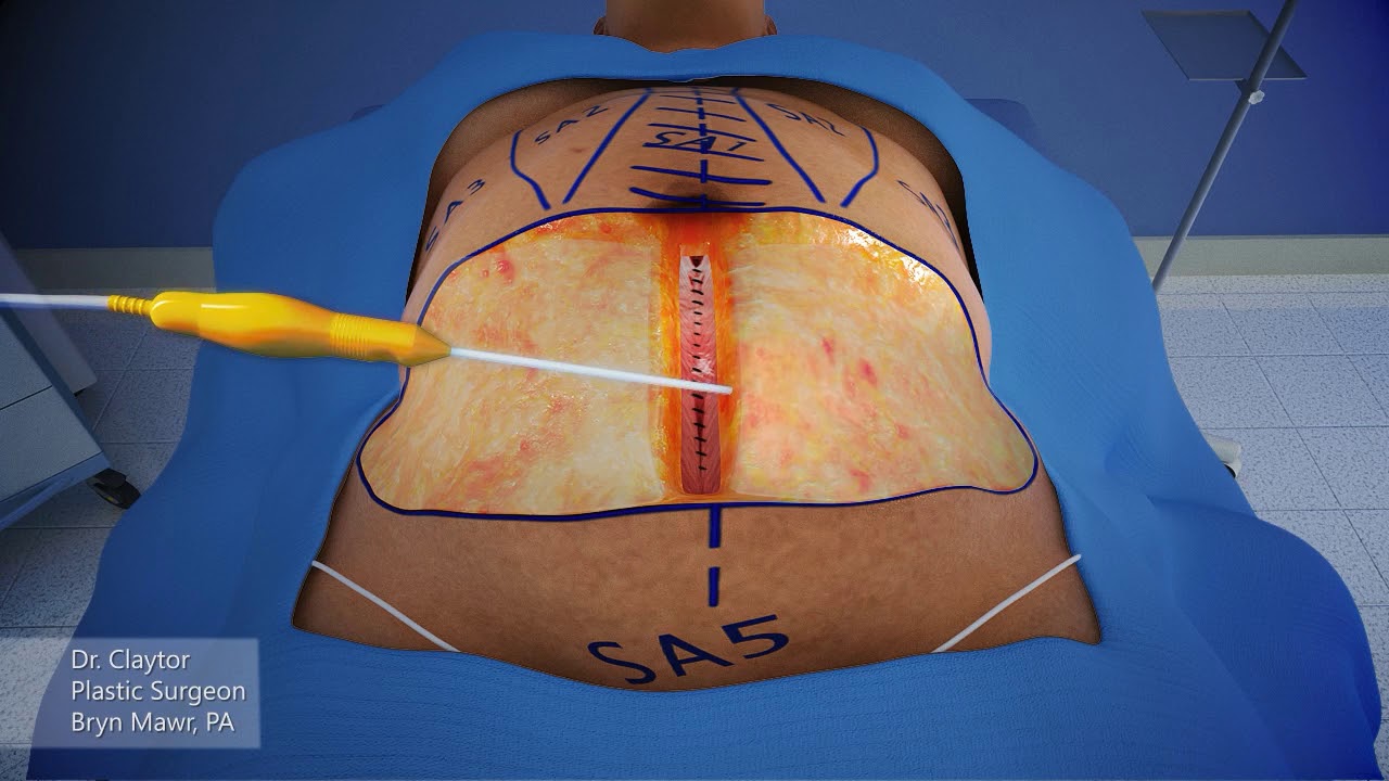
Dr. Claytor uses a 3-D animation to demonstrate how a drainless tummy tuck combined with liposuction can effectively reduce excess skin and fat on the abdomen WITHOUT the need for drains during post-op recovery!
Learn more about Dr. Claytor’s drainless tummy tucks here: https://www.cnplasticsurgery.c....om/procedures/body/t
R. Brannon Claytor, MD, FACS is a renowned double board-certified plastic surgeon and director of Claytor Noone Plastic Surgery, a premium plastic surgery practice in Bryn Mawr, PA that proudly serves the Philadelphia, Main Line, and surrounding areas. Dr. Claytor’s superb skill and results have been recognized for over a decade, earning him numerous awards in both local and national publications, including Philadelphia Magazine, Main Line Today, and Newsweek.
Together, Dr. Claytor and his experienced aesthetics team provide a variety of surgical and non-surgical procedures for the face, breasts, and body to help you look and feel your best. To learn more about how Dr. Claytor and our entire staff can help you reach your goals, please visit our website or give us a call at 610-527-4833.
About Dr. Claytor: https://www.cnplasticsurgery.c....om/our-practice/dr-r
Claytor Noone Plastic Surgery: https://www.cnplasticsurgery.com/
Essential guide to plastic surgery (procedures, costs, planning and more): https://www.cnplasticsurgery.c....om/our-practice/esse
Questions? Contact us online: https://www.cnplasticsurgery.com/contact-us/

A tummy tuck is a surgical process that removes excess fat and skin. Learn more about the procedure by watching this video!
Looking to book a consultation? Call Zuri Plastic Surgery now at 786-804-1603 or DM us today to schedule a complimentary consultation with Dr. Z.
Un tummy tuck es un procedimiento quirúrgico que elimina el exceso de grasa y piel. ¡Aprenda más sobre este procedimiento viendo este video!
¿Quiere agendar una consulta? Llame a Zuri Plastic Surgery ahora al 786-804-1603 o envíenos un DM hoy para programar una consulta gratuita con el Dr. Z.

If you’ve lost a significant amount of weight, either after pregnancy or through exercise and dietary changes, excess skin and weakened abdominal muscles can leave you self-conscious about your appearance. In this video, Dr. Catherine Hannan and Dr. Lauren Patrick, two of our Board-Certified Plastic Surgeons, are performing a Tummy Tuck (Abdominoplasty) surgery. Tummy Tuck surgery gets rid of the excess skin, as well as tightens your abdominal muscles, resulting in a flatter and smoother abdomen. The results of the surgery are permanent except in cases of large weight gain or pregnancy after surgery.
We are so excited to have taken a part in our patient's body transformation journey!
Before & After Gallery:
https://www.westendplasticsurg....ery.com/surgical/bod
To learn more, visit our website or call (202) 785-4187
http://www.westendplasticsurgery.com
~~~~~~~~~~~~~~~~~~~
Social Media:
✨ Instagram: http://www.instagram.com/westendplasticsurgery
✨ Facebook: http://www.facebook.com/westendplasticsurgery
✨ Twitter: http://www.twitter.com/weplasticsurg
✨ Blog: https://www.westendplasticsurgery.com/blog
✨ Business Inquiries: info@westendplasticsurgery.com
~~~~~~~~~~~~~~~~~~~
#TummyTuck #Abdominoplasty

The MINI tummy-tuck is a lesser variant of the classic tummy tuck. The MINI tummy-tuck always involved skin excision (often a scar revision and skin excision of the flabby skin over a C-section scar or hysterectomy or laparotomy scar) but may also involve liposuction, umbilical floating, etc. Commonly it will not include any muscle repair otherwise it it now a classic tummy tuck (aka abdominoplasty). Cost varies depending on the components involved. Here, Toronto Aesthetic Plastic Surgeon Dr Marc DuPéré describes a MINI tummy-tuck done on a patient who had a Brazilian Butt Lift before (and skin harvesting from abdomen) and a recent 20 lbs weight loss, a patient who wants more liposuction to abdomen and flanks and whose skin has now lost elasticity, hence the requirement for this small skin excision. Dr DuPéré also explains what UMBILICAL floating means. Dr DuPéré performs more than 5 different techniques of tummy-tucks in Toronto and the technique chosen reflects the patient’s expectations and anatomy. Call us if interested in learning about YOUR options for a flatter tummy! 📱 416-929-9800
Patient consent obtained. Thank you to my patient.
Visage Clinic Toronto
https://www.visageclinic.com/
(416) 929-9800
101-133 Hazelton Avenue, Toronto, ON M5R 0A6
https://www.facebook.com/VisageClinic/
https://www.instagram.com/VisageClinicDrDuPere/
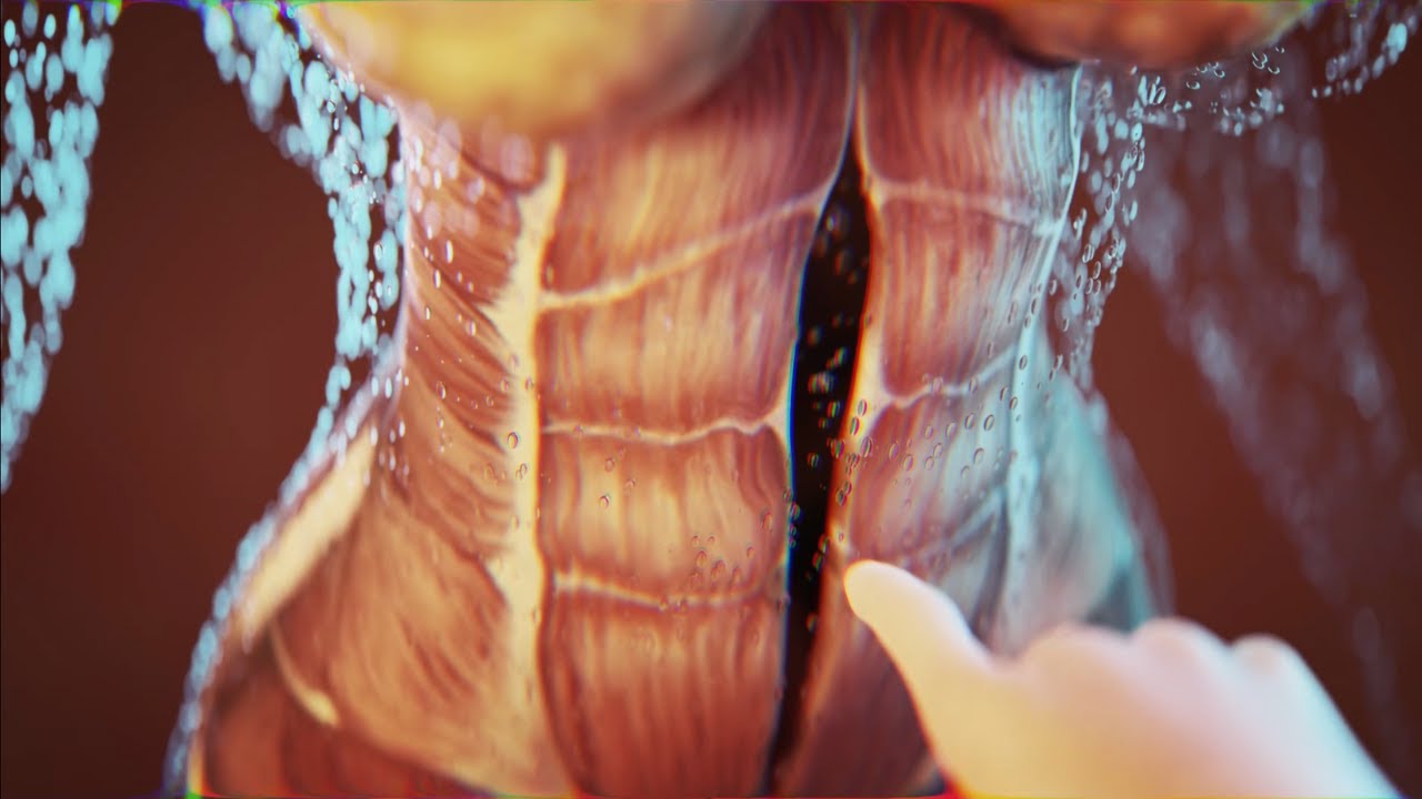
Today I'm using the best 3D animation to explain WHAT IS DIASTASIS RECTI and what you need to know about diastasis recti after pregnancy! Grab the Complete Diastasis Recti Healing Guide: https://landing.mailerlite.com..../webforms/landing/n0
If you are't sure what video to start with and you just want step-by-step daily instructions you can start my 30-day core healing program. You get a new 10-min core healing video daily for 30 days. https://pregnancyandpostpartum....tv.thinkific.com/cou
How I healed my 4-finger diastasis recti gap:
Jessica Pumple is a registered dietitian, and pre & postnatal fitness instructor and certified pregnancy and postpartum core exercise specialist (CPES). She helps pregnant women stay fit, have healthy babies, and easier labors. She helps new moms with postpartum recovery, to heal and strengthen their core and feel energized after pregnancy!
If you enjoy our content subscribe to our channel, hit the bell button, leave a comment and share with your friends so I can make you more of the videos you enjoy!
Disclaimer: This is general postnatal fitness only. Please check with your doctor or health care provider to see if this video is safe for you. Wait until you get clearance (usually 4-6 weeks or 6-8 weeks after a c-section).You are responsible for your own safety. Don’t do anything that feels unsafe for you or baby. Stop if you have any pain or discomfort, bleeding, chest pain or shortness of breath, dizziness or if you feel unwell. P&P Health Inc., Pregnancy and Postpartum TV and Jessica Pumple are not liable in any way for any injury, loss, damages, costs or expenses suffered by you in relation to this video or its content.
Copyright 2023 P&P Health Inc. All rights reserved
#diastasisrecti #whatisdiastasisrecti #3danimation
Music: Epidemic Sound

Dr. Alex Campbell and Dr. Carolina Restrepo of Premium Care Plastic Surgery in Cartagena, Colombia perform a Mommy Makeover on an international patient. Watch the procedure as Dr. Campbell and Dr. Restrepo work together to offer this patient more surgery in less time, which leads to a quicker recovery and better results.
