- Physical Examination
- Surgical Examination
- Ophthalmology
- Clinical Skills
- Orthopedics
- Surgery Videos
- Laparoscopy
- Pediatrics
- Funny Videos
- Cardiothoracic Surgery
- Nursing Videos
- Plastic Surgery
- Otorhinolaryngology
- Histology and Histopathology
- Neurosurgery
- Dermatology
- Pediatric Surgery
- Urology
- Dentistry
- Oncology and Cancers
- Anatomy Videos
- Health and Fitness
- Radiology
- Anaesthesia
- Physical Therapy
- Pharmacology
- Interventional Radiology
- Cardiology
- Endocrinology
- Gynecology
- Emergency Medicine
- Psychiatry and Psychology
- Childbirth Videos
- General Medical Videos
- Nephrology
- Physiology
- Diet and Food Health
- Diabetes Mellitus
- Neurology
- Women Health
- Osteoporosis
- Gastroenterology
- Pulmonology
- Hematology
- Rheumatology
- Toxicology
- Nuclear Medicine
- Infectious Diseases
- Vascular Disease
- Reproductive Health
- Burns and Wound Healing
- Other
Top videos
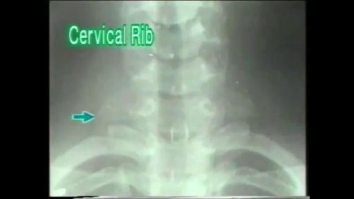
A cervical rib in humans is an extra rib which arises from the seventh cervical vertebra. Sometimes known as "neck ribs", their presence is a congenital abnormality located above the normal first rib. A cervical rib is estimated to occur in 0.2% (1 in 500 people) to 0.5% of the population.

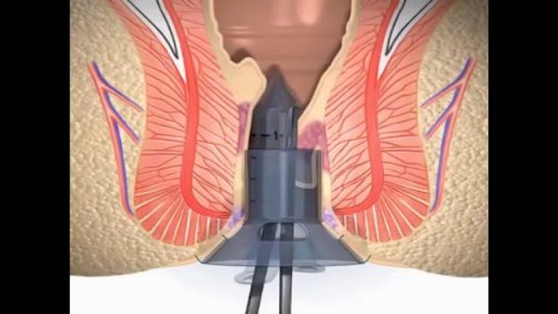
Stapling is used to treat prolapsed hemorrhoids. A surgical staple fixes the prolapsed hemorrhoid back into place inside your rectum and cuts off the blood supply so that the tissue will shrink and be reabsorbed. Stapling recovery takes less time and is less painful than recovery from a hemorrhoidectomy.

Infected Tattoo Abscess
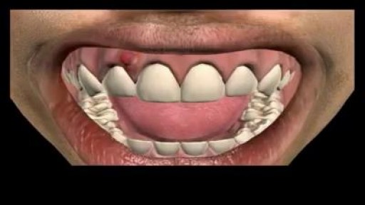
Tooth Abscess Relief
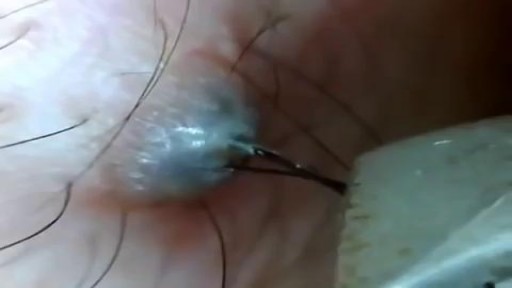
Ingrown Hair Removal Video
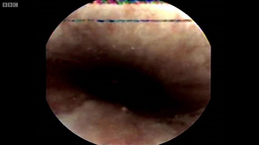
There’s a strange, mysterious world inside us, an alien-looking environment that turns the food we eat into nutrients that keep us alive. Michael Mosley swallows a camera to take a closer look.

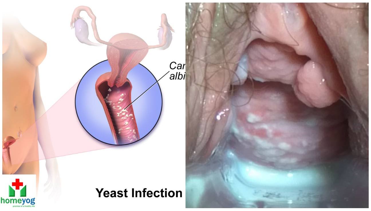
Watch that video to know the Female Genital Infections Causes and treatments.
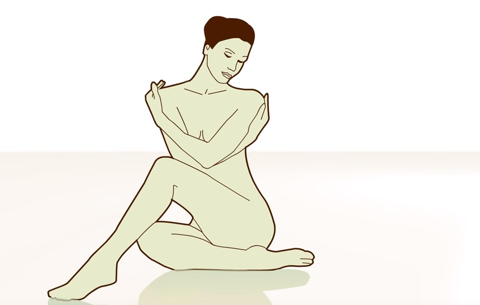
Watch that video to know everything about male and female orgasm
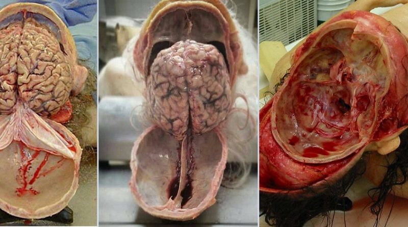
Watch that Human Brain Removal During Autopsy
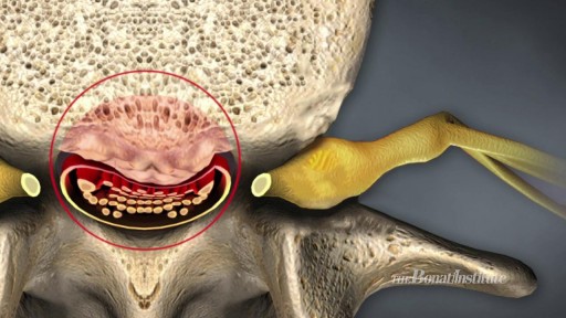
Watch Spinal Stenosis Videos Spinal stenosis occurs when the spinal cord in the neck (cervical spine) or the spinal nerve roots in the lower back (lumbar spine) are compressed. Symptoms of lumbar stenosis often include leg pain (sciatica) and leg tingling, weakness, or numbness. Arm pain is a typical symptom of cervical spinal stenosis. For cervical spinal stenosis with myelopathy, difficulty with coordination often occurs. Stenosis treatment may include non-surgical options (exercise, anti-inflammatory medication, epidural injections, and activity modification) or back surgery.

Watch that video to know the Difference Between White and Clear Sperm
Renal replacement therapies are a set of interventions and techniques aimed at replacement of the filtering function of the kidneys. These include both dialysis and renal transplant. In this presentation we will talk about the indication and modalities of dialysis. This includes hemodialysis, peritoneal dialysis and continuous renal replacement therapies. The circuit diagram of each modalities along with its procedure and complications are also discussed.
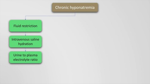
Hyponatremia is defined as a serum sodium of less than 135 Meq per litre and occurs in upto 22 % of hospitalised patients. The causes of hyponatremia may be understood based on the pre-existing volume status of the patient which may either be hypovolemic, euvolemic or hypervolemic hyponatremia. This presentation discusses in detail, the causes of these underlying conditions. Also mentioned are the clinical features and management options and therapeutic sodium targets in patients with hyponatremia. Drugs such as demeclocycline and vaptans (Tolvaptan, Conivaptan) are also mentioned as management options which may be used on a case to case basis. Finally, the all important targets of sodium correction over 24 hours are also mentioned, along with a practical formula for calculation of sodium deficit which is explained with an example.
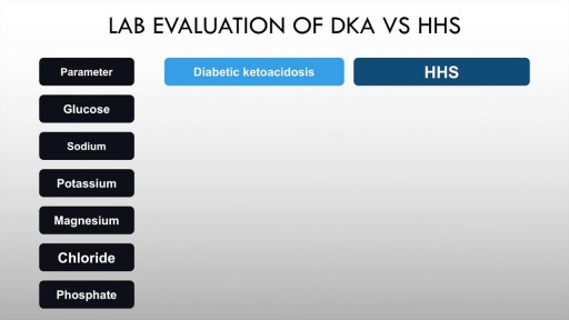
Diabetic ketoacidosis is an acute complication of uncontrolled hyperglycaemia characterised by high anion gap metabolic acidosis, dehydration and other metabolic abnormalities. Upto half of patients with Type 1 diabetes mellitus may have DKA. The incidence in T2DM is also rising. Precipitants include acute illness such as myocardial infarction, trauma and infection. Paitents of diabetic ketoacidosis may present with vomiting, pain abdomen and lethargy. Mental obtundation may also be present. Management of diabetic ketoacidosis revolves around administration of IV normal saline, insulin, replacement of potassium with frequent monitoring of sugars and electrolytes.
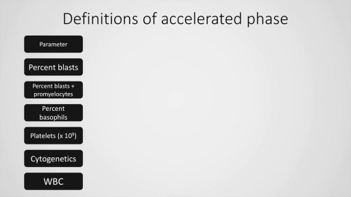
Chronic myeloid leukaemia is a common malignancy worldwide. We have come a long way from the limited treatment options and survival in this condition. Today, CML is a treatable malignancy with more than 80% patients surviving beyond 10 years after diagnosis, in absence of complications. This presentation deals with the definition, diagnostic criteria of chronic phase, accelerated and blastic phase (MD Anderson cancer centre, International bone marrow transplant registry and the WHO for the latter two) and management (first and second generation tyrosine kinase inhibitors) of this condition. Finally, a stepwise approach to chronic myeloid leukaemia is also presented including the definitive modality of treatment, allogeneic stem cell transplantation.
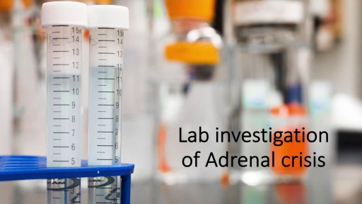
A detailed description of Adrenal insufficiency (Addison's disease) including basic physiology of the HPA axis, causes of primary and secondary insufficiency, clinical features of acute and chronic adrenal insufficiency. Lab testing for Addison's disease is also dealt with in detail. The management, both short term and long term are discussed in detail.