- Physical Examination
- Surgical Examination
- Ophthalmology
- Clinical Skills
- Orthopedics
- Surgery Videos
- Laparoscopy
- Pediatrics
- Funny Videos
- Cardiothoracic Surgery
- Nursing Videos
- Plastic Surgery
- Otorhinolaryngology
- Histology and Histopathology
- Neurosurgery
- Dermatology
- Pediatric Surgery
- Urology
- Dentistry
- Oncology and Cancers
- Anatomy Videos
- Health and Fitness
- Radiology
- Anaesthesia
- Physical Therapy
- Pharmacology
- Interventional Radiology
- Cardiology
- Endocrinology
- Gynecology
- Emergency Medicine
- Psychiatry and Psychology
- Childbirth Videos
- General Medical Videos
- Nephrology
- Physiology
- Diet and Food Health
- Diabetes Mellitus
- Neurology
- Women Health
- Osteoporosis
- Gastroenterology
- Pulmonology
- Hematology
- Rheumatology
- Toxicology
- Nuclear Medicine
- Infectious Diseases
- Vascular Disease
- Reproductive Health
- Burns and Wound Healing
- Other
Top videos

Blocked coronary arteries.

At Hutzel Women's Hospital, Dr. Giancarlo Mari performs breakthrough in-utero surgery to save the lives of high-risk twins developing with a rare "shared" circulatory problem. ~ Detroit Medical Center
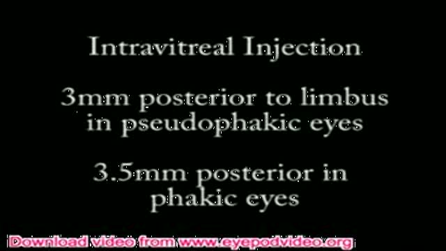
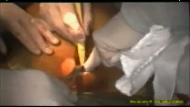
Instructional video explaining intravitreal injection technique used in endophthalmitis (a serious eye infection), macular degeneration, and other eye diseases.
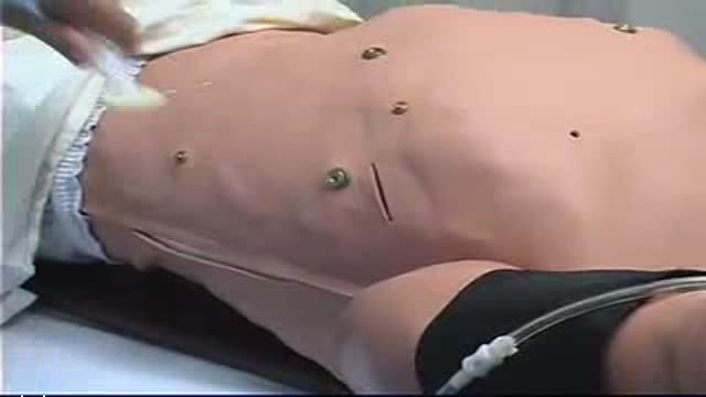
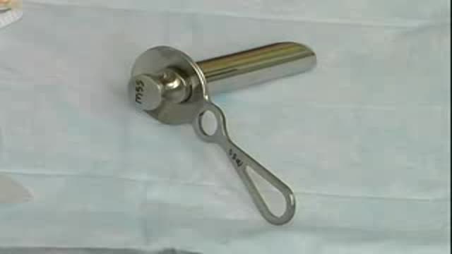
Chest Tube Placement
Ulnar Gutter Cast
39 Yr. Male with Aortic Stenosis and Incompetance and Good LV Function. The Patient is an athlete and did not want to take oral anticoagulants so opted out for a Bio-prosthesis. A 23mm Hancock II Porcine Xenograft was used in this operation. Usually central aortic and Rt. Atrial cannulation is per...formed with this procedure, however on occasions Percutaneous (Seldinger Technique) Femoro Femoral artery cannulation is used. The Kit is manufactured by DLP and consists of a 20mm Arterial cannula and a 29mm two stage Rt. Atrial Cannula.
Laparoscopic Tubal Ligation using Filshie Clips. Brought to you by http://nursing-resource.com
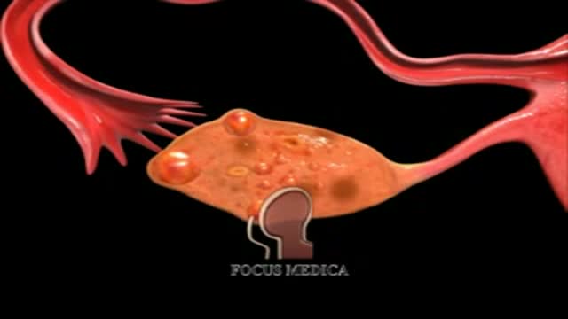
An animation showing what PCO is
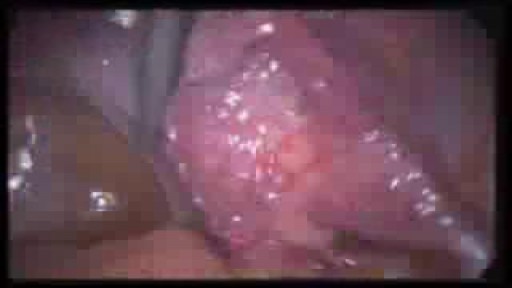
An excellent video demonstrating how a laparoscopy is performed to evaluate the uterus (note a small fibroid appearing as a bulge in the uterus), fallopian tubes and ovaries. Blue dye is injected into the uterus, entering the fallopian tubes and spilling from the end of the tubes into the abdominal cavity, confirming that both tubes are open

An overview of stomach cancer

Patient Greg Grindley communicates with host Bryant Gumbel and his wife for the first time while undergoing deep brain stimulation surgery at University Hospital's Case Medical Center in Cleveland, Ohio.
➡ Subscribe: http://bit.ly/NatGeoSubscribe
About National Geographic:
National Geographic is the world's premium destination for science, exploration, and adventure. Through their world-class scientists, photographers, journalists, and filmmakers, Nat Geo gets you closer to the stories that matter and past the edge of what's possible.
Get More National Geographic:
Official Site: http://bit.ly/NatGeoOfficialSite
Facebook: http://bit.ly/FBNatGeo
Twitter: http://bit.ly/NatGeoTwitter
Instagram: http://bit.ly/NatGeoInsta
Greg's First In-Surgery Conversation | Brain Surgery Live
https://youtu.be/zvqV_2zncNU
National Geographic
https://www.youtube.com/natgeo
CORRECTION: After review of this video, it is clear that this video is of a baby who is near full term (40 weeks) based on the size. Late trimester "abortions" are defined only to viability of a baby (24 weeks) A 24 week baby is much smaller than this baby shown and by definition this is not a late "abortion" procedure. The proper labeling of this video should be management of a deceased breech baby with "head entrapment" as this was almost certainly a naturally occuring delivery and an OB nightmare (Reviewed by Dr. Frederick Bright)

Breast Reduction Surgery video Operation مركز افارا لجراحات التجميل الخدود تكبير الشفايف

Orgasmic childbirth is a new variant of water birth delivery.
Educational video of male patient receiving an anoscopy.
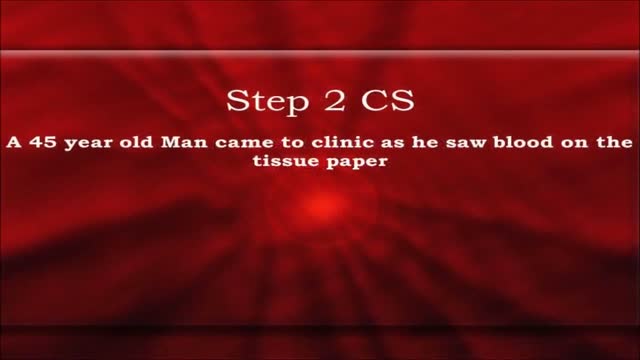
USMLE Step 2 CS - LGIB This is just preview video. To get full access please visit our website : www.usmletutoring.com

Some bodybuilders, particularly at professional level, use substances such as "site enhancement oil", commonly known as synthol, to mimic the appearance of developed muscle where it may otherwise be disproportionate or lagging. This is known as "fluffing". Synthol is 85% oil, 7.5% lidocain, and 7.5% alcohol.Use is legal and many brands are available on the internet.The use of injected oil to enhance muscle appearance had previously been used in the late 19th century before being abandoned due to health risks such as sclerosing lipogranuloma. Its use was revived more recently by bodybuilders. Use can cause pulmonary embolisms, nerve damage, infections, stroke, and the formation of oil-filled oleomas, cysts or ulcers in the muscle. Sesame oil is often used, which can cause allergic reactions such as vasculitis. An aesthetic issue is drooping of muscle under gravity. Surgical methods are also often employed to remove steroid-related gynecomastia in male bodybuilders, and breast implants in female bodybuilders who wish to retain a feminine physique, which can be compromised in terms of breast reduction by intense dieting.
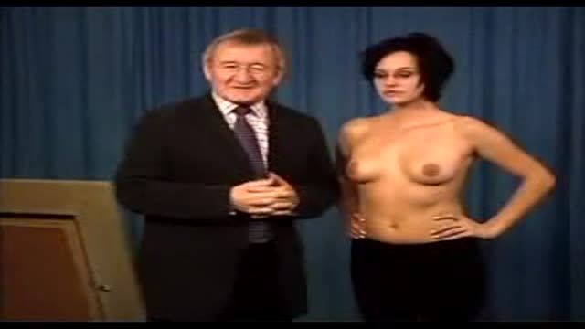
Dr Chris Steele demonstrates a breast examination on a live model. This shows how to check yourself for early signs of tumours, cysts and other symptoms of breast cancer.
Cranial nerves exam 8th to 12th from the USMLE collection
Michael La Corte MD
Ped Card
