- Physical Examination
- Surgical Examination
- Ophthalmology
- Clinical Skills
- Orthopedics
- Surgery Videos
- Laparoscopy
- Pediatrics
- Funny Videos
- Cardiothoracic Surgery
- Nursing Videos
- Plastic Surgery
- Otorhinolaryngology
- Histology and Histopathology
- Neurosurgery
- Dermatology
- Pediatric Surgery
- Urology
- Dentistry
- Oncology and Cancers
- Anatomy Videos
- Health and Fitness
- Radiology
- Anaesthesia
- Physical Therapy
- Pharmacology
- Interventional Radiology
- Cardiology
- Endocrinology
- Gynecology
- Emergency Medicine
- Psychiatry and Psychology
- Childbirth Videos
- General Medical Videos
- Nephrology
- Physiology
- Diet and Food Health
- Diabetes Mellitus
- Neurology
- Women Health
- Osteoporosis
- Gastroenterology
- Pulmonology
- Hematology
- Rheumatology
- Toxicology
- Nuclear Medicine
- Infectious Diseases
- Vascular Disease
- Reproductive Health
- Burns and Wound Healing
- Other
Top videos

Ellis demonstrates how to perform a sterile wound dressing change. It would be appropriate to perform hand hygiene between glove changes.
Our Critical Nursing Skills video tutorial series is taught by Ellis Parker MSN, RN-BC, CNE, CHS and intended to help RN and PN nursing students study for your nursing school exams, including the ATI, HESI and NCLEX.
#NCLEX #ClinicalSkills #woundcare #HESI #Kaplan #ATI #NursingSchool #NursingStudent #Nurse #RN #PN #Education #LVN #LPN #nurseeducator
00:00 What to expect
00:51 Prepping for wound dressing change
1:15 Removing the old wound dressing
1:40 Assessing a wound
2:05 Setting up sterile field
2:49 Sterile gloving
4:02 Preparing equipment for wound dressing change
5:09 Cleaning a wound
6:13 Drying a wound
6:28 Packing a wound
7:19 Covering a wound
7:47 Labeling a wound dressing
🚨 Reminder: shipping deadlines are looming 👀
🎁 Regular Shipping: Order by Friday, December 15
🚀 Expedited Shipping: Order by Monday, December 18
🔍 Still searching for last-minute gifts? Consider a Level Up RN Gift Card! 💌 It’s not only a thoughtful present but also the perfect way to share treasures like Pharmacology Flashcards OR digital treasures like Flashables Digital Nursing Flashcards & the Level Up RN membership. Give the gift of knowledge this holiday season! 🧠⚡️💖 bit.ly/LevelUpRNGC
🚪 Access our Cram Courses, Quizzes and Videos all in one ad free space with Level Up RN Membership https://bit.ly/LevelUpRNMembership
Want more ways to MASTER Clinical Skills? Check out our flashcards & videos!
👇👇👇👇👇👇👇👇👇👇
👉 https://bit.ly/clinicalnursingskills 👈
☝️👆☝️👆☝️👆☝️👆☝️👆
This is your one-stop-shop for materials to help you LEARN & REVIEW so you can PASS Nursing School.
🤔🤔🤔 DO YOU WANT TO PASS your classes, proctored exams and the NCLEX? 🤔🤔🤔 Our resources are the best you can buy. They are built with a single goal: help you pass with no fluff. Everything you need, and nothing you don’t. Don’t take our word for it, though! Check out our hundreds of ⭐️⭐️⭐️⭐️⭐️ reviews from nurses who passed their exams and the NCLEX with Level Up RN.
🗂️ Our Ultimate Nursing School Survival kit is your number 1 resource to get through nursing school and to pass the NCLEX. Whether you're just starting school or you’re already prepping for the NCLEX, this bundle of flashcards is the best you can buy. It covers all the information you need to know to pass all your exams and it has FREE shipping!
➡️ https://bit.ly/TUNSSK ⬅️
L👀king for EVEN MORE resources to survive Nursing School? Make your Nursing School experience your own! Life’s difficult enough—learning shouldn’t be.
🪅 Games https://nursesquad.com
💻 Digital resources https://bit.ly/NursingStudyCourses
📅 Organizational tools https://bit.ly/OrganizingSchool
✨Want perks? Join our channel!
https://youtube.com/leveluprn/join
🏷 Head to https://leveluprn.com/specials for all our latest deals!🥳️
📧 LOOKING FOR FREE RESOURCES TO HELP WITH YOUR EXAMS? Get exclusive tips, latest video releases and more delivered to your email!
➡️ https://leveluprn.com/signup ⬅️
⚕ 👩 LEVEL UP NURSE SQUAD 👩⚕️
All of the nurses at Level Up RN are here to help! Cathy Parkes started helping her fellow classmates back when she was in nursing school, tutoring so they could pass their exams and graduate. After she got her BSN and started working as an RN at Scripps Encinitas Hospital, she started this YouTube channel to help nursing students around the world. Since then she has built a team of top-notch dedicated nurses and nurse educators who are focused on improving nursing education and supporting career advancement for nurses everywhere. With flashcards, videos, courses, organizational tools and more, we are singularly focused on helping students and nurses Level Up on their exams and nursing careers.
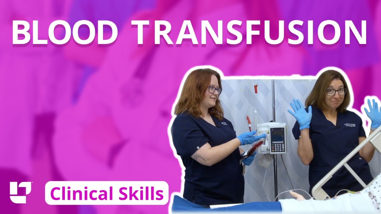
Ellis and Cathy demonstrate how to administer blood to a patient.
Our Critical Nursing Skills video tutorial series is taught by Ellis Parker MSN, RN-BC, CNE, CHS and intended to help RN and PN nursing students study for your nursing school exams, including the ATI, HESI and NCLEX.
#NCLEX #ClinicalSkills #Blood #bloodtransfusion #HESI #Kaplan #ATI #NursingSchool #NursingStudent #Nurse #RN #PN #Education #LVN #LPN
00:00 What to expect blood transfusion
00:26 First steps for a blood transfusion
1:03 Priming the tubing for blood transfusion
2:29 Confirming the blood for transfusion
4:36 Hanging the blood for transfusion
5:06 Clamping a Y-tube
5:34 Priming the blood for transfusion
7:00 Responding to a blood transfusion reaction
🚨 Reminder: shipping deadlines are looming 👀
🎁 Regular Shipping: Order by Friday, December 15
🚀 Expedited Shipping: Order by Monday, December 18
🔍 Still searching for last-minute gifts? Consider a Level Up RN Gift Card! 💌 It’s not only a thoughtful present but also the perfect way to share treasures like Pharmacology Flashcards OR digital treasures like Flashables Digital Nursing Flashcards & the Level Up RN membership. Give the gift of knowledge this holiday season! 🧠⚡️💖 bit.ly/LevelUpRNGC
🚪 Access our Cram Courses, Quizzes and Videos all in one ad free space with Level Up RN Membership https://bit.ly/LevelUpRNMembership
Want more ways to MASTER Clinical Skills? Check out our flashcards & videos!
👇👇👇👇👇👇👇👇👇👇
👉 https://bit.ly/clinicalnursingskills 👈
☝️👆☝️👆☝️👆☝️👆☝️👆
This is your one-stop-shop for materials to help you LEARN & REVIEW so you can PASS Nursing School.
🤔🤔🤔 DO YOU WANT TO PASS your classes, proctored exams and the NCLEX? 🤔🤔🤔 Our resources are the best you can buy. They are built with a single goal: help you pass with no fluff. Everything you need, and nothing you don’t. Don’t take our word for it, though! Check out our hundreds of ⭐️⭐️⭐️⭐️⭐️ reviews from nurses who passed their exams and the NCLEX with Level Up RN.
🗂️ Our Ultimate Nursing School Survival kit is your number 1 resource to get through nursing school and to pass the NCLEX. Whether you're just starting school or you’re already prepping for the NCLEX, this bundle of flashcards is the best you can buy. It covers all the information you need to know to pass all your exams and it has FREE shipping!
➡️ https://bit.ly/TUNSSK ⬅️
L👀king for EVEN MORE resources to survive Nursing School? Make your Nursing School experience your own! Life’s difficult enough—learning shouldn’t be.
🪅 Games https://nursesquad.com
💻 Digital resources https://bit.ly/NursingStudyCourses
📅 Organizational tools https://bit.ly/OrganizingSchool
✨Want perks? Join our channel!
https://youtube.com/leveluprn/join
🏷 Head to https://leveluprn.com/specials for all our latest deals!🥳️
📧 LOOKING FOR FREE RESOURCES TO HELP WITH YOUR EXAMS? Get exclusive tips, latest video releases and more delivered to your email!
➡️ https://leveluprn.com/signup ⬅️
⚕ 👩 LEVEL UP NURSE SQUAD 👩⚕️
All of the nurses at Level Up RN are here to help! Cathy Parkes started helping her fellow classmates back when she was in nursing school, tutoring so they could pass their exams and graduate. After she got her BSN and started working as an RN at Scripps Encinitas Hospital, she started this YouTube channel to help nursing students around the world. Since then she has built a team of top-notch dedicated nurses and nurse educators who are focused on improving nursing education and supporting career advancement for nurses everywhere. With flashcards, videos, courses, organizational tools and more, we are singularly focused on helping students and nurses Level Up on their exams and nursing careers.
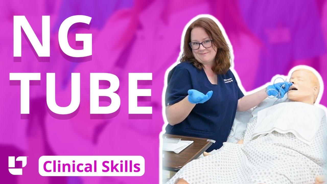
Ellis demonstrates how to insert and then remove an NG tube. This includes drawing gastric residual and checking the pH. After the demonstration, Ellis provides additional tips about clamping the NG tube and using the blue pigtail.
Our Critical Nursing Skills video tutorial series is taught by Ellis Parker MSN, RN-BC, CNE, CHS and intended to help RN and PN nursing students study for your nursing school exams, including the ATI, HESI and NCLEX.
#NCLEX #HESI #Kaplan #ATI #NursingSchool #NursingStudent #Nurse #RN #PN #Education #LVN #LPN #ClinicalSkills #NGTube #nurseeducator
00:00 What to expect
00:30 Preparing NG tube patient
00:56 Preparing NG tube equipment
1:29 Measuring the NG tube
2:02 Preparing for NG tube insertion
2:28 Inserting the NG tube
3:17 Checking placement with pH
4:23 Anchoring with split-tape
5:32 Connecting to suction
6:05 Disconnecting from suction
6:17 What to do before removal?
7:03 Removing NG tube
7:40 Additional tips on clamping
8:31 The blue pigtail
🚨 Reminder: shipping deadlines are looming 👀
🎁 Regular Shipping: Order by Friday, December 15
🚀 Expedited Shipping: Order by Monday, December 18
🔍 Still searching for last-minute gifts? Consider a Level Up RN Gift Card! 💌 It’s not only a thoughtful present but also the perfect way to share treasures like Pharmacology Flashcards OR digital treasures like Flashables Digital Nursing Flashcards & the Level Up RN membership. Give the gift of knowledge this holiday season! 🧠⚡️💖 bit.ly/LevelUpRNGC
🚪 Access our Cram Courses, Quizzes and Videos all in one ad free space with Level Up RN Membership https://bit.ly/LevelUpRNMembership
Want more ways to MASTER Clinical Skills? Check out our flashcards & videos!
👇👇👇👇👇👇👇👇👇👇
👉 https://bit.ly/clinicalnursingskills 👈
☝️👆☝️👆☝️👆☝️👆☝️👆
This is your one-stop-shop for materials to help you LEARN & REVIEW so you can PASS Nursing School.
🤔🤔🤔 DO YOU WANT TO PASS your classes, proctored exams and the NCLEX? 🤔🤔🤔 Our resources are the best you can buy. They are built with a single goal: help you pass with no fluff. Everything you need, and nothing you don’t. Don’t take our word for it, though! Check out our hundreds of ⭐️⭐️⭐️⭐️⭐️ reviews from nurses who passed their exams and the NCLEX with Level Up RN.
🗂️ Our Ultimate Nursing School Survival kit is your number 1 resource to get through nursing school and to pass the NCLEX. Whether you're just starting school or you’re already prepping for the NCLEX, this bundle of flashcards is the best you can buy. It covers all the information you need to know to pass all your exams and it has FREE shipping!
➡️ https://bit.ly/TUNSSK ⬅️
L👀king for EVEN MORE resources to survive Nursing School? Make your Nursing School experience your own! Life’s difficult enough—learning shouldn’t be.
🪅 Games https://nursesquad.com
💻 Digital resources https://bit.ly/NursingStudyCourses
📅 Organizational tools https://bit.ly/OrganizingSchool
✨Want perks? Join our channel!
https://youtube.com/leveluprn/join
🏷 Head to https://leveluprn.com/specials for all our latest deals!🥳️
📧 LOOKING FOR FREE RESOURCES TO HELP WITH YOUR EXAMS? Get exclusive tips, latest video releases and more delivered to your email!
➡️ https://leveluprn.com/signup ⬅️
⚕ 👩 LEVEL UP NURSE SQUAD 👩⚕️
All of the nurses at Level Up RN are here to help! Cathy Parkes started helping her fellow classmates back when she was in nursing school, tutoring so they could pass their exams and graduate. After she got her BSN and started working as an RN at Scripps Encinitas Hospital, she started this YouTube channel to help nursing students around the world. Since then she has built a team of top-notch dedicated nurses and nurse educators who are focused on improving nursing education and supporting career advancement for nurses everywhere. With flashcards, videos, courses, organizational tools and more, we are singularly focused on helping students and nurses Level Up on their exams and nursing careers.

Vital signs help us assess patients in the nursing profession, and there are six common vital signs that we assess as nurses:
1. Heart Rate (Pulse)
2. Respiration Rate
3. Temperature
4. Blood Pressure
5. Pain Rating
6. Oxygen Saturation
This video will demonstrate how to check vital signs (live) on a patient, along with normal rates for each assessment. I also give you a few tips for taking vital signs as a nurse, CNA, or other healthcare profession.
🟣ABG eBook: https://registerednursern.creator-spring.com/
🟣ABG physical book: https://amzn.to/3EsF0Mc (affiliate link)
More nursing skills: https://www.youtube.com/watch?v=G5-Rp-6FMCQ&list=PLQrdx7rRsKfUhd_qQYEbp0Eab3uUKhgKb
Website: https://www.registerednursern.com/
More Videos: https://www.youtube.com/watch?v=R2XMro13dD0&list=UUPyMN8DzkFl2__xnTEiGZ1w
Nursing Gear: https://teespring.com/stores/registerednursern
Instagram: https://www.instagram.com/registerednursern_com/
Facebook: https://www.facebook.com/RegisteredNurseRNs
Twitter: https://twitter.com/NursesRN
Popular Playlists:
NCLEX Reviews: https://www.youtube.com/playli....st?list=PLQrdx7rRsKf
Fluid & Electrolytes: https://www.youtube.com/playli....st?list=PLQrdx7rRsKf
Nursing Skills: https://www.youtube.com/playli....st?list=PLQrdx7rRsKf

Vial medication administration nursing skill. Learn techniques to withdraw medication from a vial using a syringe with a needle.
Medications can come in different forms, such as ampules, vials, tablets, capsules, and so forth. When withdrawing medication from a vial, there are a few things you'll want to know as a nursing student or nurse.
First, there are different needles that can be attached to the syringe. You can use a traditional needle with a beveled tip; you can use a blunt-tip needle to reduce the risk of needle sticks; or you can use a filter needle, which is sometimes required or recommended when drawing medication from a vial, particularly in cases of reconstituted medication.
When withdrawing from a vial, you'll want to do these things (assuming they fit with the protocols and manufacturer's instructions):
NOTE: Some medications or vaccines may require a different technique, so always consult with the manufacturer's instructions.
-gather your supplies
-perform hand hygiene
-clean the vial's top with alcohol prep
-attach the appropriate needle
-stick the needle using a technique to prevent coring of the rubber on the vial (start with 45 degree angle, and as you puncture the vial, rotate the needle to a 90 degree angle in one smooth motion).
-push air into the vial equal to the amount of medication you plan to draw
-invert the vial to withdraw medication
-remove air bubbles
-and much more
See more Nursing Skills: https://www.youtube.com/playli....st?list=PLQrdx7rRsKf
Notes: https://www.registerednursern.....com/how-to-withdraw-
Website: https://www.registerednursern.com/
More Videos: https://www.youtube.com/watch?v=R2XMro13dD0&list=UUPyMN8DzkFl2__xnTEiGZ1w
Nursing Gear: https://teespring.com/stores/registerednursern
Instagram: https://www.instagram.com/registerednursern_com/
Facebook: https://www.facebook.com/RegisteredNurseRNs
Twitter: https://twitter.com/NursesRN
Popular Playlists:
NCLEX Reviews: https://www.youtube.com/playli....st?list=PLQrdx7rRsKf
Fluid & Electrolytes: https://www.youtube.com/playli....st?list=PLQrdx7rRsKf

Pass your tests and improve your grades with the below FREE resources:
1) A FREE 140 Must Know Meds book
Click here to get your FREE copy of the 140 Must Know Meds Book: https://bit.ly/41rxSt0
2) A FREE test-taking tips webinar
Join us for our free test-taking tips webinar to boost your exam scores: https://bit.ly/nursingtesttaking
You can now test your knowledge with a free lesson quiz on NURSING.com!
Click here for your free quiz: https://bit.ly/3uyTWEu
Learn what's working for other Nursing Students! Check out our Top 10 Most Popular Lessons Here: https://bit.ly/3nda5u3
Dressing Changes- Wet to Dry (Nursing Skills)
FREE Nursing School Cheat Sheets at: http://www.NURSING.com
Get the full Dressing Change lesson here: https://nursing.com/lesson/ski....lls-05-04-wound-care
Click here for the related lesson on Wound Assessment: https://nursing.com/lesson/ski....lls-05-02-wound-care
Welcome to the NURSING Family, we call it the most supportive nursing cohort on the planet.
At NURSING.com, we want to help you remove the stress and overwhelm of nursing school so that you can focus on becoming an amazing nurse.
Check out our freebies and learn more at: (http://www.nursing.com)
Dressing Changes- Wet to Dry (Nursing Skills):
In this video we’re going to look at how to do a wet to dry dressing change. Wound care and dressing changes should be performed at least daily or more often depending on orders. Dressing changes should be sterile to avoid introducing any new bacteria to the wound and to promote wound healing.
Bookmarks:
0:05 Introduction
0:10 Wound Assessment link above
0:24 Dressing Change Prep
1:24 Wet vs Dry Gauze
1:37 Soaking Gauze
2:00 Gauze Ring Out
2:25 Packing the wound
3:00 Covering the wound bed
3:37 Dry gauze barrier
4:00 ABD pad application
4:46 Documentation
4:54 Outro
Visit us at https://nursing.com/medical-disclaimer/ for disclaimer information.
NCLEX®, NCLEX-RN® are registered trademarks of the National Council of State Boards of Nursing, INC. and hold no affiliation with NURSING.com.

Glass ampules are often used to store medication, and as a nurse, you'll need to know how to use them.
In this video, I demonstrate how to clean an ampule using alcohol prep, how to open (or break) an ampule, as well as how to dispose of the ampule.
In addition, I show how to use an ample filter straw while drawing up (withdrawing) medication, how to use the syringe, and how to remove the air bubbles in the syringe.
This is another video in our series on clinical nursing skills.
Notes: https://www.registerednursern.....com/how-to-withdraw-
Website: https://www.registerednursern.com/
More Videos: https://www.youtube.com/watch?v=R2XMro13dD0&list=UUPyMN8DzkFl2__xnTEiGZ1w
Nursing Gear: https://teespring.com/stores/registerednursern
Instagram: https://www.instagram.com/registerednursern_com/
Facebook: https://www.facebook.com/RegisteredNurseRNs
Twitter: https://twitter.com/NursesRN
Popular Playlists:
NCLEX Reviews: https://www.youtube.com/playli....st?list=PLQrdx7rRsKf
Fluid & Electrolytes: https://www.youtube.com/playli....st?list=PLQrdx7rRsKf
Nursing Skills: https://www.youtube.com/playli....st?list=PLQrdx7rRsKf

Full Tummy Tuck 3D Video - http://drlandsman.com
Look great... feel great
•Smart Liposuction + Liposculpture
•Abdominplasty (Tummy Tuck)
+ Full Mini Modified
•Brazilian Lift with Fat Transfer
•Vaginal Aesthetics & Rejuvenation
•Laser Hair Removal
•Full Body Lift
•Thigh lift
•Brachioplasty (Arm Lift) + Short Scar
Expertise in Body Contouring
Board Certified Plastic Surgeon
Expertise in body contouring combines skin excision techniques and advanced fat contouring technology
Weight control personalized training and smoking cessation results in a healthier lifestyle improved shape and longer lasting results
With over 2 decades of experience Dr Lloyd Landsman provides state of the art cosmetic and plastic surgery
Dr Landsman integrates the finest and safest products with the newest procedures
A customized treatment plan is created for each patient utilizing classic surgical and minimally invasive techniques for optimal results
Call for your complimentary consultation to learn how Dr Landsman can help you look your very best
Visit http://drlandsman.com Call 631 864 4111
Main Office 994 W Jericho Tpke Smithtown NY 11787
Affiliates East Islip • Westbury • Jackson Heights • Manhattan

Dr.Young Cho explains what happens during a tummy tuck procedure, and what he does to get that hourglass shape.

Tummy Tuck Surgery | Immediate Before and After Results | Abdominoplasty
Here are the stunning before and after results of a tummy tuck surgery performed at Divine Cosmetic Surgery.
#tummytuck #tummytuckdelhi #fatreduction #fatreductionlipo #liposuctiondelhi #liposuctionIndia #bodyreshaping
Know more
https://www.divinecosmeticsurg....ery.com/tummy-tuck.p
Tummy Tuck Before & After,
Tummy Tuck Surgery in Delhi,
Abdominoplasty surgeon,
tummy tuck results before and after,
Dr. Amit Gupta,
tummy tuck immediate result,
Tummy fat reduction,
tummy loose skin removal,
𝗗𝗿. 𝗔𝗺𝗶𝘁 𝗚𝘂𝗽𝘁𝗮 (𝗙𝗼𝘂𝗻𝗱𝗲𝗿 & 𝗗𝗶𝗿𝗲𝗰𝘁𝗼𝗿) of Divine Cosmetic Surgery
Skin removal Step 2 of Tummy Tuck - https://www.youtube.com/watch?v=cc9xsk9T_AU
------------------------------------------------------------------------------
A to Z of Tummy Tuck - https://youtu.be/5i6zD0xBkwA
------------------------------------------------------------------------------
Differences between Liposuction & Tummy tuck - https://www.youtube.com/watch?v=jzgeiz4mvc8
------------------------------------------------------------------------------
Tummy tuck surgery with Vaser (A to Z Steps) - https://www.youtube.com/watch?v=6abeUkb1ZcA&t=15s
------------------------------------------------------------------------------
For more details about Tummy tuck Visit - https://www.divinecosmeticsurgery.com/
------------------------------------------------------------------------------
Dr. Amit Gupta
MBBS, M.S., DNB (Plastic & Cosmetic Surgery)
Divine Cosmetic Surgery | Call us at +91 9811994417
info@divinecosmeticsurgery.com | 01141828787
Delhi | Mumbai | Gurgaon
𝗦𝗼𝗰𝗶𝗮𝗹 𝗠𝗲𝗱𝗶𝗮 𝗮𝗻𝗱 𝗬𝗼𝘂𝘁𝘂𝗯𝗲 𝘃𝗶𝗱𝗲𝗼 𝗰𝗵𝗮𝗻𝗻𝗲𝗹 : -
🎦 http://www.youtube.com/c/DrAmi....tGuptaBestPlasticCos
👍🏻 https://www.facebook.com/dramitguptaplasticsurgeon
📷 https://www.instagram.com/divineaesthetics_delhi/
🐥 https://twitter.com/dramitguptajee
🖇️ https://www.linkedin.com/compa....ny/divinecosmeticsur
📌 https://pinterest.com/divinesurgery
#tummytuck #TummyTuckResult #TummyTuckResultBeforeandAfter #dramitgupta #divinecosmeticsurgery #fatreduction #tummytuckdelhi #shorts
Disclaimer: The information on our videos & social media is provided for informational purposes only and is not meant for the advice provided by your surgeon.
We are not responsible for any harm if anyone misguides you from our name. Our all-social media official handles are linked up on our website. All images & content used on our videos & social media are for illustrative concerns only, original results and processes may vary.

A tummy tuck is a surgical process that removes excess fat and skin. Learn more about the procedure by watching this video!
Looking to book a consultation? Call Zuri Plastic Surgery now at 786-804-1603 or DM us today to schedule a complimentary consultation with Dr. Z.
Un tummy tuck es un procedimiento quirúrgico que elimina el exceso de grasa y piel. ¡Aprenda más sobre este procedimiento viendo este video!
¿Quiere agendar una consulta? Llame a Zuri Plastic Surgery ahora al 786-804-1603 o envíenos un DM hoy para programar una consulta gratuita con el Dr. Z.

It’s not tummy tuck procedure.. it’s liposuction only.. don’t get confused with both procedure..
#beforeandafter #kmc #nose #aesthetic #antiaging #beauty #drhabibhairtransplant #peshawar #nose #islamabad #swat #kohat #nowshehra #karakin #mardan

If you’ve lost a significant amount of weight, either after pregnancy or through exercise and dietary changes, excess skin and weakened abdominal muscles can leave you self-conscious about your appearance. In this video, Dr. Catherine Hannan and Dr. Lauren Patrick, two of our Board-Certified Plastic Surgeons, are performing a Tummy Tuck (Abdominoplasty) surgery. Tummy Tuck surgery gets rid of the excess skin, as well as tightens your abdominal muscles, resulting in a flatter and smoother abdomen. The results of the surgery are permanent except in cases of large weight gain or pregnancy after surgery.
We are so excited to have taken a part in our patient's body transformation journey!
Before & After Gallery:
https://www.westendplasticsurg....ery.com/surgical/bod
To learn more, visit our website or call (202) 785-4187
http://www.westendplasticsurgery.com
~~~~~~~~~~~~~~~~~~~
Social Media:
✨ Instagram: http://www.instagram.com/westendplasticsurgery
✨ Facebook: http://www.facebook.com/westendplasticsurgery
✨ Twitter: http://www.twitter.com/weplasticsurg
✨ Blog: https://www.westendplasticsurgery.com/blog
✨ Business Inquiries: info@westendplasticsurgery.com
~~~~~~~~~~~~~~~~~~~
#TummyTuck #Abdominoplasty
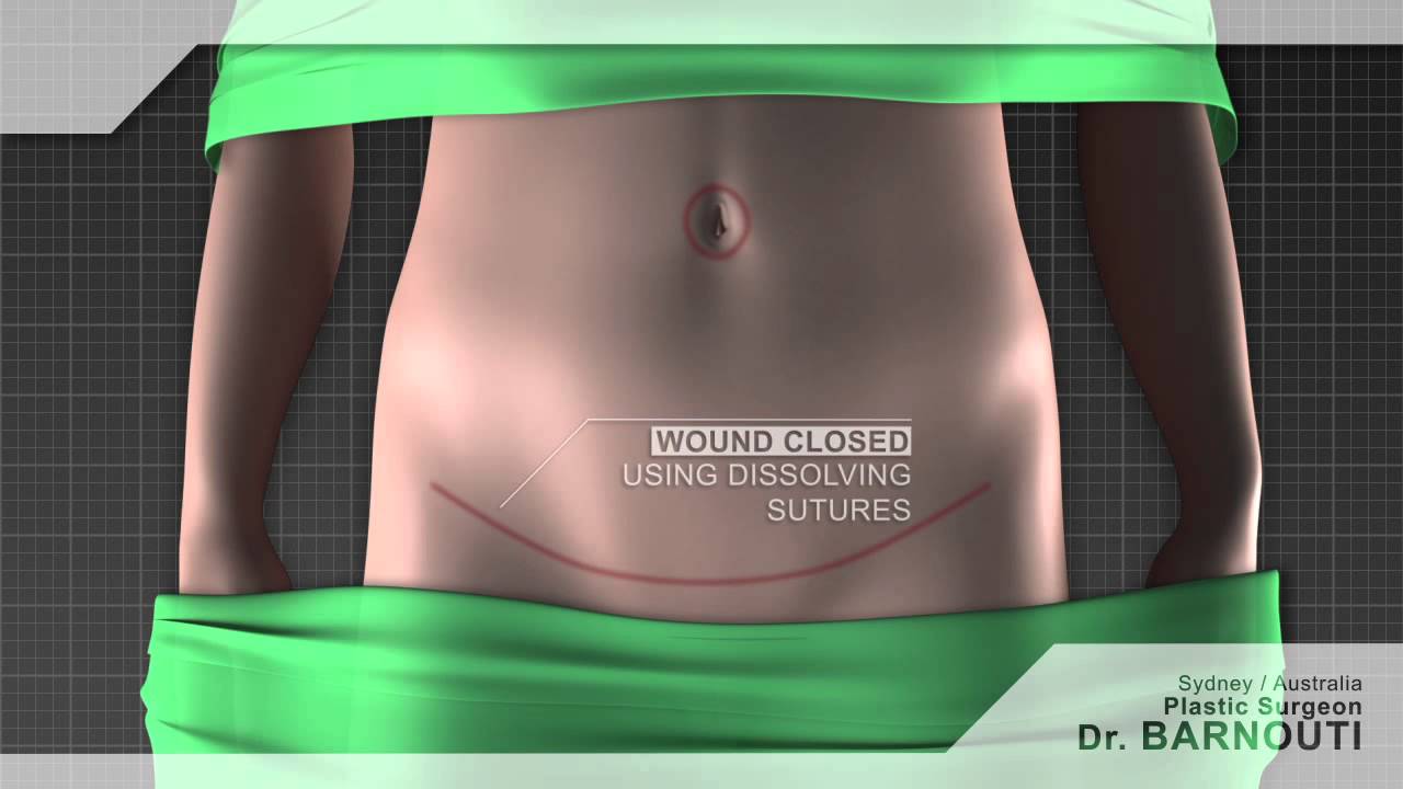
Tummy tuck Sydney Dr Barnouti. Call us on 02-9561 0222 or 1300 002 006
Broadway, Chatswood, Burwood NSW Australia
email:drbarnouti@australiaplasticsurgery.com.au
https://www.plasticsurgery-syd....ney.com.au/abdominop
What is a tummy tuck?
A tummy tuck operation is also known as abdominoplasty. It involves removing excess skin and fat from the stomach area, mainly the lower part of the tummy through surgical procedure. A tummy tuck operation is intended to leave the patient with a flatter tummy and to remove any signs of an 'apron' stomach or an overhang which is sometimes visible above underwear. The skin on this area tends to be stretched and of poor quality. A tummy tuck operation will usually focus on the lower part of the stomach, below the belly button and may require the belly button to be repositioned in some cases. The procedure is often carried out on women or men who have suffered from stretched skin in the stomach area after pregnancy, giving birth, excess fat deposition or weight loss.
What happens during a tummy tuck?
During a tummy tuck procedure the aim of the surgeon is to cut away fat and excess skin. To do this Dr Barnouti will make in incision on the lowest part of the stomach, where a fold will be visible above the pubic bone. He will take out as much excess fat as can be removed and will then cut the skin to fit back over the place where the fat has been removed from. It is important to have realistic expectations of a tummy tuck. Taking too much fat and skin away can result in folds at each end of the resulting scar which are sometimes referred to as "dog ears". Dr Barnouti will make sure you will not have this problem.
Who should have a tummy tuck?
Tummy tucks are recommended for either men or women who have an excess of fat and skin around their abdomen which cannot be removed by weight loss, exercise or liposuction. Tummy tuck operations in women are usually reserved for those who are not likely to have children as it is inadvisable to get pregnant again after having skin removed, this can cause the wound to stretch and scar.
The cost of a tummy tuck in Sydney Australia
The total cost is $7,900 if the patient's health fund cover the hospital's fees. In case the health fund does not cover the hospital's fee, the total cost will be around $12,000 inclusive of the Surgeon, assistant surgeon, Anaesthetist, hospital, operating theatre and follow ups visit.
Payment plans are alos available from Dr Barnouti's office in Chatswood, Burwood or Broadway.
A tummy tuck is a cosmetic procedure that removes excess skin and fatty tissue in order to give a flatter appearance to the stomach. Tummy tucks, also known as abdominoplasties, are ideal for patients who are not excessively overweight but suffer from an overhang of skin around the abdomen.
Performed under general anaesthetic, tummy tucks involve a horizontal incision being made just above the pubic area between the hip bones. Skin and fatty tissue is separated from the muscle and the area is tightened, with the excess skin and fatty tissues then being pulled downwards and removed.
Following your tummy tuck, there will be a scar present across the lower abdomen, but this will gradually fade. You may experience moderate tissue swelling for several months, but this will disappear with time. There may also be a sensation reduction just above the pubic area.
Once your tummy tuck recovery is complete however, you'll benefit from a more attractive figure and the ability to wear a wider selection of clothes.

Full Tummy Tuck 3D Video - http://drlandsman.com
Look great... feel great
•Smart Liposuction + Liposculpture
•Abdominplasty (Tummy Tuck)
+ Full Mini Modified
•Brazilian Lift with Fat Transfer
•Vaginal Aesthetics & Rejuvenation
•Laser Hair Removal
•Full Body Lift
•Thigh lift
•Brachioplasty (Arm Lift) + Short Scar
Expertise in Body Contouring
Board Certified Plastic Surgeon
Expertise in body contouring combines skin excision techniques and advanced fat contouring technology
Weight control personalized training and smoking cessation results in a healthier lifestyle improved shape and longer lasting results
With over 2 decades of experience Dr Lloyd Landsman provides state of the art cosmetic and plastic surgery
Dr Landsman integrates the finest and safest products with the newest procedures
A customized treatment plan is created for each patient utilizing classic surgical and minimally invasive techniques for optimal results
Call for your complimentary consultation to learn how Dr Landsman can help you look your very best
Visit http://drlandsman.com Call 631 864 4111
Main Office 994 W Jericho Tpke Smithtown NY 11787
Affiliates East Islip • Westbury • Jackson Heights • Manhattan

#anatomy #histology #biology #bytesizemed
✨If you would like my help studying the structure of bones, check out my long-form video on it.
🔅Structure of Bone : https://youtu.be/MYInVEnnS_I
💫 For more videos like this, subscribe to my channel!
Byte Size Med: https://youtube.com/channel/UC....ZghvlgylH3r_CWfA18eF
📚Factual References & for Further Reading:
- DiFiore's Atlas of Histology
- Junqueira's Basic Histology
- Gartner's Concise Histology
- Openstax Anatomy and Physiology
https://openstax.org/details/b....ooks/anatomy-and-phy
- Openstax Biology
https://openstax.org/details/books/biology-2e
(The last two are links to open-source references. They are NOT affiliate links)
🌤 Note:
These are just a collection of my notes. So use them the way you would use borrowed notes from a friend. 📝
The images in this video are hand-drawn for illustration and explanation only.✍️ Hence, they may not be anatomically accurate. I am just one person making these videos. If there are any errors, that is unintentional. I try super hard to avoid them. Please let me know if you find any, so it gets clarified for other viewers. Science constantly evolves and changes. New discoveries are made everyday. So some of the information in these videos may become outdated. If you notice that, please let me know so I can update them.
⚡️Disclaimer:
These videos are NOT a substitute for a medical textbook. Textbooks are written by experts (which I do not claim to be), edited, proofread and referenced. Please use them.
The information has been sourced from multiple references as mentioned above. I draw all the pictures myself. But if I have inadvertently infringed on any copyright, that is completely unintentional. I only make these videos to impart education. If I have accidentally violated copyright in any way, do let me know so I can make the necessary changes or give credit to anyone who is owed the same.
These videos are NOT intended for patient education. They are NOT a substitute for diagnosis and treatment by a licensed medical professional. Always seek the advice of a qualified health care provider for any questions you may have regarding any medical condition, so that they can address your individual needs.
🔅They are ONLY meant to help students of medicine and health sciences with studying, and should be used for just that purpose and absolutely nothing else.
Byte Size Med. All Rights Reserved.
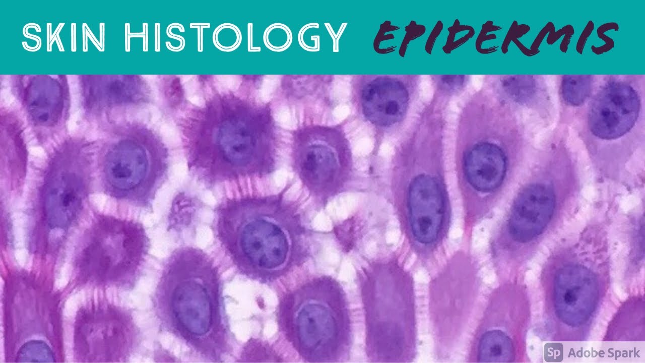
Excerpt from my Normal Skin Histology video: https://kikoxp.com/posts/3660.
A complete organized library of all my videos, digital slides, pics, & sample pathology reports is available here: https://kikoxp.com/posts/5084 (dermpath) & https://kikoxp.com/posts/5083 (bone/soft tissue sarcoma pathology).
Please check out my Soft Tissue Pathology & Dermatopathology survival guide textbooks: http://bit.ly/2Te2haB
Also, in the past I used "keratinocyte" and "squamous cell" interchangeably (this is because in dermatopathology, we see and talk about squamous cell carcinomas all the time, and those tumors are composed of keratinocytes). But technically, in normal skin histology, "squamous cell" refers only to the flattened keratinocytes in the superficial epidermis. Thankfully, a histology PhD colleague pointed this out to me and corrected my lazy nomenclature!
This video is geared towards medical students, pathology or dermatology residents, or practicing pathologists or dermatologists. Of course, this video is for educational purposes only and is not formal medical advice or consultation.
Presented by Jerad M. Gardner, MD. Please subscribe to my channel to be notified of new pathology teaching videos.
Follow me on:
Snapchat: JMGardnerMD
Twitter: @JMGardnerMD
Instagram: @JMGardnerMD
Kiko: https://kikoxp.com/profile/jer....ad_gardner1/content?
Facebook: https://www.facebook.com/JMGardnerMD/
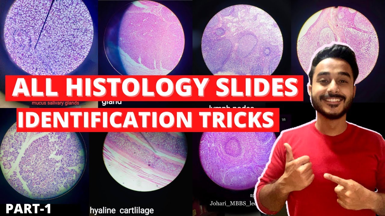
| MBBS मतलब JOHARI MBBS I
Download Johari MBBS APP ( For Online LIVE Classes, Notes, Books PDFs, Test Series )
Johari MBBS ( iPhone IOS Users ) LINK { FOR Online LIVE Classes }
https://apps.apple.com/in/app/....johari-mbbs/id647466
JOHARI MBBS APP ( Android ) LINK { FOR Online LIVE Classes }
https://play.google.com/store/....apps/details?id=co.d
CRASH COURSE LINK ( Anatomy in 30Days with Biochemistry In 7Days Series )
https://zczob.on-app.in/app/oc/389813/zczob
IMPORTANT LINKS :-
1) ORDER Anatomy Next Edition Module , Biochemistry in 7Days & Physiology MODULE
https://joharimbbs.com/
2) Join INSTAGRAM ( For Notes, Revision REELs, Updates )
https://www.instagram.com/johari_mbbs_lectures/
3) INSTA Broadcast Channel ( FOR Daily VLOGS, Life Update )
https://ig.me/j/Abal9xRcXcUyrYpT/
4) Telegram ( For FREE BOOKS PDFs )
https://t.me/joharimbbsofficial
5) Whatsapp Channel ( Daily Update )
https://whatsapp.com/channel/0....029VaEeWKWHAdNb0xOzl
6) Follow On Twitter ( For Latest Updates )
https://twitter.com/JohariMbbs
CRASH COURSE LINK ( Anatomy in 30Days with Biochemistry In 7Days Series )
https://zczob.on-app.in/app/oc/389813/zczob
#mbbs #joharimbbs #anaatomy #biochemistry #physiology #medico #doctors
histology slide identification tricks
histology slides identification tricks
histology slide
histology slides
histology slides identification
histology slide preparation
histology slides identification epithelium
histology slides identification connective tissue
histology slides identification tricks
histology slides of epithelium
histology slide identification
#anatomy#clinicalanatomy #MBBS #neroanatomy #bdc #medsudent #medicalcollege
For notes IG - johari_mbbs_lectures
link- https://www.instagram.com/p/CNOwFgEJmJL/
Join Telegram Channel ( JOhari MBBS Public 20x )
https://t.me/JohariMBBS
For Tag :-
histology , histology slides , histology slide identification , histology slides identification , histology slides preparation , tongue histology slide , histology slides asked in exam , histology slide identification trick , histology slides of connective tissue , mbbs histology , histology slides identification epithelium , trachea histology slide , slides , duodenum histology slide , histology slides tricks , histology slides review , examine histology slides

An animated description of the composition of bones.
Visit www.orthofilms.com for more videos and info.
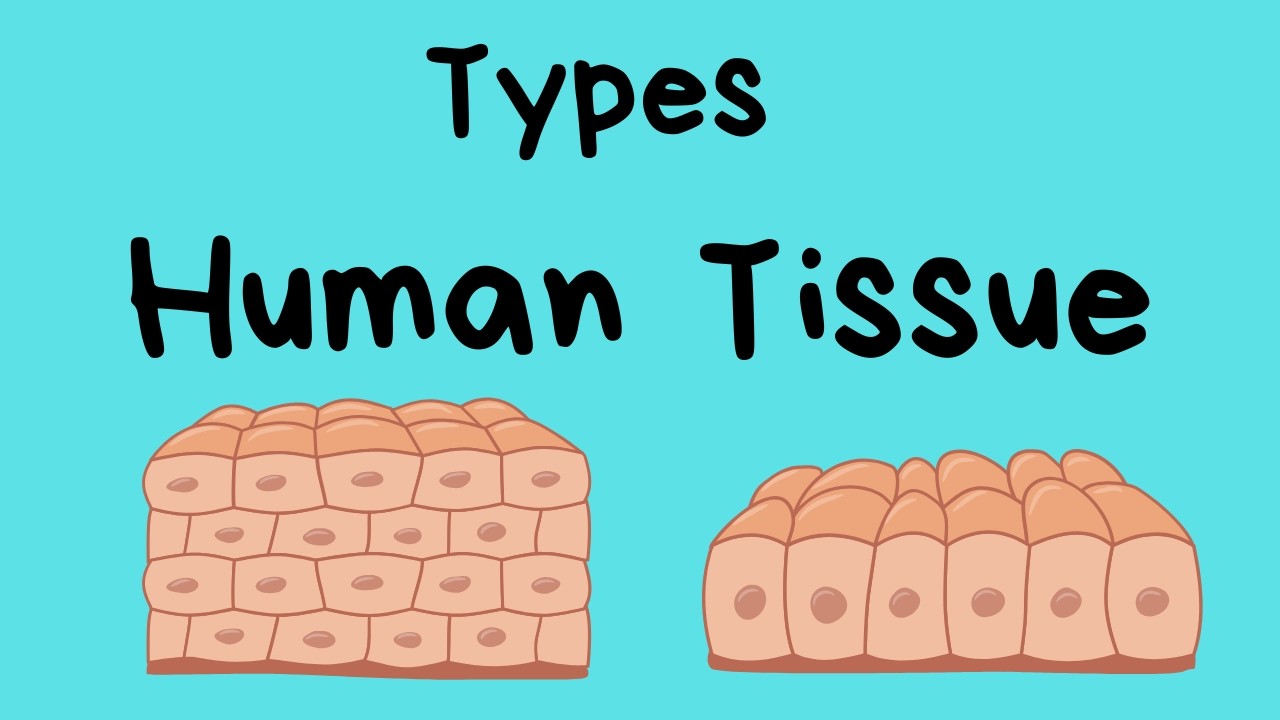
Types of Human Body Tissue
In this video, I review four types of tissue.
Connective tissue, epithelial tissue, muscle tissue, and nerve tissue.
Tissues are made up of cells working together.
*
*
For more Life Science videos and summaries see,
http://www.moomoomath.com/Midd....le-School-Science-an
