- Physical Examination
- Surgical Examination
- Ophthalmology
- Clinical Skills
- Orthopedics
- Surgery Videos
- Laparoscopy
- Pediatrics
- Funny Videos
- Cardiothoracic Surgery
- Nursing Videos
- Plastic Surgery
- Otorhinolaryngology
- Histology and Histopathology
- Neurosurgery
- Dermatology
- Pediatric Surgery
- Urology
- Dentistry
- Oncology and Cancers
- Anatomy Videos
- Health and Fitness
- Radiology
- Anaesthesia
- Physical Therapy
- Pharmacology
- Interventional Radiology
- Cardiology
- Endocrinology
- Gynecology
- Emergency Medicine
- Psychiatry and Psychology
- Childbirth Videos
- General Medical Videos
- Nephrology
- Physiology
- Diet and Food Health
- Diabetes Mellitus
- Neurology
- Women Health
- Osteoporosis
- Gastroenterology
- Pulmonology
- Hematology
- Rheumatology
- Toxicology
- Nuclear Medicine
- Infectious Diseases
- Vascular Disease
- Reproductive Health
- Burns and Wound Healing
- Other
Top videos
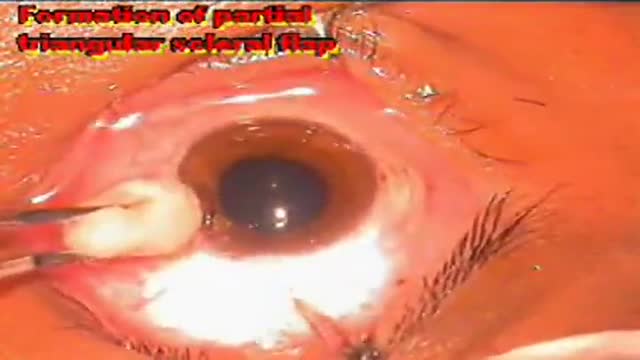
Eye Surgery Trabeculectomy

-Traumatic amputation of a body part requires rapid transport of the appendage, which should be wrapped in a saline-moistened gauze, placed in a plastic bag, and transported in a container filled with ice mixed with either saline or sterile water to best preserve the body part and attempt replantation.
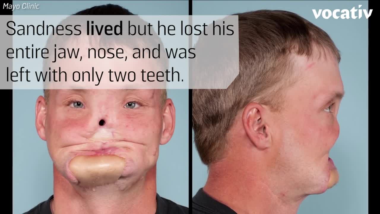
Doctors at the Mayo Clinic used 3D-printed models to prepare for their first-ever face transplant.
The male reproductive system includes the scrotum, testes, spermatic ducts, sex glands, and penis. These organs work together to produce sperm, the male gamete, and the other components of semen.

Cervical Mucus
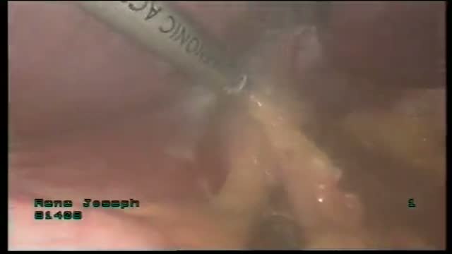
Laparoscopic Hiatus Hernia Repair in Qatar by Dr. Al-Emadi

Going to the dentist is not a very fun experience for most. In fact, let's face it, most of us dread it.
http://www.dentistmaps.com/
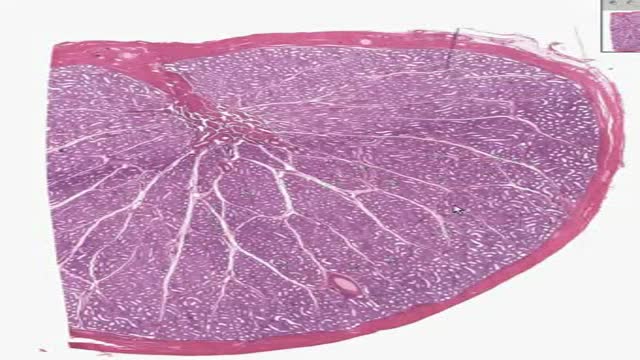
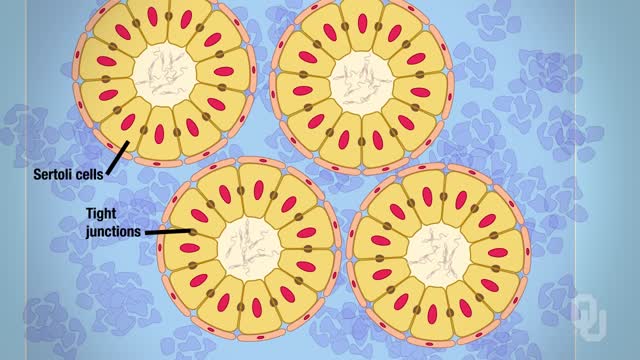
Histology of Testis
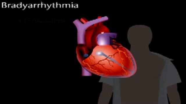
Bradyarrythmias
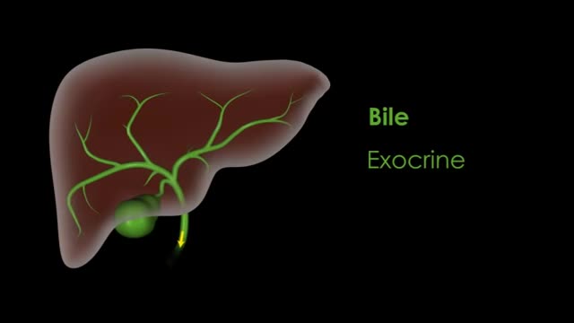
The liver also detoxifies chemicals and metabolizes drugs. As it does so, the liver secretes bile that ends up back in the intestines. The liver also makes proteins important for blood clotting and other functions. First, for those impatient, short answers to the mini-questions (if you're reading this in the news feed, you may want to click through for the question details): No one knows why we evolved 2 kidneys and one liver.
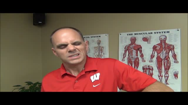
Shoulder pain and exercises Milwaukee WI
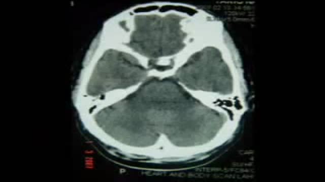
Right PCA Aneurysm Clipping Posterior Cerebral Artery
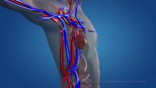
The circulatory system consists of three independent systems that work together: the heart (cardiovascular), lungs (pulmonary), and arteries, veins, coronary and portal vessels (systemic). The system is responsible for the flow of blood, nutrients, oxygen and other gases, and as well as hormones to and from cells

Angiogenesis and Cancer
Glaucoma is called the silent thief of sight. It does not have symptoms during the early stages of the diseases and can make a patient blind over several years
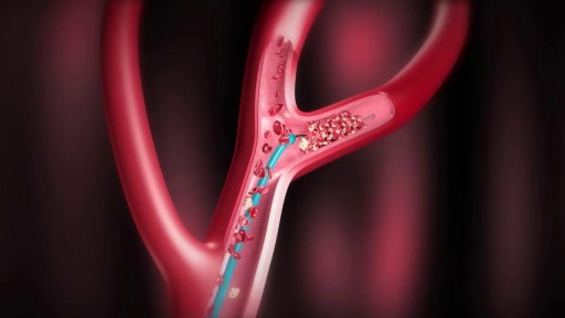
Diagnosis To determine the most appropriate treatment for your stroke, your emergency team needs to evaluate the type of stroke you're having and the areas of your brain affected by the stroke. They also need to rule out other possible causes of your symptoms, such as a brain tumor or a drug reaction. Your doctor may use several tests to determine your risk of stroke, including: Stroke consultation Stroke consultation Stroke consultation at Mayo Clinic Brain tissue damaged by stroke CT scan of brain tissue damaged by stroke Cerebral angiogram Cerebral angiogram Physical examination. Your doctor will ask you or a family member what symptoms you've been having, when they started and what you were doing when they began. Your doctor then will evaluate whether these symptoms are still present. Your doctor will want to know what medications you take and whether you have experienced any head injuries. You'll be asked about your personal and family history of heart disease, transient ischemic attack and stroke. Your doctor will check your blood pressure and use a stethoscope to listen to your heart and to listen for a whooshing sound (bruit) over your neck (carotid) arteries, which may indicate atherosclerosis. Your doctor may also use an ophthalmoscope to check for signs of tiny cholesterol crystals or clots in the blood vessels at the back of your eyes. Blood tests. You may have several blood tests, which tell your care team how fast your blood clots, whether your blood sugar is abnormally high or low, whether critical blood chemicals are out of balance, or whether you may have an infection. Managing your blood's clotting time and levels of sugar and other key chemicals will be part of your stroke care. Computerized tomography (CT) scan. A CT scan uses a series of X-rays to create a detailed image of your brain. A CT scan can show a hemorrhage, tumor, stroke and other conditions. Doctors may inject a dye into your bloodstream to view your blood vessels in your neck and brain in greater detail (computerized tomography angiography). There are different types of CT scans that your doctor may use depending on your situation. Magnetic resonance imaging (MRI). An MRI uses powerful radio waves and magnets to create a detailed view of your brain. An MRI can detect brain tissue damaged by an ischemic stroke and brain hemorrhages. Your doctor may inject a dye into a blood vessel to view the arteries and veins and highlight blood flow (magnetic resonance angiography, or magnetic resonance venography). Carotid ultrasound. In this test, sound waves create detailed images of the inside of the carotid arteries in your neck. This test shows buildup of fatty deposits (plaques) and blood flow in your carotid arteries. Cerebral angiogram. In this test, your doctor inserts a thin, flexible tube (catheter) through a small incision, usually in your groin, and guides it through your major arteries and into your carotid or vertebral artery. Then your doctor injects a dye into your blood vessels to make them visible under X-ray imaging. This procedure gives a detailed view of arteries in your brain and neck. Echocardiogram. An echocardiogram uses sound waves to create detailed images of your heart. An echocardiogram can find a source of clots in your heart that may have traveled from your heart to your brain and caused your stroke. You may have a transesophageal echocardiogram. In this test, your doctor inserts a flexible tube with a small device (transducer) attached into your throat and down into the tube that connects the back of your mouth to your stomach (esophagus). Because your esophagus is directly behind your heart, a transesophageal echocardiogram can create clear, detailed ultrasound images of your heart and any blood clots. Treatment Emergency treatment for stroke depends on whether you're having an ischemic stroke blocking an artery — the most common kind — or a hemorrhagic stroke that involves bleeding into the brain. Ischemic stroke To treat an ischemic stroke, doctors must quickly restore blood flow to your brain. Emergency treatment with medications. Therapy with clot-busting drugs must start within 4.5 hours if they are given into the vein — and the sooner, the better. Quick treatment not only improves your chances of survival but also may reduce complications. You may be given: Intravenous injection of tissue plasminogen activator (tPA). This injection of recombinant tissue plasminogen activator (tPA), also called alteplase, is considered the gold standard treatment for ischemic stroke. An injection of tPA is usually given through a vein in the arm. This potent clot-busting drug ideally is given within three hours. In some instances, tPA can be given up to 4.5 hours after stroke symptoms begin. This drug restores blood flow by dissolving the blood clot causing your stroke, and it may help people who have had strokes recover more fully. Your doctor will consider certain risks, such as potential bleeding in the brain, to determine if tPA is appropriate for you. Emergency endovascular procedures. Doctors sometimes treat ischemic strokes with procedures performed directly inside the blocked blood vessel. These procedures must be performed as soon as possible, depending on features of the blood clot: Medications delivered directly to the brain. Doctors may insert a long, thin tube (catheter) through an artery in your groin and thread it to your brain to deliver tPA directly into the area where the stroke is occurring. This is called intra-arterial thrombolysis. The time window for this treatment is somewhat longer than for intravenous tPA, but is still limited. Removing the clot with a stent retriever. Doctors may use a catheter to maneuver a device into the blocked blood vessel in your brain and trap and remove the clot. This procedure is particularly beneficial for people with large clots that can't be completely dissolved with tPA, though this procedure is often performed in combination with intravenous tPA. Several large and recent studies suggest that, depending on the location of the clot and other factors, endovascular therapy might be the most effective treatment. Endovascular therapy has been shown to significantly improve outcomes and reduce long-term disability after ischemic stroke. Other procedures. To decrease your risk of having another stroke or transient ischemic attack, your doctor may recommend a procedure to open up an artery that's narrowed by plaque. Doctors sometimes recommend the following procedures to prevent a stroke. Options will vary depending on your situation: Carotid endarterectomy. In a carotid endarterectomy, a surgeon removes plaques from arteries that run along each side of your neck to your brain (carotid arteries). In this procedure, your surgeon makes an incision along the front of your neck, opens your carotid artery and removes plaque that blocks the carotid artery. Your surgeon then repairs the artery with stitches or a patch made from a vein or artificial material (graft). The procedure may reduce your risk of ischemic stroke. However, a carotid endarterectomy also involves risks, especially for people with heart disease or other medical conditions. Angioplasty and stents. In an angioplasty, a surgeon usually accesses your carotid arteries through an artery in your groin. Here, your surgeon can gently and safely navigate to the carotid arteries in your neck. A balloon is then inflated to expand the narrowed artery. Then a stent can be inserted to support the opened artery. Hemorrhagic stroke Emergency treatment of hemorrhagic stroke focuses on controlling your bleeding and reducing pressure in your brain. You might also need surgery to help reduce future risk. Emergency measures. If you take warfarin (Coumadin, Jantoven) or anti-platelet drugs such as clopidogrel (Plavix) to prevent blood clots, you may be given drugs or transfusions of blood products to counteract the blood thinners' effects. You may also be given drugs to lower pressure in your brain (intracranial pressure), lower your blood pressure, prevent vasospasm or prevent seizures. Once the bleeding in your brain stops, treatment usually involves supportive medical care while your body absorbs the blood. Healing is similar to what happens while a bad bruise goes away. If the area of bleeding is large, your doctor may perform surgery to remove the blood and relieve pressure on your brain. Surgical blood vessel repair. Surgery may be used to repair blood vessel abnormalities associated with hemorrhagic strokes. Your doctor may recommend one of these procedures after a stroke or if an aneurysm or arteriovenous malformation (AVM) or other type of vascular malformation caused your hemorrhagic stroke: Surgical clipping. A surgeon places a tiny clamp at the base of the aneurysm, to stop blood flow to it. This clamp can keep the aneurysm from bursting, or it can prevent re-bleeding of an aneurysm that has recently hemorrhaged. Coiling (endovascular embolization). A surgeon inserts a catheter into an artery in your groin and guides it to your brain using X-ray imaging. Tiny detachable coils are guided into the aneurysm (aneurysm coiling). The coils fill the aneurysm, which blocks blood flow into the aneurysm and causes the blood to clot. Surgical AVM removal. Surgeons may remove a smaller AVM if it's located in an accessible area of your brain, to eliminate the risk of rupture and lower the risk of hemorrhagic stroke. However, it's not always possible to remove an AVM if its removal would cause too large a reduction in brain function, or if it's large or located deep within your brain. Stereotactic radiosurgery. Using multiple beams of highly focused radiation, stereotactic radiosurgery is an advanced minimally invasive treatment used to repair vascular malformations. Stroke recovery and rehabilitation Brain hemisphere connections Brain hemisphere connections After emergency treatment, stroke care focuses on helping you recover as much function as possible and return to independent living. The impact of your stroke depends on the area of the brain involved and the amount of tissue damaged. If your stroke affected the right side of your brain, your movement and sensation on the left side of your body may be affected. If your stroke damaged the brain tissue on the left side of your brain, your movement and sensation on the right side of your body may be affected. Brain damage to the left side of your brain may cause speech and language disorders. In addition, if you've had a stroke, you may have problems with breathing, swallowing, balancing and vision. Most stroke survivors receive treatment in a rehabilitation program. Your doctor will recommend the most rigorous therapy program you can handle based on your age, overall health and degree of disability from your stroke. Your doctor will take into consideration your lifestyle, interests and priorities, and the availability of family members or other caregivers. Your rehabilitation program may begin before you leave the hospital. After discharge, you might continue your program in a rehabilitation unit of the same hospital, another rehabilitation unit or skilled nursing facility, an outpatient unit, or your home. Every person's stroke recovery is different. Depending on your condition, your treatment team may include: Doctor trained in brain conditions (neurologist) Rehabilitation doctor (physiatrist) Nurse Dietitian Physical therapist Occupational therapist Recreational therapist Speech pathologist Social worker Case manager Psychologist or psychiatrist Chaplain Speech therapy session Speech therapy is often a part of stroke rehabilitation. Treatment outcomes One way to evaluate the care of patients diagnosed with stroke is to look at the percentage of patients receiving the timely and effective care measures that are appropriate. The goal is 100 percent. The graphs below display the percentage of eligible Mayo Clinic patients diagnosed with stroke receiving all of the appropriate care measures.
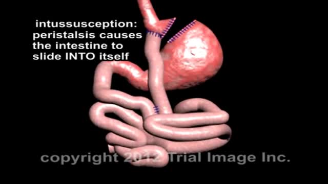
In cases when the presentation is unclear, ultrasonography is the imaging methodology of choice. The characteristic finding is the presence of a "target sign". Ultrasonography is not required in patients with obvious clinical diagnosis (as seen in this patient). Such patients can proceed directly to treatment with diagnostic and therapeutic air (pneumatic) or water-soluble (hydrostatic contrast) enema.

Robyn Benincasa, an extreme sports adventure racer, marathoner and firefighter maintains her active lifestyle following a hip replacement at St. Vincent Medical Center's Joint Replacement Institute with Dr. Thomas Schmalzried in Los Angeles, California. For more information, please visit: www.jri-docs.com
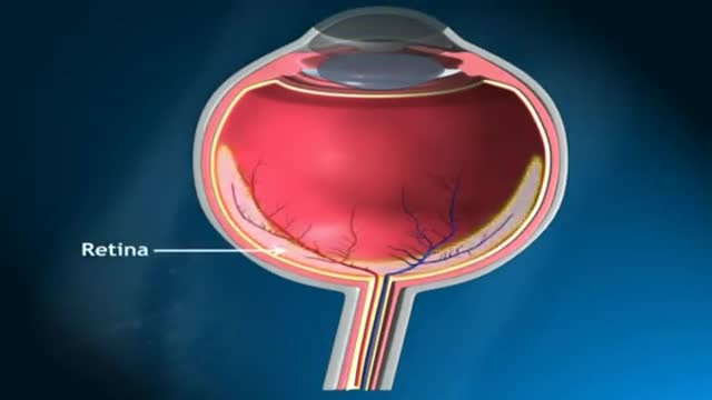
Anatomy of The Orbit
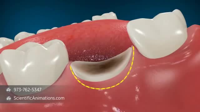
A palatal view of a maxillary premolar during a crown lengthening procedure. Crown lengthening is a surgical procedure performed by a dentist to expose a greater amount of tooth structure for the purpose of subsequently restoring the tooth prosthetically.
