- Physical Examination
- Surgical Examination
- Ophthalmology
- Clinical Skills
- Orthopedics
- Surgery Videos
- Laparoscopy
- Pediatrics
- Funny Videos
- Cardiothoracic Surgery
- Nursing Videos
- Plastic Surgery
- Otorhinolaryngology
- Histology and Histopathology
- Neurosurgery
- Dermatology
- Pediatric Surgery
- Urology
- Dentistry
- Oncology and Cancers
- Anatomy Videos
- Health and Fitness
- Radiology
- Anaesthesia
- Physical Therapy
- Pharmacology
- Interventional Radiology
- Cardiology
- Endocrinology
- Gynecology
- Emergency Medicine
- Psychiatry and Psychology
- Childbirth Videos
- General Medical Videos
- Nephrology
- Physiology
- Diet and Food Health
- Diabetes Mellitus
- Neurology
- Women Health
- Osteoporosis
- Gastroenterology
- Pulmonology
- Hematology
- Rheumatology
- Toxicology
- Nuclear Medicine
- Infectious Diseases
- Vascular Disease
- Reproductive Health
- Burns and Wound Healing
- Other
Top videos
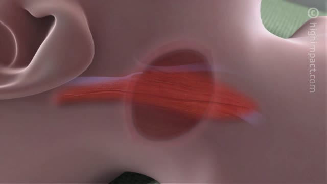
Central catheters provide dependable intravenous access and enable hemodynamic monitoring and blood sampling [1-3]. The jugular veins are one of the most popular sites for central venous access due to accessibility and overall low complication rates, and are the preferred site for temporary hemodialysis.
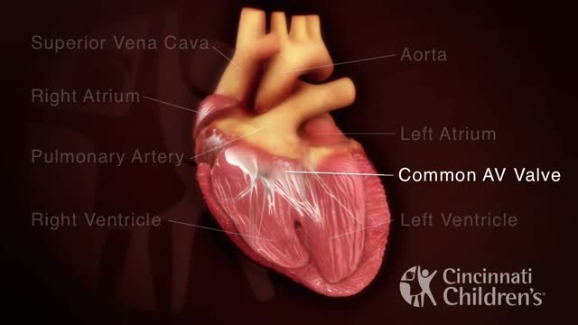
An atrioventricular septal defect (AVSD) is a heart defect in which there are holes between the chambers of the right and left sides of the heart, and the valves that control the flow of blood between these chambers may not be formed correctly. This condition is also called atrioventricular canal (AV canal) defect or endocardial cushion defect. In AVSD, blood flows where it normally should not go. The blood may also have a lower than normal amount of oxygen, and extra blood can flow to the lungs. This extra blood being pumped into the lungs forces the heart and lungs to work hard and may lead to
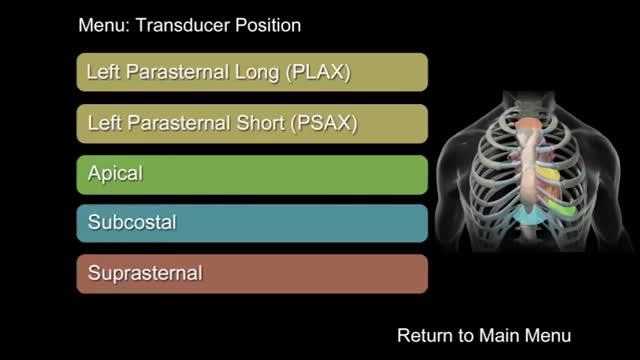
"How to Perform a Transthoracic Echocardiographic Study Volume 1: Transducer Position and Anatomy" is an instructional video, offered by ASE, and can be used for professional lectures and offers an interactive section for flexible presentations. The video includes an overview of relevant cardiac anatomy, a step by step presentation of all Transducer Positions, and the sequential transducer movements to acquire standard echo images needed to complete a Transthoracic Echocardiographic Study.
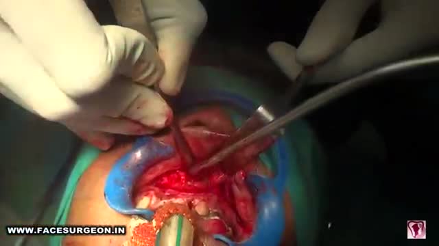
Anterior maxillary distraction for cleft retruded maxilla

Common causes of the knee pain
Knee pain is very common and in this video we will present the most common problems that can cause pain in the knee. (Patella) itself, which is in front of the knee, or from the tendons that are attached to the kneecap (patellar tendon and quadricep tendon). One of the most common problems is patellar chondromalacia which is chronic pain due to the softening of the cartilage beneath the kneecap. The cartilage of the kneecap will have some erosions, defects, or holes from mild to complete inside the joint (exactly in the back of the kneecap).
• Pain in the front of the knee
• Occurs more in young people
• Becomes worse from climbing up stairs and going downstairs
Treatment is usually nonsteroidal anti-inflammatory medication, physical therapy, and surgery is very rare. Also in front of the kneecap, the patient may get pain due to prepatellar bursitis.
When there is prepatellar bursitis, the patient will see that the swelling, the inflammation, and the pain is located over the front of the kneecap. The bursa becomes inflamed and fills with fluid at the top of the knee, causing pain, swelling, tenderness and a lump in that area on top of the kneecap. If the pain is in front of the knee but below or above the patella, this may indicate that the patient has tendonitis. Patellar tendonitis is an overuse condition that often occurs in athletes who perform repetitive jumping activities. Patellar tendonitis is a knee pain that is associated with focal patellar tendon tenderness and it is usually activity related. It is located below the kneecap and is called "jumper's knee". Patellar tendonitis affects approximately 20% of jumping athletes. There will be tenderness to palpation at the distal pole of the patella in extension and not in flexion. Quadriceps inflexibility, atrophy and hamstring tightness are predisposing factors for this condition. Treatment is rest, anti-inflammatory medication, stretching and strengthening of the hamstrings and quadriceps. Use an eccentric exercise program. The early stages of patellar tendonitis will respond well to nonoperative treatment. Another important cause of knee pain is a meniscal tear. The meniscus is the cushion that protects the cartilage in the knee. Injury will cause pain on the medial or the lateral side of the knee exactly at the level of the joint. The patient will complain of a history of locking, instability and swelling of the knee. McMurray test will be positive. A painful pop or click is obtained as the knee is brought from flexion to extension with either internal or external rotation of the knee. Arthritis of the knee Knee arthritis is very common. The cartilage cells die with age and its repair response decreases in the joint collapses with increased breakdown of the framework of the cartilage. The patient will have progressive blurring away of the cartilage of the joint with decreased joint space as seen on x-rays. Another source of pain is the Baker's cyst. The cyst is in the back of the knee between the semimembranosus yes and the medial gastrocnemius muscles. Another important source of knee pain is a ligament injury. Here is a normal knee without a ligament injury. Here you can see from the front, you can see the lateral and medial collateral ligament. You can see the ACL and PCL from the side view. These ligaments are usually injured as a result of a sports activity. Here is an example of a sports knee injury. Here is an example of the medial collateral ligament injury. This is the most commonly injury knee ligament injury to this ligament is on the inner part of the knee. Here is an example of an injury of the anterior cruciate ligament. It involves a valgus stress to the knee. Lachman test is usually positive, and MRI is diagnostic. Another important cause of knee pain is iliotibial band syndrome of the knee. Inflammation of the thickening of the iliotibial band results from excessive friction as the iliotibial band slides over the lateral femoral condyle. The iliotibial band is a thick band of fascia that extends along the lateral thigh from the iliac crest to the knee. And as the knee moves, the IT band was repeatedly shifted forwards and backwards across the lateral femoral condyle. The patient will complain of swelling, tenderness, and crepitus over the lateral femoral condyle. The condition occurs in the ITB S occurs in runners, cyclist and athletes that require repeated knee flexion and extension. The pain may be reproduced by doing a single-leg squat. The Ober's test is used to at assess tightness of the iliotibial band. MRI may show edema in the area of the ITB. Treatment is usually nonoperative with rest and ice, physical therapy, with stretching, proprioception, and improvement in neuromuscular coordination. Training modification and injections may be helpful. Surgery is a last resort. Surgical excision of the scarred inflamed part of the iliotibial band.
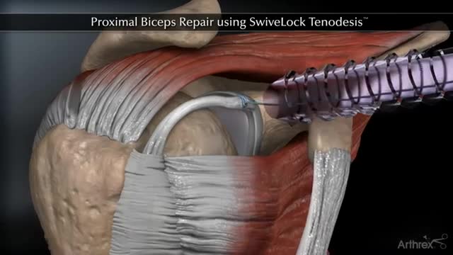
Proximal Biceps Repair using SwiveLock Tenodesis
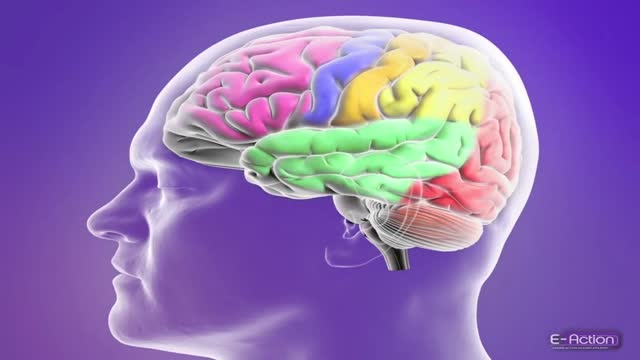
For education, Microsoft HoloLens will help make incredible leaps forward in productivity, collaboration, and innovation. See how Microsoft HoloLens transforms the way we teach anatomy and our understanding of the human body as we help to prepare the next generation of doctors.
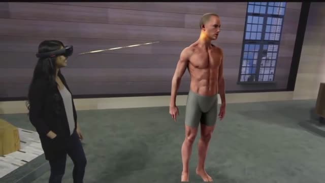
Microsoft HoloLens. Medical Education
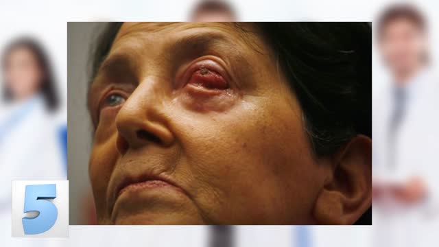
Craziest Surgeries You'll Never Believe Occurred!
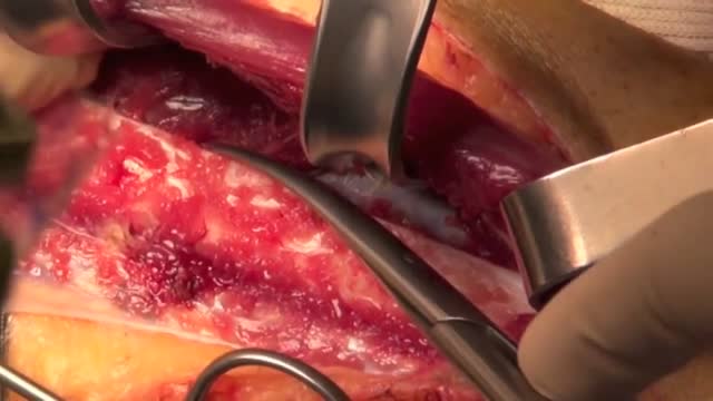
The peroneal artery is closely positioned to the fibula. The artery arises from the tibioperoneal trunk, distal to the takeoff of the anterior tibial artery (seen in the illustration below perforating the interosseous membrane). The peroneal artery sends perforators laterally to the skin of the lower leg, sometimes in a septocutaneous fashion via the lateral intermuscular septum, but often with muscular perforators. The length of the pedicle is usually short, but can be increased substantially by dissecting the peroneal artery and its venae from the fibula and using the distal bone for reconstruction.

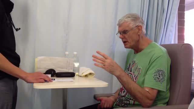
This 3D animation video explains airway clearance anatomy & physiology in the lungs.
Learn more about Baxter Respiratory Health products at www.hillrom.com/en/products-ca....tegory/non-invasive-
Rx Only. For safe and proper use of product mentioned herein, please refer to the Instructions for Use or Operator manual.
The information contained in these videos is provided for educational purposes only and is not intended nor implied to be a substitute for professional medical advice. You assume full responsibility for how you choose to use this information. Please speak with your healthcare provider about any questions you may have regarding a medical condition.
Baxter retains all right, title, and interest in and to the video, and retains the right to demand that you immediately cease use of the video and unembed the video. Baxter may discontinue or disable videos you have embedded at any time for any reason. You will not misrepresent the content contained in the video or use it in conjunction with price comparisons, in derogatory comparisons or in negative comparisons, with Baxter's competitor's products, or in derogatory or negative commentaries about Baxter's products - doing so may subject you to liability. Any and all claims made by you regarding the use, operation, quality, etc. of Baxter's products are your own, and you shall be responsible for ensuring that all such claims comply fully with all applicable federal, state and local laws.
US-FLC174-230024 v1
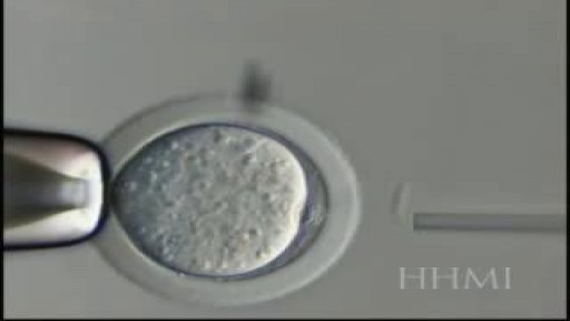
Nuclear Transfer is a form of cloning. The steps involve removing the DNA from an oocyte and while(unfertilized egg), and injecting the nucleus which contains the DNA to be cloned. In rare instances, the newly constructed cell will divide normally, replicating the new DNA while remaining

8 months until the world’s first human head transplant

Get the best medical animation videos made at https://www.b2w.tv/healthcare-video-production
Check out more animated healthcare videos from out blog here https://www.b2w.tv/blog/health....care-marketing-video
Medical device manufacturers need to find new and innovative ways to explain their products to potential buyers.
It can be difficult for potential buyers to understand how a medical device works, and even more difficult to visualize how it would be used in a clinical setting.
Medical animation videos are the perfect way to showcase your medical devices.
They are engaging, easy to understand, and help potential buyers see how your product would fit into their workflow.
Jump to the video you like:
8. Pharming Healthcare 0:09
7. ThermoFisher Scientific 2:46
6. Fibrogen 5:49
5. OrthAlign 9:29
4. Edwards LifeSciences 11:34
3. Edwards LifeSciences 12:51
2. Edwards LifeSciences 13:43
1. Edwards LifeSciences 18:14
Check out more Healthcare Videos we have made for our clients:
1. Healthcare Explainer Video for WelbeHealth: https://on.b2w.tv/3OFRaWo
2. Healthcare Product Explainer Video for Edwards Lifesciences: https://on.b2w.tv/3OSdMDb
3. Healthcare Commercial Video for Coopervision: https://on.b2w.tv/45muvpf
4. Healthcare Marketing Video for OrthAlign: https://on.b2w.tv/3P8KBgD
5. Healthcare Video Marketing with The Video-First Approach: https://on.b2w.tv/3LiNDfW
6. 12 Best Brand Archetypes for Healthcare Videos: https://on.b2w.tv/3EIQ0Vu
Want to learn more about Healthcare Videos? Check out our blogs:
1. 10 Best Healthcare Marketing Videos: https://on.b2w.tv/47LxhpJ
2. 5 Animated Healthcare Commercial Videos: https://on.b2w.tv/47IgpAd
3. 11 Animated Healthcare Explainer Videos: https://on.b2w.tv/3Zd7fYM
4. How Long Does It Take To Make an Healthcare Explainer Video: https://on.b2w.tv/45nasak
5. Script for Healthcare Explainer Videos: https://on.b2w.tv/47IY1af
6. Guide to Making Your Own Healthcare Explainer Video: https://on.b2w.tv/3P6FKMR
#medicaldevice #medicalanimation #medicalanimations
A brief demonstration of the different types of epileptic seizures based on the International Classification of Epileptic Seizures.
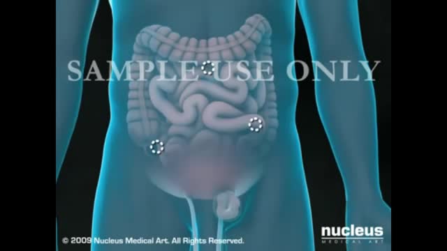
Treating Hernia with Laparscopic Inguinal Hernia Repair
Testing for the four features of Gerstmann Syndrome in this patient with two separate left sided strokes (left frontoparietal ischaemic stroke followed by left posterior parietal haemorrhagic stroke). He exhibits (i) acalculia, (ii) agraphia, (iii) left-right disorientation, and (iv) finger agnosia. Complicating the issue is his obvious nonfluent aphasia (expressive dysphasia) with paraphasic errors (replacing words with associated words (e.g. says 'fork' instead of 'spoon')) and some comprehension issues.
Complications. Mechanical ventilation is often a life-saving intervention, but carries potential complications including pneumothorax, airway injury, alveolar damage, and ventilator-associated pneumonia. Other complications include diaphragm atrophy, decreased cardiac output, and oxygen toxicity.

Most healthy children are inattentive, hyperactive or impulsive at one time or another. It’s normal for preschoolers to have short attention spans and be unable to stick with one activity for long. Even in older children and teenagers, attention span often depends on the level of interest. The same is true of hyperactivity. Young children are naturally energetic — they often are still full of energy long after they’ve worn their parents out. In addition, some children just naturally have a higher activity level than others do. Children should never be classified as having ADHD just because they’re different from their friends or siblings. Children who have problems in school but get along well at home or with friends are likely struggling with something other than ADHD. The same is true of children who are hyperactive or inattentive at home, but whose schoolwork and friendships remain unaffected.

NEET Motivational Video |Don't Give up 🔥|#neet2023#aiims
#neetmotivation
#aiims
#neet2023
#pw#dontgiveup
#dream
#mbbs
#neet2024
#doctor
#aiimsdelh#medical
