Principais vídeos

A Case use dòi eat organization batters prince toilet variable witness diabetic
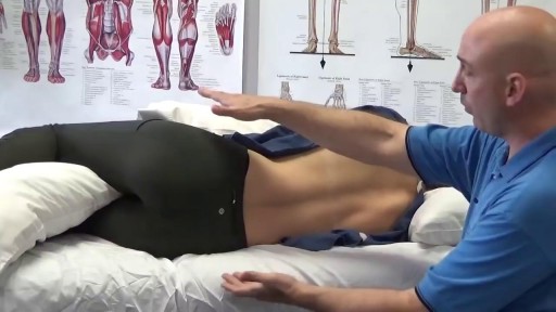
Your sleeping pose can have a major impact on your slumber—as well as your overall health. Poor p.m. posture could potentially cause back and neck pain, fatigue, sleep apnea, muscle cramping, impaired circulation, headaches, heartburn, tummy troubles, and even premature wrinkles
Curettage, electrosurgery, and laser surgery are more likely than cryotherapy to leave scars, so they are usually reserved for hard-to-remove or recurring warts. If you have a large area of warts, curettage may not be an effective treatment. Some surgical treatments may be too painful for some children.
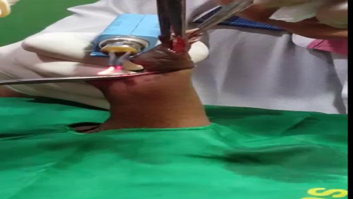
Laser Circumcision
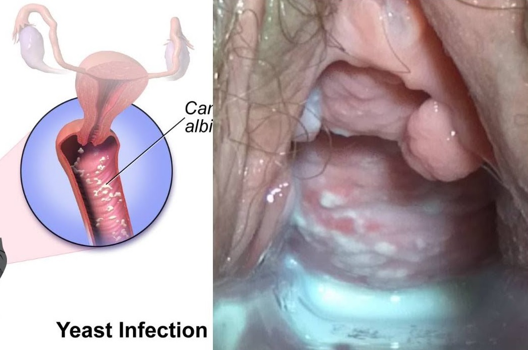
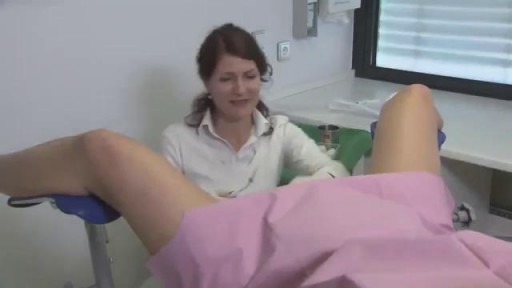
Watch that video to know Types and Causes of Vaginal Infection Yeast or Candidiasis, Trichomoniasis or Bacterial ?
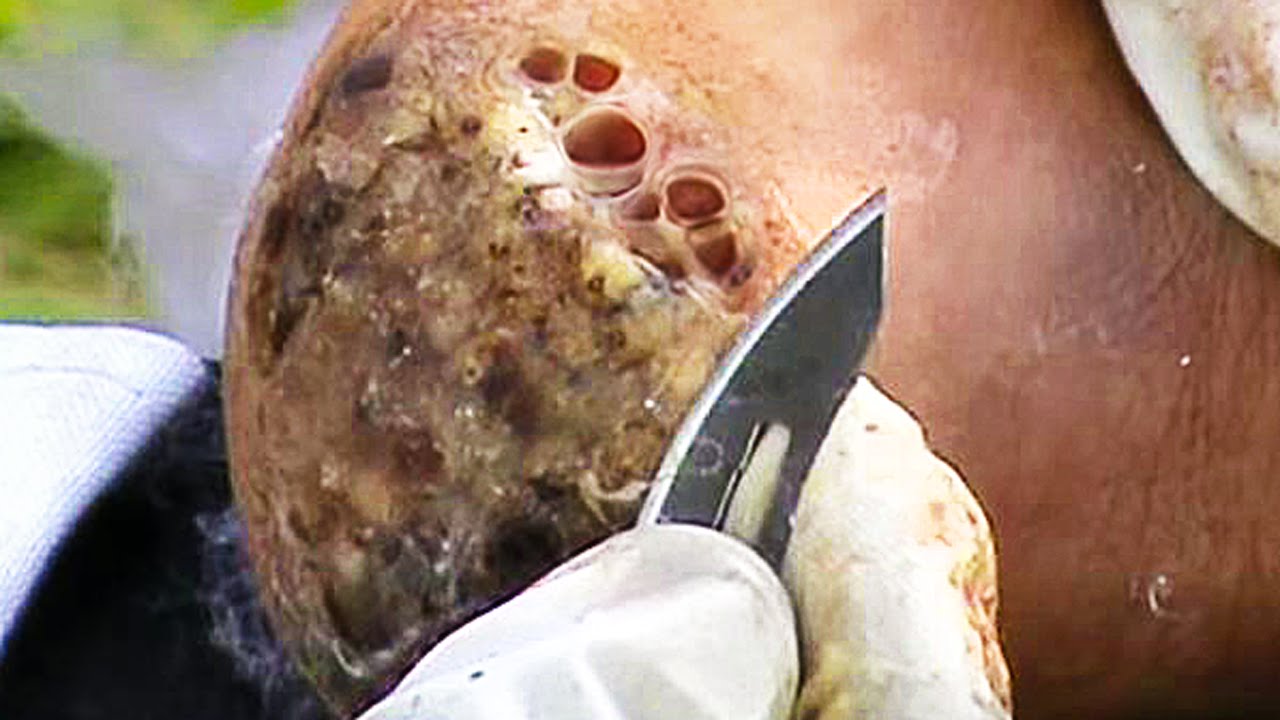
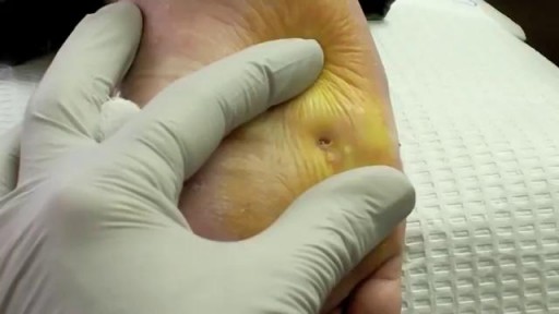
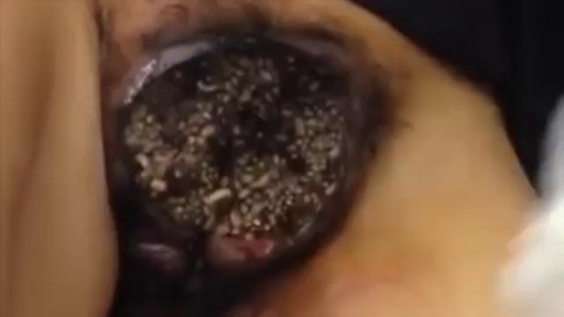
Watch that Skin Jiggers Removal Procedure
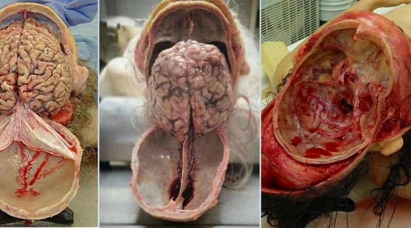
Watch that video of Human Skull Opening and Brain Removal During Autopsy
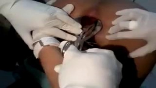
Watch that IUD Female Birth Control Insertion Surgery

Watch that video to know How to Get Pregnant With Twins

Watch that Full Human Body Medical Anatomy Autopsy
Watch that video of Stuck Sex Toy Medical Removal Surgery

Watch that video to know about the Health Benefits from KISSING
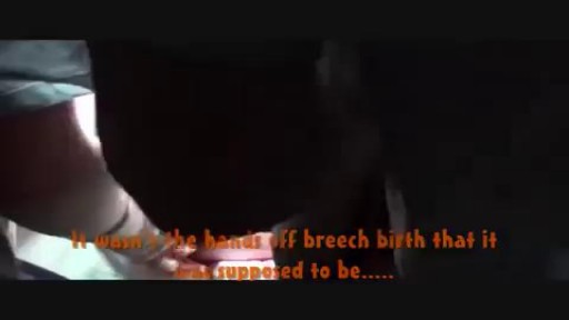
Watch that video of a Woman Giving Triplets Natural Vaginal Birth
Watch that video of Nasty Female Genital Infection

Pulsatile Tinnitus Cure, Constant Ear Ringing, Ear Wax Tinnitus, Whistling In Ear, Ringing In Ears. http://tinnitus-solution.info-pro.co First the good news - we know what causes tinnitus. And now the bad news - conventional medical science cannot cure it. Not permanently at least. Sure enough, your doctor would suggest a few remedies, and it may seem to you that the noises you hear are going down. As a result, you begin to relax believing that a pesky problem has been resolved. But suddenly the sounds return again. This is a very common problem actually. So let us turn to the causes instead, and see whether we can try to solve the issue from this end. Here Are Some of the Most Common Causes of Tinnitus Exposure to noise - Did your mom always tell you in your younger days to turn down the volume? She was right. Exposure to loud noise can give you tinnitus. In fact, rock musicians, and those who work with them, or in night clubs often have it. Those who work in construction sites also have tinnitus. So turn down that volume while you still can. You could begin to hear all kinds of noises if you have been exposed to just a single high-pitched noise. Or it could be due to a continuous attack of loud noises close to your ear. This is what happens. Prolonged exposure to noise can damage the Cochlea and cause tinnitus. So if you cannot simply stay away from all that noise, at least get some protection. Use an ear plug when you can. Head injury - Take care of your head because a severe blow or a slight bang could make you hear the tinnitus noises. The head is of course one of the most sensitive parts of the human body. But some people cannot live without an injury, such as those who are into sports - boxers and football players. That's why athletes are more prone to a tinnitus attack. Even a dental surgery could make you hear them. Ear infections and other ear problems - An ear infection, and even sinus can lead to tinnitus as well. When there is an allergy or a sinus infection, the mucous thickens within the inner ear, and this causes more pressure. The extra pressure can lead to tinnitus. Meniere's disease, where the fluid level goes up inside the middle ear is another reason. It could even cause hearing loss. Prescription medications - Conventional drugs often cause side effects, and tinnitus is one of them. Actually, all kinds of drugs have been blamed for instigating this condition. Such as antibiotics like Aminoglycosides, Erythromycin and Vancomycin, Aspirin or medicines containing it. Anti inflammatory drugs like Advil, Aleve, Anaprox, Clinoril, Feldene, Indocin, Lodine and Motrin have also been blamed. Sometimes people heard noises after taking chemotherapy agents such as Cisplatin, Nitrogen Mustard and Vincristine. And some others have even blamed quinine and loop diuretics for this. or even the result of a virus or infection. but is in fact far more shocking that you’ve been led to believe. You’ll finally be able to concentrate on your life, rather that the incessant noise. You’ll be able to no longer live in fear of loud noises, of music, of cinemas. of having fun. The Tinnitus Scandal Revealed, A cure DOES exist. click here: http://tinnitus-solution.info-pro.co
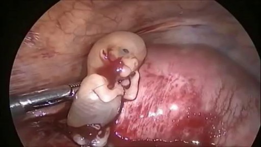
Watch that Ectopic Pregnancy Baby Abortion Surgery

Watch that Male Catheter Insertion Procedure

Watch that Terrible Skin Jiggers Removal Video

Watch that Human Baby Medical Abortion Surgery
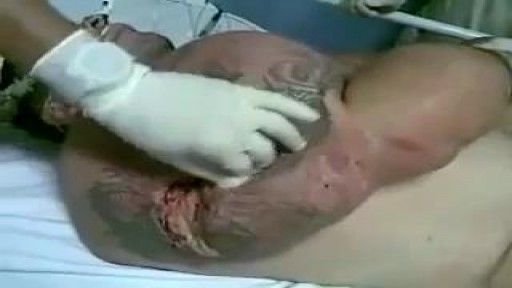
Watch that video of a Man's Arm Exploded Due to Illegal Muscles Injections
