- Physical Examination
- Surgical Examination
- Ophthalmology
- Clinical Skills
- Orthopedics
- Surgery Videos
- Laparoscopy
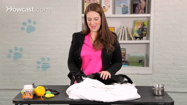
- Pediatrics
- Funny Videos
- Cardiothoracic Surgery
- Nursing Videos
- Plastic Surgery
- Otorhinolaryngology
- Histology and Histopathology
- Neurosurgery
- Dermatology
- Pediatric Surgery
- Urology
- Dentistry
- Oncology and Cancers
- Anatomy Videos
- Health and Fitness
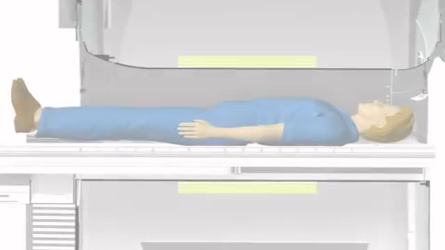
- Radiology
- Anaesthesia
- Physical Therapy
- Pharmacology
- Interventional Radiology
- Cardiology
- Endocrinology
- Gynecology
- Emergency Medicine
- Psychiatry and Psychology
- Childbirth Videos
- General Medical Videos
- Nephrology
- Physiology
- Diet and Food Health
- Diabetes Mellitus
- Neurology
- Women Health
- Osteoporosis
- Gastroenterology
- Pulmonology
- Hematology
- Rheumatology
- Toxicology
- Nuclear Medicine
- Infectious Diseases
- Vascular Disease
- Reproductive Health
- Burns and Wound Healing
- Other
Top videos
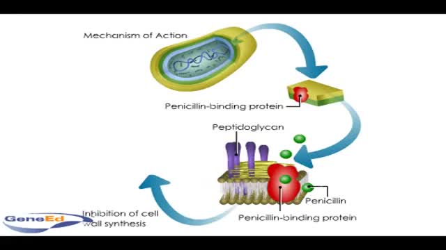
Penicllin mechanism of action
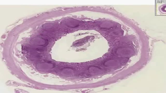
Histology of Appendix
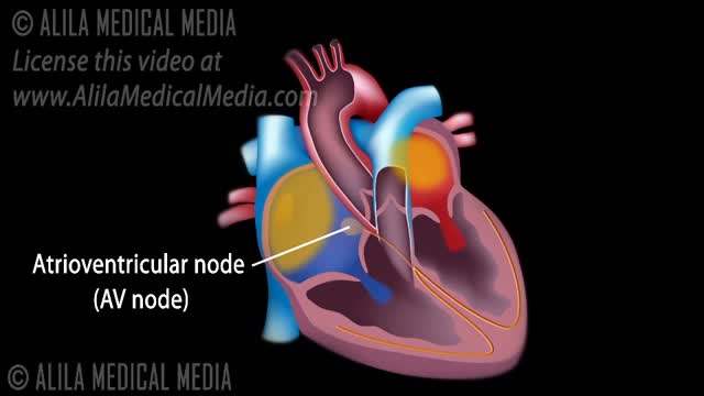
The normal electrical conduction in the heart allows the impulse that is generated by the sinoatrial node (SA node) of the heart to be propagated to (and stimulate) the cardiac muscle (myocardium). The myocardium contracts after stimulation.
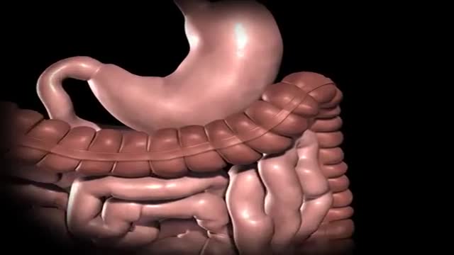
Gastric bypass is surgery that helps you lose weight by changing how your stomach and small intestine handle the food you eat. After the surgery, your stomach will be smaller. You will feel full with less food. The food you eat will no longer go into some parts of your stomach and small intestine that absorb food. Because of this, your body will not get all of the calories from the food you eat.
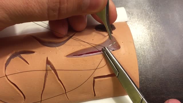
The two biggest considerations when choosing a suture are the location and tension of the wound. Other important considerations are tensile strength, knot strength, handling, and tissue reactivity. Sutures are divided into two major groups: Absorbable – lose the majority of their tensile strength in less than 60 days. They are generally used for buried sutures and do not require removal. Non-absorbable – maintain the majority of their tensile strength for more than 60 days. They are generally used for skin surface sutures and do require removal postoperatively. Suture needles also come in a variety of shapes and sizes. Curved needles are almost exclusively used in dermatological surgery. Cutting needles move through the tissue more easily and may have their primary cutting edge on the inside of the curve (conventional cutting) or outside of the curve (reverse cutting). The benefit of reverse cutting is that the tapered puncture left by the suture is directed away from the wound edge and therefore tissue tearing is less common. Non-cutting round needles cause even less tissue tearing and may be especially useful in delicate areas and fascia.
Tests. This test tracks electrical signals from the brain. There are a number of blood tests that may be recommended as part of your epilepsy diagnosis and treatment. A positron emission tomography (PET) scan may be used to locate the part of the brain that is causing seizures.
The foods for your child are easily digestible foods, such as rice cereal, pasta, breads, cooked beans, mashed potatoes, cooked carrots, applesauce, and bananas. Pretzels or salty crackers can help your child replace the salt lost from diarrhea. Foods containing large amounts of sugar or fat should be avoided.
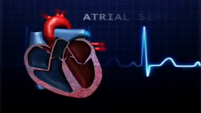
atrial septal defect (ASD) is a hole in the wall between the two upper chambers of your heart (atria). The condition is present from birth (congenital). Small atrial septal defects may close on their own during infancy or early childhood. Large and long-standing atrial septal defects can damage your heart and lungs. Small defects may never cause a problem and may be found incidentally. An adult who has had an undetected atrial septal defect for decades may have a shortened life span from heart failure or high blood pressure that affects the arteries in the lungs (pulmonary hypertension). Surgery may be necessary to repair atrial septal defects to prevent complications

The arm and leg muscles are affected later. Myasthenia gravis (MG) is an autoimmune disease — a disease that occurs when the immune system attacks the body's own tissues. In MG, that attack interrupts the connection between nerve and muscle — the neuromuscular junction.
Controlled studies on treatment of catscratch disease (CSD) are lacking. Thus, treatment recommendations are based on case reports, reviews, a single controlled trial, and anecdotal data. Practice guidelines for the diagnosis and management of skin and soft-tissue infections, including CSD, have been established.Oct 19, 2016
Given the common presentation of onychodystrophy, physicians should have a firm grasp of common presentations of conditions like onychomycosis, trachyonychia and psoriasis. Accordingly, this author reviews keys to effective diagnosis and pertinent treatment considerations. Nail cosmesis and discomfort are the main motivators for most of our patients to schedule a podiatric consultation. During that patient visit, it is important for the podiatric practitioner to delve into the cause of the problematic nail change, known as onychodystrophy. Onychodystrophy, which is any alteration of nail morphology, encompasses a wide spectrum of nail disorders. Caused by either exogenous or endogenous factors, nail dystrophy may manifest as a misshapen, damaged, infected or discolored nail unit that may affect the toenails, fingernails or both.
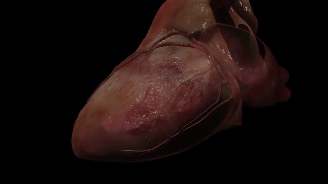
The heart is the body's engine room, responsible for pumping life-sustaining blood via a 60,000-mile-long (97,000-kilometer-long) network of vessels. The organ works ceaselessly, beating 100,000 times a day, 40 million times a year—in total clocking up three billion heartbeats over an average lifetime. It keeps the body freshly supplied with oxygen and nutrients, while clearing away harmful waste matter.

Cosmetic Eye and Eyelid Surgery
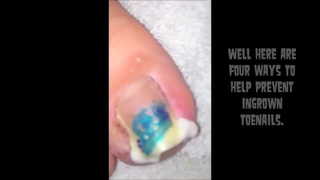
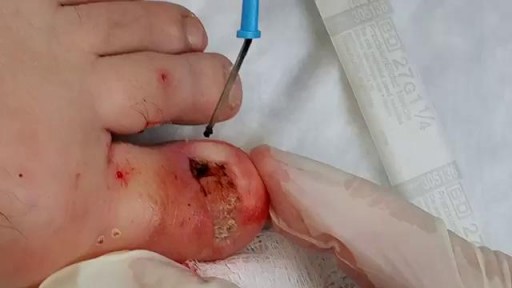
irregular, curved toenails. footwear that places a lot of pressure on the big toes, such as socks and stockings that are too tight or shoes that are too tight, narrow, or flat for your feet. toenail injury, including stubbing your toe, dropping something heavy on your foot, or kicking a ball repeatedly. poor posture. How can ingrowing toenails be prevented? Cut your nails straight across; do not cut them too short or too low at the sides. ... Keep your feet clean and dry. ... Avoid tight shoes and use cotton socks rather than synthetic. If you have diabetes, you should take extra care when cutting your nails:
Magnetic resonance imaging (MRI) can be an important tool in the diagnosis of multiple sclerosis (MS). MRI can also be used to monitor the progression of the disease in people living with MS. How does it work? MRI uses very strong magnets, radio signals, and computer software to take 3-dimensional pictures of the inside of the body. Will I need contrast material? Maybe. Contrast material is a substance that temporarily changes the way imaging tools interact with the body. They are often used to visualize certain types of MS disease activity on the MRI. If your doctor thinks your scan requires this contrast material, you may get an injection before you get in the MRI machine. How long will it take? The time may vary based on the type of MRI. Be sure to discuss with your doctor in advance so he or she can provide you with exact timing. But don’t worry, you won’t have to stay still the whole time. The technician will let you know when they’re starting a new image.
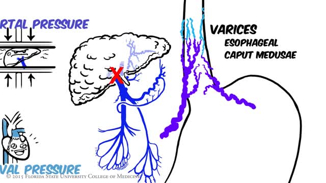
Portal hypertension is an increase in the blood pressure within a system of veins called the portal venous system. Veins coming from the stomach, intestine, spleen, and pancreas merge into the portal vein, which then branches into smaller vessels and travels through the liver.

Hemorrhoids repair: Disposable hemorrhoidal stapler
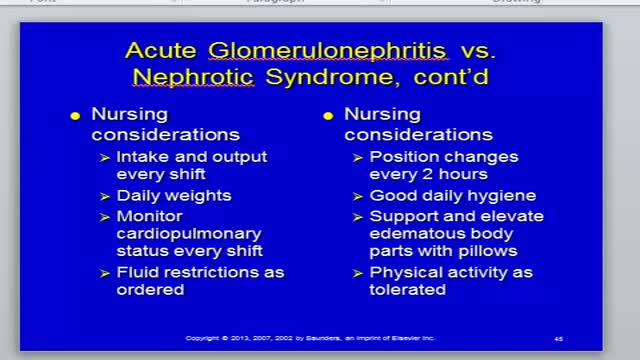
Nephritis and Nephrotic Syndrome

Father & Mom feel their baby the same

Major signs and symptoms include enlargement of the liver and spleen (hepatosplenomegaly), a low number of red blood cells (anemia), easy bruising caused by a decrease in blood platelets (thrombocytopenia), lung disease, and bone abnormalities such as bone pain, fractures, and arthritis.
