- Physical Examination
- Surgical Examination
- Ophthalmology
- Clinical Skills
- Orthopedics
- Surgery Videos
- Laparoscopy
- Pediatrics
- Funny Videos
- Cardiothoracic Surgery
- Nursing Videos
- Plastic Surgery
- Otorhinolaryngology
- Histology and Histopathology
- Neurosurgery
- Dermatology
- Pediatric Surgery
- Urology
- Dentistry
- Oncology and Cancers
- Anatomy Videos
- Health and Fitness
- Radiology
- Anaesthesia
- Physical Therapy
- Pharmacology
- Interventional Radiology
- Cardiology
- Endocrinology
- Gynecology
- Emergency Medicine
- Psychiatry and Psychology
- Childbirth Videos
- General Medical Videos
- Nephrology
- Physiology
- Diet and Food Health
- Diabetes Mellitus
- Neurology
- Women Health
- Osteoporosis
- Gastroenterology
- Pulmonology
- Hematology
- Rheumatology
- Toxicology
- Nuclear Medicine
- Infectious Diseases
- Vascular Disease
- Reproductive Health
- Burns and Wound Healing
- Other
Top videos
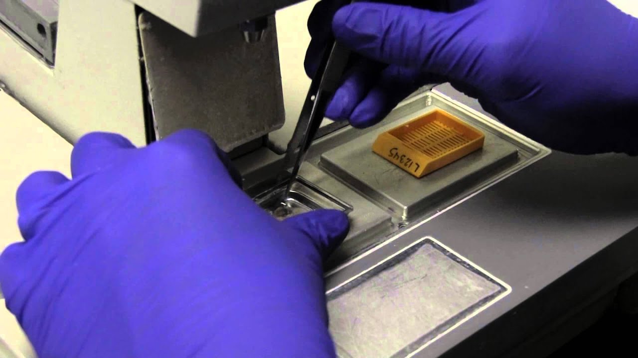

Histology of all the different types of cartilage. This video is a part of our Histology Video Course (https://youtube.com/playlist?l....ist=PLnr1l7WuQdDynxT
All Histology Videos: https://youtube.com/playlist?l....ist=PLnr1l7WuQdDynxT
Thank you to our sponsor Doc2Doc Lending, the Personal Lending platform designed for Doctors, by Doctors. Check out https://doc2doclending.com/davinci to learn more today.
Additional YouTube Content
Biochemistry videos: https://youtube.com/playlist?l....ist=PLnr1l7WuQdDzCUC
Anatomy Videos: https://youtube.com/playlist?l....ist=PLnr1l7WuQdDz2dK
DaVinci Cases Videos: https://youtube.com/playlist?l....ist=PLnr1l7WuQdDyJUl
The DaVinci Hour Podcast: https://youtube.com/playlist?l....ist=PLnr1l7WuQdDwSm9
DaVinci Academy Website: https://www.dviacademy.com/

Doctors try to find a way for their patient suffering from a rare skin condition that causes her skin to blister and bleed if it touches anything, to attend her senior prom.
From Pure Genius Season 1 Episode 11 'Touch and Go' - James and Zoe try radical treatments on a girl with a rare skin disorder in an attempt to heal her in time for her prom; James considers an experimental cure for Louis Keating's GSS condition; Malik is jealous of James and Zoe's special connection.
Pure Genius (2016) A young tech-titan from Silicon Valley decides to build a hospital with a new-school approach to medicine and enlists a veteran surgeon who has a controversial past.
Watch full episodes of Pure Genius here: https://www.justwatch.com/uk/tv-series/pure-genius
Welcome to MD TV! A channel dedicated to your favourite medical dramas! Featuring iconic moments from House M.D., Chicago Med and more. Follow the professional and personal lives of the hospital staff, as you go a journey right from the very first doctor's call to the E.R and beyond. MD TV is packed full of drama, intrigue, and plenty of medical emergencies!
#MDTV #medicaldrama #medicaltvshow

https://bit.ly/3HIStRc #shorts
Coloscopy | Colon Polyp Resection | Polypectomy
Colonoscopies are essential for detecting colorectal abnormalities, including colon polyps. Polypectomy, the surgical removal of these growths, can prevent them from becoming cancerous. This article offers a brief overview of colonoscopies, colon polyps, and polypectomy procedures.
A colonoscopy is an endoscopic examination allowing healthcare providers to visualize the colon and rectum using a colonoscope. The colonoscope, a flexible tube with a camera and light source, helps detect abnormalities, including polyps or tumors.
Colon polyps are abnormal growths arising from the colon's inner lining. While most polyps are benign, some can become malignant. Adenomatous polyps have a higher potential to become cancerous, whereas hyperplastic and inflammatory polyps pose a lower risk.
Polypectomy involves removing colon polyps during a colonoscopy. Two primary techniques include snare polypectomy, using a wire loop to cut the polyp, and cold forceps polypectomy, which employs forceps to grasp and remove smaller polyps.
Following a polypectomy, patients may experience mild discomfort or bleeding. Regular surveillance is crucial to minimize colorectal cancer risk. The frequency of surveillance colonoscopies depends on the number, size, and type of polyps found, as well as the patient's overall risk factors.
Colonoscopies and polypectomies play vital roles in detecting and removing colon polyps, reducing the risk of colorectal cancer, and maintaining optimal colon health.
Do you want to learn more about colon polyps and colonoscopy? check our:
Article @ https://bit.ly/41w5Ooq
For more free resources, find us on Pinterest & Facebook pages:
https://www.pinterest.ca/medicalartsofficial/
https://www.facebook.com/Medicalartsofficial
https://www.youtube.com/@medic....alarts?sub_confirmat
https://www.instagram.com/medicalartsofficial/
https://www.tiktok.com/@medicalarts
#endoscopicsurgery #digestivesystem
coloscopy
polyp
colon polyp
polypectomy
colonoscopy
colon polyp animation
gi endoscopy
@MedicalArts , 2023.

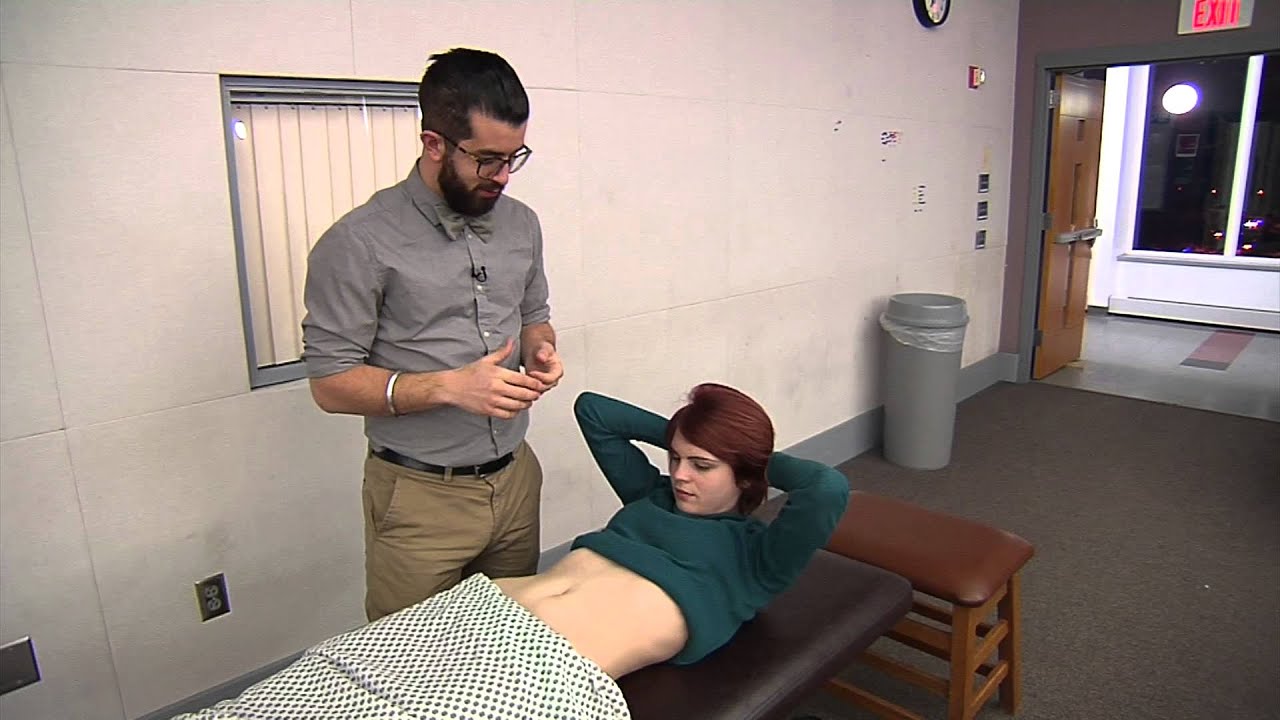
The cardiac examination is one of the earliest clinical skills that medical students learn. As a junior doctor, the examination of the cardiovascular system can be almost a dreaded examination, as cardiac murmurs can literally take years of exposure in order to gain confidence with their identification through cardiac auscultation.
This video demonstrates not merely the examination of the heart, but the complete cardiovascular system including peripheries.
I hope these clinical skill revision videos are helpful, please like and subscribe and join the community so that we can create more effective videos to help with your journey through medical school
#CardiacExam #ClinicalExamination #asmr

Surgeons at St Mary's Hospital, part of Imperial College Healthcare have come up with a new surgical procedure that cures heartburn with a device called RefluxStop.
Mr Ahmed Ahmed, a consultant surgeon, says surgery should now be seen as an alternative to life-long drug treatment - as Sky's Thomas Moore reports.
Read more: https://news.sky.com/story/new....-nhs-heartburn-surge
#heartburncure #surgery #skynews
SUBSCRIBE to our YouTube channel for more videos: http://www.youtube.com/skynews
Follow us on Twitter: https://twitter.com/skynews
Like us on Facebook: https://www.facebook.com/skynews
Follow us on Instagram: https://www.instagram.com/skynews
Follow us on TikTok: https://www.tiktok.com/@skynews
For more content go to http://news.sky.com and download our apps: Apple https://itunes.apple.com/gb/ap....p/sky-news/id3163919 Android https://play.google.com/store/apps/details?id=com.bskyb.skynews.android&hl=en_GB
Sky News Daily podcast is available for free here: https://podfollow.com/skynewsdaily/
Sky News videos are now available in Spanish here/Los video de Sky News están disponibles en español aquí: https://www.youtube.com/channe....l/UCzG5BnqHO8oNlrPDW
To enquire about licensing Sky News content, you can find more information here: https://news.sky.com/info/library-sales
When your child needs surgery, it can be overwhelming and sometimes scary. At Mayo Clinic Children’s Center, our highly skilled surgeons apply deep experience and specialized training to offer individualized care for your child and your family.
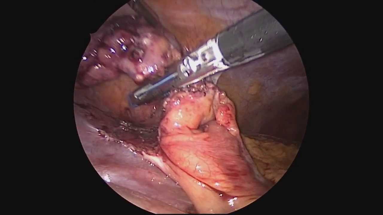
Dr. Celia Divino, Chief, Division of General Surgery at The Mount Sinai Hospital, performs a laparoscopic appendectomy. Visit the Division of General Surgery at http://bit.ly/18z944M. Click here to learn more about Dr. Celia Divino http://bit.ly/12RF0ee
We are looking for 5 patients with knee pain who want to get significantly better in the next 30 days. Click this link to let me know you're interested and learn more.
https://www.drdavidgeier.com/work-with-me/contact/
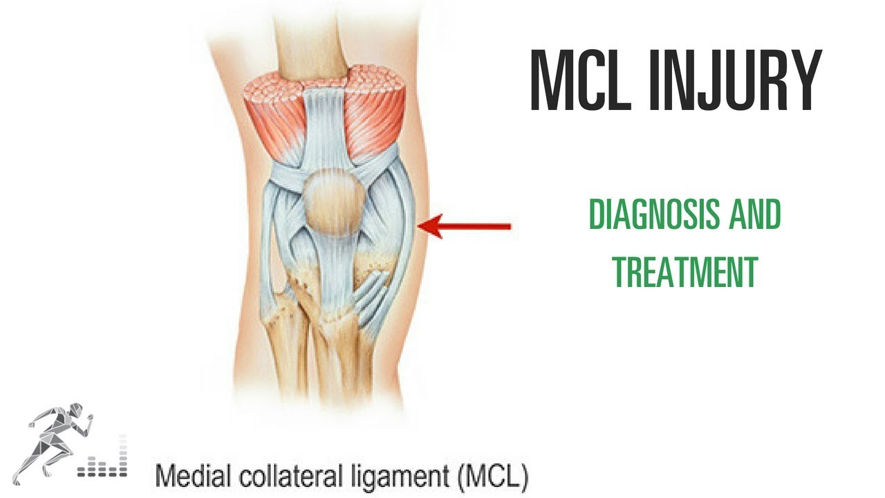
One of the most common knee injuries in contact and collision sports is a medial collateral ligament (MCL) injury. This is a ligament on the medial (side closest to the midline) side of your knee that provides stability against side-to-side stress to the knee. You might injure it by cutting maneuvers in sports like soccer or hockey. You can also suffer an MCL injury if another player hits you on the outside of your knee.
Please note: I don't respond to questions and requests for specific medical advice left in the comments to my videos. I receive too many to keep up (several hundred per week), and legally I can't offer specific medical advice to people who aren't my patients (see below). If you want to ask a question about a specific injury you have, leave it in the comments below, and I might answer it in an upcoming Ask Dr. Geier video. If you need more detailed information on your injury, go to my Resources page: https://www.drdavidgeier.com/resources/
The content of this YouTube Channel, https://www.youtube.com/user/drdavidgeier (“Channel”) is for INFORMATIONAL PURPOSES ONLY. The Channel may offer health, fitness, nutritional and other such information, but such information is intended for educational and informational purposes only. This content should not be used to self-diagnose or self-treat any health, medical, or physical condition. The content does not and is not intended to convey medical advice and does not constitute the practice of medicine. YOU SHOULD NOT RELY ON THIS INFORMATION AS A SUBSTITUTE FOR, NOR DOES IT REPLACE, PROFESSIONAL MEDICAL ADVICE, DIAGNOSIS, OR TREATMENT. You should consult with your healthcare professional before doing anything contained on this Channel. You agree that Dr. Geier is not responsible for any actions or inaction on your part based on the information that is presented on the Channel. Dr. David Geier Enterprises, LLC makes no representations about the accuracy or suitability of the content. USE OF THE CONTENT IS AT YOUR OWN RISK.
Unlike tears of the ACL, MCL injuries most often heal without surgery. You might need to wear a hinged knee brace for 2-6 weeks. The length of time you miss from sports or exercise varies depending on the location and severity of the injury.
In this video, I share my thoughts on the nature of an MCL injury, the diagnosis, the treatment options and return to sports.
Please remember, while I appreciate your questions, I cannot and will not offer specific medical advice by email, online, on my show, or in the comments at the end of these posts. My responses are meant to provide general medical information and education. Please consult your physician or health care provider for your specific medical concerns.

BioDigital Systems created this 3D animation of a knee replacement surgery.
---
BioDigital is happy to share helpful health information, but we do not offer medical advice. For medical advice, please contact your healthcare provider directly.
For more information on the content of this video, you can:
Access these 3D visuals: human.biodigital.com
Learn more about BioDigital: www.biodigital.com
Subscribe: www.youtube.com/c/BioDigital?sub_confirmation=1
Facebook: www.facebook.com/BioDigitalHuman
Instagram: www.instagram.com/biodigital.human
Twitter: twitter.com/biodigitalhuman
LinkedIn: www.linkedin.com/company/biodigital/ Medical disclaimer:
BioDigital, Inc is not a health care provider and we do not provide medical advice. You should not rely on the information provided on our sites or services as a substitute for, nor does it replace, professional medical advice, diagnosis, or treatment. The services are not intended to be used by consumers or clinicians in making treatment decisions. You are encouraged to seek professional medical diagnosis and treatment for any medical condition, and to discuss information from the sites and services with your healthcare provider. Information provided on the sites and media is provided for informational purposes and is in no way intended to substitute consulting a medical professional. Nothing stated or posted by BioDigital is intended to be, and must not be taken to be, the practice of medicine, the provision of medical care, or a tool relied on by patients or clinicians. If you rely on any of the information provided by BioDigital, you do so solely at your own risk.

Knee pain can happen at any age, but some doctors say they're seeing more people with osteoarthritis who are still young and active.
Subscribe to WCVB on YouTube for more: http://bit.ly/2526UpS
Get more Boston news: http://www.wcvb.com
Like us: https://www.facebook.com/wcvb5
Follow us: https://twitter.com/WCVB
Google+: https://plus.google.com/+wcvb
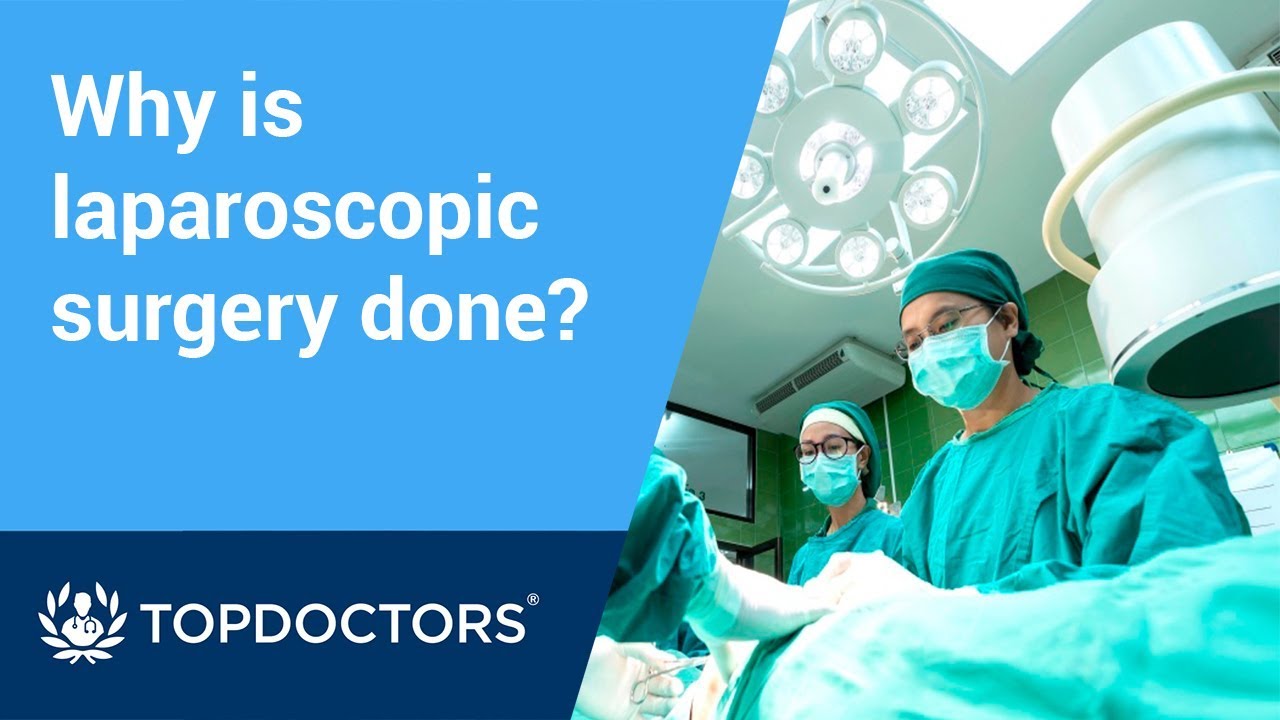
Laparoscopic surgery is minimally-invasive (keyhole) surgery and it is performed through very small incisions, using a camera to guide the surgeon during the procedure. Miss Sarah Mills, a top colorectal surgeon, explains why laparoscopic surgery is performed over alternative methods.
Make an appointment with Miss Sarah Mills here: https://www.topdoctors.co.uk/doctor/sarah-mills

For more information, visit https://ukhealthcare.uky.edu/doctors.

While in residency, Marc Pelletier, MD, helped in a bypass surgery and knew it was the field in which he would excel. Watch as the Chief of Cardiac Surgery for University Hospitals Harrington Heart & Vascular Institute in Cleveland, Ohio explains, in detail, what happens in preparation for heart surgery, in the operating room and the feeling he experiences after surgery.
How does a heart-lung machine work? What is 'efficiency of motion'? These questions and more are answered in this compelling, dramatic look at heart surgery.
To learn more about heart surgery at University Hospitals: https://www.uhhospitals.org/fo....r-clinicians/special
University Hospitals is one of the nation’s leading health care systems, providing patient-centered care that meets the highest standards for quality and patient safety and have received numerous awards and recognitions from some of the most prestigious institutions in the country for our leadership and exceptional patient outcomes. As an accountable care organization, we foster long-term patient-provider relationships that help promote preventive care, increase wellness and healthy behaviors, decrease emergency episodes, and prevent hospitalizations. To learn more: https://www.uhhospitals.org

Mr Brian MacCormack talking about Paediatric Surgery Emergencies. This talk is part of the Paediatric Emergencies 2022 event. To get your CME certificate for watching the video please visit https://www.paediatricemergenc....ies.com/conference/p
#PaediatricEmergencies #PaediatricEmergencies2022 #PaediatricSurgery
Female heart surgeons are rare, but pediatric female surgeons are even more so.

Get a 60-day free trial at https://shipstation.com/doctormike. Thanks to ShipStation for sponsoring the show!
I’ll teach you how to become to media’s go-to expert in your field. Enroll in The Professional’s Media Academy now: https://www.professionalsmediaacademy.com/
Listen to my podcast, @DoctorMikeCheckup, here:
Spotify: https://go.doctormikemedia.com..../spotify/CheckUpSpot
Apple Podcasts: https://go.doctormikemedia.com..../applepodcast/AppleP
Body Bizarre is a TLC show with a name I'm not too wild about, but with stories that are nonetheless fascinating. Today we look at separating conjoined twins, a girl with ants crawling out of her ears, a man who nearly lost his hand in a factory accident, a family that all has 6 fingers, and more.
Help us continue the fight against medical misinformation and change the world through charity by becoming a Doctor Mike Resident on Patreon where every month I donate 100% of the proceeds to the charity, organization, or cause of your choice! Residents get access to bonus content, an exclusive discord community, and many other perks for just $10 a month. Become a Resident today:
https://www.patreon.com/doctormike
Let’s connect:
IG: https://go.doctormikemedia.com..../instagram/DMinstagr
Twitter: https://go.doctormikemedia.com/twitter/DMTwitter
FB: https://go.doctormikemedia.com/facebook/DMFacebook
TikTok: https://go.doctormikemedia.com/tiktok/DMTikTok
Reddit: https://go.doctormikemedia.com/reddit/DMReddit
Contact Email: DoctorMikeMedia@Gmail.com
Executive Producer: Doctor Mike
Production Director and Editor: Dan Owens
Managing Editor and Producer: Sam Bowers
Editor and Designer: Caroline Weigum
Editor: Juan Carlos Zuniga
* Select photos/videos provided by Getty Images *
** The information in this video is not intended nor implied to be a substitute for professional medical advice, diagnosis or treatment. All content, including text, graphics, images, and information, contained in this video is for general information purposes only and does not replace a consultation with your own doctor/health professional **
Click here to get 2 free filet mignons and $15 off your first ButcherBox: https://butcherbox.com/doctormike
Includes FREE Shipping. Be sure to enter your email to access the deal. Thanks to ButcherBox for sponsoring this video.
I’ll teach you how to become to media’s go-to expert in your field. Enroll in The Professional’s Media Academy now: https://www.professionalsmediaacademy.com
Listen to my podcast, @DoctorMikeCheckup here:
YouTube: https://go.doctormikemedia.com..../youtube/channel/The
Spotify: https://go.doctormikemedia.com..../spotify/CheckUpSpot
Apple Podcasts: https://go.doctormikemedia.com..../applepodcast/AppleP
Survivor is coming up on its 43rd season this fall (whaaat??), and with all that reality TV goodness in the can already I knew there would be some medical moments to react to. Turns out, I was right, in that there have been a bunch of ailments on the show over the years! These injuries span the entire length of the whole series, so if you're a long time Survivor and Jeff Probst fan, this one is for you!
I LOVE reading your comments and take your suggestions seriously. If there’s a subject you want me to discuss or something you’d like for me to react to, leave a comment down below. Many of my videos have been born out of suggestions directly from you, so don’t hold back!
-Doctor Mike Varshavski
Help us continue the fight against medical misinformation and change the world through charity by becoming a Doctor Mike Resident on Patreon where every month I donate 100% of the proceeds to the charity, organization, or cause of your choice! Residents get access to bonus content, an exclusive discord community, and many other perks for just $10 a month. Become a Resident today:
https://www.patreon.com/doctormike
Please SUBSCRIBE for new videos every Wednesday afternoon and Sunday morning! https://goo.gl/87kYq6
Let’s connect:
IG https://goo.gl/41ZS7w - Doctor Mike
Reddit https://www.reddit.com/r/DoctorMike/
Twitter https://goo.gl/kzmGs5 - Real Doctor Mike
Facebook https://goo.gl/QH4nJS - Real Doctor Mike
Contact Email: DoctorMikeMedia@Gmail.com
Executive Producer: Doctor Mike
Production Director and Editor: Dan Owens
Managing Editor and Producer: Sam Bowers
Editor and Designer: Caroline Weigum
* Select photos/videos provided by Getty Images *
** The information in this video is not intended nor implied to be a substitute for professional medical advice, diagnosis or treatment. All content, including text, graphics, images, and information, contained in this video is for general information purposes only and does not replace a consultation with your own doctor/health professional **

An animation of blood flow inside the hollow fiber of a hemofilter, or a dialyzer, and the flow of the dialysate in an opposite direction with increased extraction of waste and small molecules from the blood as the concentration of these molecules is reduced downstream and exposed to new dialysate.
To learn about Hemodialysis..
https://www.thevirtualnephrolo....gist.com/specialties
The Virtual Nephrologist is your gateway to optimal health.
To learn more about Hypertension, Kidney Disease and Dialysis:
https://thevirtualnephrologist.com/
About Dr. Rifai:
Dr. Ahmad Oussama Rifai is certified by the American Board of Internal Medicine (ABIM) in the specialty of Internal Medicine and the sub-specialty of Nephrology.
MEET DR. RIFAI
https://www.thevirtualnephrologist.com/rifai/
Follow The Virtual Nephrologist on SOCIAL MEDIA:
-Facebook: https://www.facebook.com/thevirtualnephrologist
-Instagram: https://www.instagram.com/thevirtualnephrologist/
-Twitter: https://twitter.com/VNephrologist
-TikTok: https://www.tiktok.com/@thevirtualnephrologist
Schedule a virtual consult:
https://www.thevirtualnephrolo....gist.com/schedule-a-
Best wishes for great health | The Virtual Nephrologist
