- Physical Examination
- Surgical Examination
- Ophthalmology
- Clinical Skills
- Orthopedics
- Surgery Videos
- Laparoscopy
- Pediatrics
- Funny Videos
- Cardiothoracic Surgery
- Nursing Videos
- Plastic Surgery
- Otorhinolaryngology
- Histology and Histopathology
- Neurosurgery
- Dermatology
- Pediatric Surgery
- Urology
- Dentistry
- Oncology and Cancers
- Anatomy Videos
- Health and Fitness
- Radiology
- Anaesthesia
- Physical Therapy
- Pharmacology
- Interventional Radiology
- Cardiology
- Endocrinology
- Gynecology
- Emergency Medicine
- Psychiatry and Psychology
- Childbirth Videos
- General Medical Videos
- Nephrology
- Physiology
- Diet and Food Health
- Diabetes Mellitus
- Neurology
- Women Health
- Osteoporosis
- Gastroenterology
- Pulmonology
- Hematology
- Rheumatology
- Toxicology
- Nuclear Medicine
- Infectious Diseases
- Vascular Disease
- Reproductive Health
- Burns and Wound Healing
- Other
Top videos

TUMMY TUCK 🤩 Immediate OR Results
This patient wanted to get her abs back, but unfortunately NO diet or workout can tighten muscles that have been stretched apart from carrying a baby 👀 But we can fix that at Lemmon Avenue Plastic Surgery & Laser Center!
To learn more about the #tummytuck click here: https://drdeuber.com/procedures/tummy-tuck/
For #mommymakeover, click here: https://drdeuber.com/procedures/mommy-makeover/
👙
#MarkDeuberMD

Dr. Shaun Kunisaki is an Associate Professor of Surgery at The Johns Hopkins University and Associate Chief of Strategy and Integration in the Division of General Pediatric Surgery at the Johns Hopkins Children's Center. His clinical practice spans the full breadth of pediatric general surgery, but he is recognized both regionally and nationally for this expertise in complex thoracic surgical problems in the fetus and young child. As Director of Pediatric Esophageal Surgery, he specializes in the management of long-gap esophageal atresia. In this role within the Johns Hopkins Children Center Fetal Program, he helps counsel parents with pregnancies complicated by fetal anomalies.
Learn more about Dr. Kunisaki at https://www.hopkinsmedicine.or....g/profiles/results/d

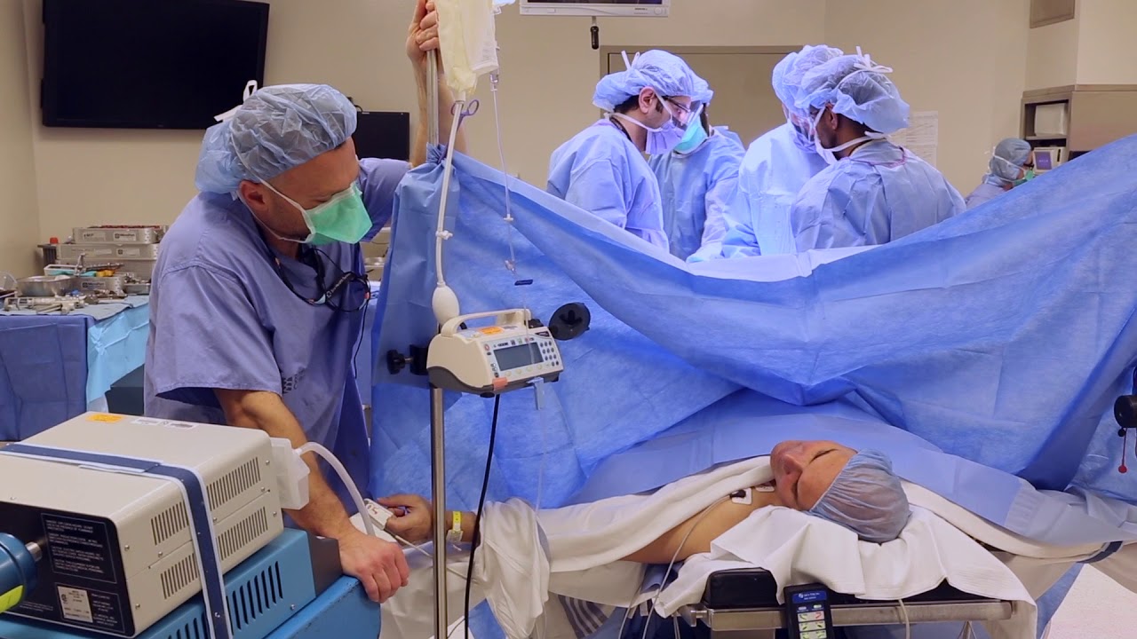
Emory has one of the few heart and vascular centers nationally performing robotic cardiac surgery using the daVinci Surgical System. Emory's robotic surgeons have completed numerous cases and are recognized in Atlanta, the Southeast and across the country for their expertise in cardiac surgery. Some of the cardiac and thoracic conditions treated by Emory cardiac surgeons include mitral valve repair and replacement, atrial septal defect repair, atrial myxoma and thrombi, coronary bypass (LIMA to LAD), mediastinal mass excision, thymectomy, epicardial lead placement and pericardial window.

Mini-Laparoscopic Cholecystectomy with Intraoperative Cholangiogram for Symptomatic Cholelithiasis (Gallstones) - Standard
Authors: Brunt LM1, Singh R1, Yee A2
Published: September 26, 2017
AUTHOR INFORMATION
1 Department of Surgery, Washington University, St. Louis, Missouri
2 Division of Plastic and Reconstructive Surgery, Washington University, St. Louis, Missouri
DISCLOSURE
No authors have a financial interest in any of the products, devices, or drugs mentioned in this production or publication.
ABSTRACT
Minimal invasive laparoscopic cholecystectomy is the typical surgical treatment for cholelithiasis (gallstones), where patients present with a history of upper abdominal pain and episodes of biliary colic. The classic technique for minimal invasive laparoscopic cholecystectomy involves four ports: one umbilicus port, two subcostal ports, and a single epigastric port. The Society of American Gastrointestinal and Endoscopic Surgeons (SAGES) has instituted a six-step strategy to foster a universal culture of safety for cholecystectomy and minimize risk of bile duct injury. The technical steps are documented within the context of the surgical video for (1) achieving a critical view of safety for identification of the cystic duct and artery, (2) intraoperative time-out prior to management of the ductal structures, (3) recognizing the zone of significant risk of injury, and (4) routine intraoperative cholangiography for imaging of the biliary tree. In this case, the patient presented with symptomatic biliary colic due to a gallstone seen on the ultrasound in the gallbladder. The patient was managed a mini-laparoscopic cholecystectomy using 3mm ports for the epigastric and subcostal port sites with intraoperative fluoroscopic cholangiogram. Specifically, the senior author encountered a tight cystic duct preventing the insertion of the cholangiocatheter and the surgical video describes how the author managed the cystic duct for achieving a cholangiogram, in addition to the entire technical details of laparoscopic cholecystectomy.

If you’ve suffered a sporting knee injury, how do you know when it’s serious? In this short video, Yorkshire Knee Clinic’s Dave Duffy reveals the two key tests that tell you whether your knee needs urgent, specialist attention.
𝗡𝗼𝘁𝗲𝘀 𝗳𝗼𝗿 𝘁𝗵𝗲 𝘀𝗾𝘂𝗲𝗮𝗺𝗶𝘀𝗵: This video features only features a model of the knee. There is no live footage from operations.
Discover more about sports knee injuries: https://yorkshirekneeclinic.com/sports-injuries/
Discover more about Dave Duffy: https://yorkshirekneeclinic.com/about/dave-duffy/

Dr. Rowe shows an easy exercise that can give knee pain relief within seconds.
This exercise will help traction open the knee, relieving pressure and tension. It can be done throughout the day at home, and only requires only a small towel.
Let us know how it works for you!
***************************
Dr. Michael Rowe
St. Joseph, Michigan chiropractor
If you are looking for effective neck, back, or sciatica pain relief, contact us at 269-408-8439 or visit us at https://www.BestSpineCare.com
Facebook: https://www.facebook.com/bestspinecare
Twitter: https://www.twitter.com/stjoechiro
Instagram: https://www.instagram.com/stjoechiro
Your local St. Joseph | Benton Harbor | Stevensville Michigan chiropractor
SpineCare Decompression and Chiropractic Center
3134 Niles Rd
Saint Joseph, MI 49085
**MEDICAL DISCLAIMER**
All information, content, and material of this video or website is for informational and demonstration purposes only. It is not intended to serve as a substitute for the consultation, diagnosis, and/or medical treatment of a qualified physician or healthcare provider.
Don’t use this content as a replacement for treatment and advice given by your doctor or health care provider. Consult with your doctor or healthcare professional before doing anything contained in this content.
By watching this video, you agree to indemnify and hold harmless SpineCare Decompression and Chiropractic Center (and its representatives) for any and all losses, injuries, or damages resulting from any and all claims that arise from your use or misuse of this content. SpineCare Decompression and Chiropractic Center makes no representations about the accuracy or suitability of this content.
USE OF THIS CONTENT IS AT YOUR OWN RISK.
#kneepain #kneepainrelief #kneepainexercise

To learn more about licensing this video for content marketing or patient education purposes, visit: http://www.nucleushealth.com/?utm_source=youtube&utm_medium=video-description&utm_campaign=hiv-112513
This video, created by Nucleus Medical Media, shows the function of white blood cells in normal immunity. It also portrays how the human immunodeficiency virus (HIV) affects the immune system and causes acquired immunodeficiency syndrome (AIDS). Common types of antiretroviral medications used to treat HIV and AIDS are also shown.
#HIV #AIDS #HumanImmunodeficiencyVirus
ANH13111
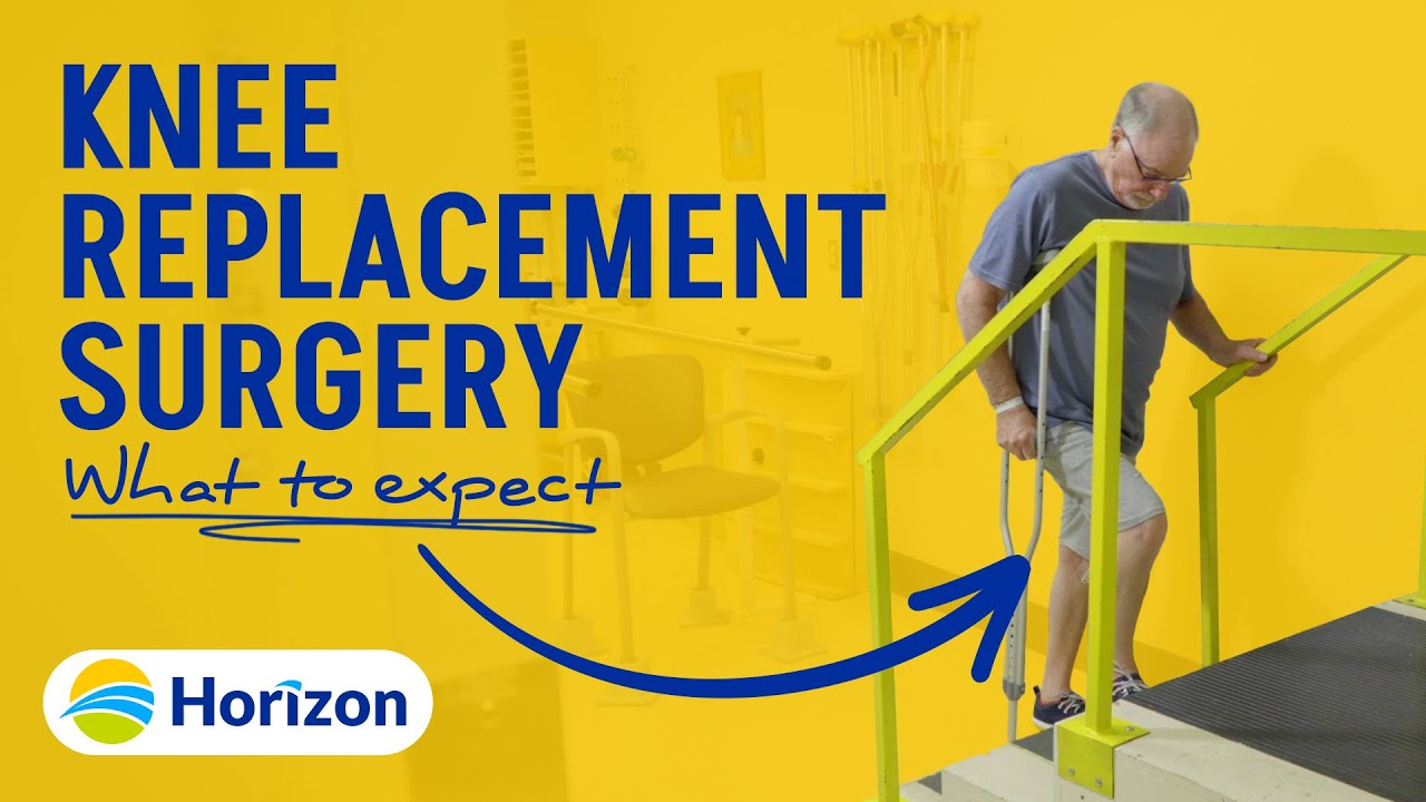
The purpose of this video is to help you learn what to expect while you are in hospital, and how to care for yourself after surgery so that you can have the best recovery possible.
----------------------------------
At Horizon Health Network, we are helping people be healthy! Chez le Réseau de santé Horizon, nous aidons les gens à être en santé!
For more information, visit/Pour plus d'information, cliquez-ici:
https://news.horizonnb.ca
Facebook: https://facebook.com/HorizonNB
Twitter: https://twitter.com/HorizonHealthNB
Instagram: https://instagram.com/horizonhealthnb/
Linkedin: https://linkedin.com/company/h....orizon-health-networ
Women's College Hospital is revolutionizing the way knee-replacement surgery is done. It is starting to provide the procedure as an ambulatory service. Patients can go home from hospital four hours after having the surgery. In some other hospitals knee replacement surgery patients have to stay as long as 4 days.
Read an excerpt from Theresa Boyle's story:
It’s been less than four hours since Greg Nemez underwent knee-replacement surgery and the 56-year-old Mississauga man is already on his way home from hospital.
This past Monday, he became the fifth patient at Toronto’s Women’s College Hospital to undergo the outpatient procedure, which normally requires a hospital stay of two or three days.
“I’m happy ... You have that freedom of movement from before. It’s like wow,” he said on the elevator as he was leaving the hospital.
After years of being unable to hold his leg straight, the real-estate agent can finally do so. A 20-year-old football injury had left him with severe arthritis and pain.
Read the full story:
https://www.thestar.com/news/g....ta/2018/04/11/he-got
Follow the Toronto Star on social media:
Facebook: https://www.facebook.com/torontostar/
Twitter: https://twitter.com/TorontoStar
Instagram: https://www.instagram.com/thetorontostar/

Mohs Surgeon Dr. Leslie Christenson shows entire Mohs Surgery from start to finish. This is the full procedure and includes the entire surgery. Dr. Christenson talks about the procedure as she removes the skin cancer.
Learn more about Dr. Christenson: https://www.mcfarlandclinic.co....m/doctors/leslie-chr
Learn more about Mohs Surgery: https://www.mcfarlandclinic.co....m/doctors/specialtie

1. What is hemodialysis?
2. Why do you do hemodialysis?
3. How does hemodialysis remove body waste?
4. What are the symptoms and side effects of hemodialysis?
5. How should I eat food when I do hemodialysis?
6. What are some precautions for patients during hemodialysis?
► If you have any health issues contact us anytime, we here to help at CloudHospital – https://icloudhospital.com/
► Subscribe: https://www.youtube.com/channel/UCmk5... to learn more about various health and beauty topics.
► Find us on Facebook: https://www.facebook.com/icloudhospital/
► On Instagram: https://www.instagram.com/cloudhospit...
► On LinkedIn: https://www.linkedin.com/company/clou...
► On Twitter: https://twitter.com/CloudhospitalI
#hemodialysis #cloudhospital #koreahospital
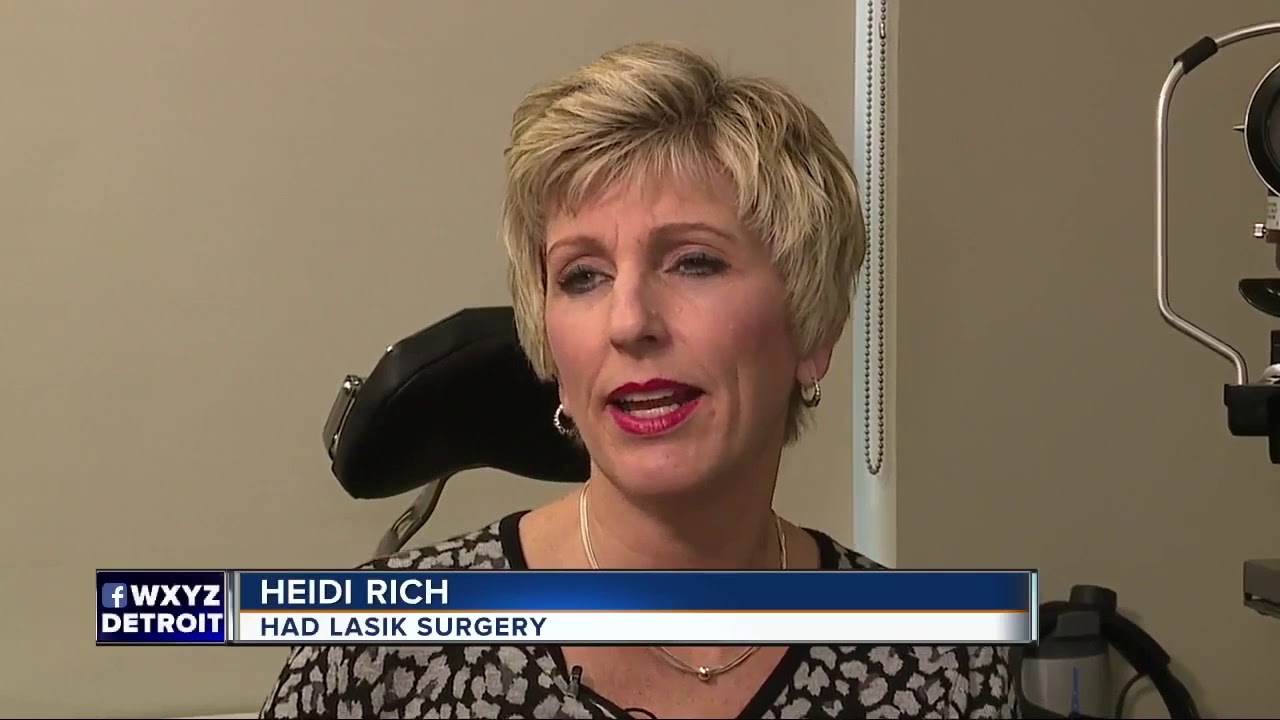
If you go to research LASIK eye surgery online, you may get conflicting messages. Some articles rave about it, but in some cases, others link it to severe pain or even suicide. 7 Action News' Carolyn Clifford sat down with one of the area's biggest providers of eye surgery to try and separate fact from fiction, so if you go under the laser, you know the risk.

Runners Knee Overview:
Welcome to our Patello-Femoral Rehab video. The goal of this video is to minimize pain around the kneecap and maximize recovery. This video should not be used as a substitute for regular physical therapy visits and guidance from your physician
Visit http://www.matthewboesmd.com/p....atello-femoral-rehab for more information

After Sammyra’s knee injury, Marvin Smith, MD, orthopaedic surgeon at Memorial Sports Medicine Center, helped her get back on the volleyball court and playing pain free. Following a thorough examination, meniscus surgery and rehabilitation got Sammyra back to playing with her college team within two months. Learn more about how Memorial Sports Medicine Center helps athletes move forward at MHS.net/SportsMedicine.
To learn more about Dr. Smith, visit his physician profile page at: https://www.mhs.net/physicians/s/smith-marvin-k

We are looking for 5 patients with knee pain who want to get significantly better in the next 30 days. Click this link to let me know you're interested and learn more.
https://www.drdavidgeier.com/work-with-me/contact/
If you suffer a knee injury, what do you do about it? Should you be concerned? On one hand, you could give it a few days to see if it gets better on its own. How can you know if you risk making the problem worse? In this Ask Dr. Geier video, I offer four signs you have a serious knee injury that could require surgery.
Please note: I don't respond to questions and requests for specific medical advice left in the comments to my videos. I receive too many to keep up (several hundred per week), and legally I can't offer specific medical advice to people who aren't my patients (see below). If you want to ask a question about a specific injury you have, leave it in the comments below, and I might answer it in an upcoming Ask Dr. Geier video. If you need more detailed information on your injury, go to my Resources page: https://www.drdavidgeier.com/resources/
The content of this YouTube Channel, https://www.youtube.com/user/drdavidgeier (“Channel”) is for INFORMATIONAL PURPOSES ONLY. The Channel may offer health, fitness, nutritional and other such information, but such information is intended for educational and informational purposes only. This content should not be used to self-diagnose or self-treat any health, medical, or physical condition. The content does not and is not intended to convey medical advice and does not constitute the practice of medicine. YOU SHOULD NOT RELY ON THIS INFORMATION AS A SUBSTITUTE FOR, NOR DOES IT REPLACE, PROFESSIONAL MEDICAL ADVICE, DIAGNOSIS, OR TREATMENT. You should consult with your healthcare professional before doing anything contained on this Channel. You agree that Dr. Geier is not responsible for any actions or inaction on your part based on the information that is presented on the Channel. Dr. David Geier Enterprises, LLC makes no representations about the accuracy or suitability of the content. USE OF THE CONTENT IS AT YOUR OWN RISK.
SWELLING
If you have a knee that is much more swollen than the opposite knee, especially if the swelling developed soon after you got hurt, then it could be a sign of structural damage. While a small amount of swelling could be normal or a sign of inflammation in the knee, a knee that is really swollen and much larger than the other knee could represent a fracture, torn ACL or other ligament or some other damaged structure.
INABILITY TO FULLY STRAIGHTEN YOUR KNEE
Pain can make it difficult for you to straighten your knee. But being unable to get your knee completely flat can be a sign of an injury like an ACL tear or meniscus tear.
LOCKING OF THE KNEE
Locking is a term orthopedic surgeons use to describe a knee that gets stuck in a certain position. Maybe you can straighten it to a certain point, but it gets stuck around 30 degrees short of fully straight. Or you can’t bend it past a certain point because something inside the knee is blocking it. Catching is a milder form of the same problem, where you can get to the point that the motion stops, but you can twist or rotate your knee past that point.
BUCKLING OR GIVING WAY
Having the tibia (shin bone) shift out from under the femur (thigh bone) usually represents an ACL injury. If your knee buckled during the traumatic injury and keeps happening in the days after the injury, you risk doing more damage to the other structures inside the knee.
This list is not comprehensive, so if you are concerned about your knee injury or knee pain, it never hurts to see your doctor or an orthopedic surgeon.

Live TV is so exciting because anything can happen, and sometimes that means injuries. Today I'm reacting to injuries and medical emergencies that happened on live tv. We're talking America's Got Talent, American Idol, newscasters having strokes, dehydration, Wendy Williams overheating, swallowing swords, being hit with a motorcycle, vasovagal syncope, drowning, Dan Harris, and magical tricks like David Blaine's needle in going wrong. Which clips did I miss? Let me know down below.
I LOVE reading your comments and take your suggestions seriously. If there’s a subject you want me to discuss or something you’d like for me to react to, leave a comment down below. Many of my videos have been born out of suggestions directly from you, so don’t hold back!
-Doctor Mike Varshavski
Help us continue the fight against medical misinformation and change the world through charity by becoming a Doctor Mike Resident on Patreon where every month I donate 100% of the proceeds to the charity, organization, or cause of your choice! Residents get access to bonus content, an exclusive discord community, and many other perks for just $10 a month. Become a Resident today:
https://www.patreon.com/doctormike
Please SUBSCRIBE for new videos every Wednesday afternoon and Sunday morning! https://goo.gl/87kYq6
Let’s connect:
IG https://goo.gl/41ZS7w - Doctor Mike
Reddit https://www.reddit.com/r/DoctorMike/
Twitter https://goo.gl/kzmGs5 - Real Doctor Mike
Facebook https://goo.gl/QH4nJS - Real Doctor Mike
Contact Email: DoctorMikeMedia@Gmail.com
* Select photos/videos provided by Getty Images *
** The information in this video is not intended nor implied to be a substitute for professional medical advice, diagnosis or treatment. All content, including text, graphics, images, and information, contained in this video is for general information purposes only and does not replace a consultation with your own doctor/health professional **

How Do I Exercise An Injured Knee?
Bob and Brad discuss how to exercise an injured knee.
Check out other products Bob and Brad Love:
https://www.amazon.com/shop/ph....ysicaltherapyvideo?l
Visit us on our other social media platforms:
Website: https://bobandbrad.com/
Facebook: https://www.facebook.com/BobandBrad/
Instagram: https://www.instagram.com/officialbobandbrad/
Twitter: https://twitter.com/ptfamous
Bob and Brad’s Products:
Grip and Forearm Strengthener: https://store.bobandbrad.com/
Wall Anchor: https://store.bobandbrad.com/
Booyah Stik: https://amzn.to/2URbgjq
Knee Glide: https://amzn.to/2WnSPU1
Check out our shirts, mugs, bags and more in our Bob and Brad merchandise shop here:
https://shop.spreadshirt.com/bob-brad
Check out other products Bob and Brad Love:
https://www.amazon.com/shop/ph....ysicaltherapyvideo?l
Check out Alex's & Liz's Product Review Channel on YouTube by clicking here:
https://www.youtube.com/user/BobSchrupp
Want to help translate our videos? We would so love the help!
http://www.youtube.com/timedtext_cs_panel?c=UCmTe0LsfEbpkDpgrxKAWbRA&tab=2
Medical Disclaimer
All information, content, and material of this website is for informational purposes only and are not intended to serve as a substitute for the consultation, diagnosis, and/or medical treatment of a qualified physician or healthcare provider.
Affiliate disclaimer:
Keep in mind that we may receive commissions when you click our links and make purchases. However, this does not impact our reviews and comparisons. We are highly selective in our products and try our best to keep things fair and balanced in order to help you make the best choice for you.


Orthopedic surgeon Donald Polakoff, MD describes recovery time from knee replacement surgery.
SUBSCRIBE FOR MORE VIDEOS AND NEWS!
http://www.youtube.com/subscri....ption_center?add_use
CONNECT WITH US!
Google+ ► http://gplus.to/spuh
Facebook ► http://www.fb.com/SaintPetersUniversityHospital
Twitter ► http://twitter.com/sphcs_news
Pinterest ► http://www.pinterest.com/sphcs
Saint Peter's Healthcare System ► http://www.saintpetershcs.com
