- Physical Examination
- Surgical Examination
- Ophthalmology
- Clinical Skills
- Orthopedics
- Surgery Videos
- Laparoscopy
- Pediatrics
- Funny Videos
- Cardiothoracic Surgery
- Nursing Videos
- Plastic Surgery
- Otorhinolaryngology
- Histology and Histopathology
- Neurosurgery
- Dermatology
- Pediatric Surgery
- Urology
- Dentistry
- Oncology and Cancers
- Anatomy Videos
- Health and Fitness
- Radiology
- Anaesthesia
- Physical Therapy
- Pharmacology
- Interventional Radiology
- Cardiology
- Endocrinology
- Gynecology
- Emergency Medicine
- Psychiatry and Psychology
- Childbirth Videos
- General Medical Videos
- Nephrology
- Physiology
- Diet and Food Health
- Diabetes Mellitus
- Neurology
- Women Health
- Osteoporosis
- Gastroenterology
- Pulmonology
- Hematology
- Rheumatology
- Toxicology
- Nuclear Medicine
- Infectious Diseases
- Vascular Disease
- Reproductive Health
- Burns and Wound Healing
- Other
Top videos
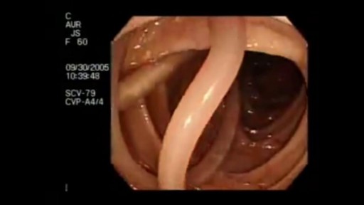
Parasites Accidentally Seen During Colonoscopy

The first stage of labor is the longest and involves three phases: Early Labor Phase –The time of the onset of labor until the cervix is dilated to 3 cm. Active Labor Phase – Continues from 3 cm. until the cervix is dilated to 7 cm.
No Scalpel Vasectomy
Abscess drainage in neck
Rare condition disorder known as Diprosopus, also known as craniofacial duplication. Diprosopus is a congenital defect also known as craniofacial duplication. The exact description of diprosopus refers to a fetus with a single trunk, normal limbs, and facial features that are duplicated to a certain degree. A less severe instance is when the fetus has a duplicated nose and the eyes are spaced far apart. In the most extreme instances, the entire face is duplicated, hence the name diprosopus, which is Greek for two-faced. Fetuses with diprosopus often also lack brains (anencephaly), have neural tube defects, or heart malformations. In some cases, if the brain is formed, it may have duplicated structures. Most infants with diprosopus are stillborn and there are fewer than fifty cases documented since 1864.

***SUBSCRIBE WITHIN THE NEXT 28 DAYS FOR A CHANCE TO WIN $1,000!***
Did you know only 20% of our video content is on YouTube? Try out our membership for FREE today! → https://bit.ly/3yRDykI
Try our NCLEX Prep FREE → https://bit.ly/3sRRjvY
Head to https://bit.ly/3yRDykI to get access to the other 80%, along with 800+ study guides, customizable quiz banks with 3,000+ test-prep questions, and answer rationales!
This video illustrates an IM injection for deltoid muscle.
Note that vaccines and other medications can be administered through the deltoid muscle. I will give you some tips through this video.
It is important to check your client’s details such as their medication, time, dose, and the route to be used. Different research works are subject to change the protocols for insertion thus, it is necessary to be up to date with the current changes.
Assemble all the supplies and conduct hand sanitation. Usually, I wear gloves before giving any injection in as much as the CDC may state it is optional unless the patient has an open lesion and contact of body fluids is likely to happen.
Use the acromion process landmark to locate the deltoid muscle. Move your fingers about two widths below the landmark. The patient’s adipose tissue determines the choice of needle length. Note that the needle gauge is determined by the type of medication you plan to give to the patient.
The Z-track technique is recommended rather than pinching the patient’s skin. Pull the patient’s skin to the side using one hand. Use a 90 degree angle to insert the needle to the patient’s skin. At the rate of 10 seconds per mL gently depress the plunger.
Remove the needle carefully and engage the safety precautions then dispose of the needle appropriately in the sharps container. Gauzing helps to cover the injection site.
Nursing School Membership - Try it FREE → https://bit.ly/3yRDykI
New NCLEX Prep - Try it FREE → https://bit.ly/3sRRjvY
Popular Playlists:
NCLEX Fluid & Electrolytes: https://bit.ly/39BSHXs
Heart Failure (CHF): https://bit.ly/2u5zfDm
Myocardial Infarction (MI): https://bit.ly/3bN9AAk
Addison’s vs. Cushing: https://bit.ly/2STvute
Diabetes Mellitus & DKA vs HHNS: https://bit.ly/37D8nbs
Cardiomyopathy: https://bit.ly/38CwcSg
IV Fluids: Hypertonic, Hypotonic & Isotonic: https://bit.ly/2P45BWx
SIADH vs Diabetes Insipidus: https://bit.ly/2wq6Bhb
Follow us on social media for more EXCLUSIVE content 👋
More Videos: https://bit.ly/37CRttH
Instagram: https://www.instagram.com/simplenursing.com_
TikTok: https://www.tiktok.com/simplenursing
Thank you for the support & for tuning in!
Remember… don’t be scared, BE PREPARED!

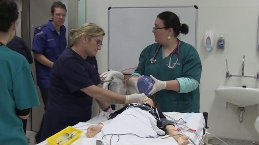
Ellis will be demonstrating how to complete an occupied bed change. It would be appropriate to wear gloves during this skill to avoid contact with bodily fluids.
Our Critical Nursing Skills video tutorial series is taught by Ellis Parker MSN, RN-BC, CNE, CHS and intended to help RN and PN nursing students study for your nursing school exams, including the ATI, HESI and NCLEX.
#NCLEX #ClinicalSkills #HESI #Kaplan #ATI #NursingSchool #NursingStudent #Nurse #RN #PN #Education #LVN #LPN #bedmaking #nurseeducator
00:00 What to expect
00:53 Initial patient position
1:50 Tucking soiled linens
2:20 Placing initial clean linen
3:30 Rolling patient
3:40 Removing soiled linen
4:05 Completing bottom layer
4:33 Changing pillow case
4:50 Top sheet and blanket
7:23 Mitered corner
🚨 Reminder: shipping deadlines are looming 👀
🎁 Regular Shipping: Order by Friday, December 15
🚀 Expedited Shipping: Order by Monday, December 18
🔍 Still searching for last-minute gifts? Consider a Level Up RN Gift Card! 💌 It’s not only a thoughtful present but also the perfect way to share treasures like Pharmacology Flashcards OR digital treasures like Flashables Digital Nursing Flashcards & the Level Up RN membership. Give the gift of knowledge this holiday season! 🧠⚡️💖 bit.ly/LevelUpRNGC
🚪 Access our Cram Courses, Quizzes and Videos all in one ad free space with Level Up RN Membership https://bit.ly/LevelUpRNMembership
Want more ways to MASTER Clinical Skills? Check out our flashcards & videos!
👇👇👇👇👇👇👇👇👇👇
👉 https://bit.ly/clinicalnursingskills 👈
☝️👆☝️👆☝️👆☝️👆☝️👆
This is your one-stop-shop for materials to help you LEARN & REVIEW so you can PASS Nursing School.
🤔🤔🤔 DO YOU WANT TO PASS your classes, proctored exams and the NCLEX? 🤔🤔🤔 Our resources are the best you can buy. They are built with a single goal: help you pass with no fluff. Everything you need, and nothing you don’t. Don’t take our word for it, though! Check out our hundreds of ⭐️⭐️⭐️⭐️⭐️ reviews from nurses who passed their exams and the NCLEX with Level Up RN.
🗂️ Our Ultimate Nursing School Survival kit is your number 1 resource to get through nursing school and to pass the NCLEX. Whether you're just starting school or you’re already prepping for the NCLEX, this bundle of flashcards is the best you can buy. It covers all the information you need to know to pass all your exams and it has FREE shipping!
➡️ https://bit.ly/TUNSSK ⬅️
L👀king for EVEN MORE resources to survive Nursing School? Make your Nursing School experience your own! Life’s difficult enough—learning shouldn’t be.
🪅 Games https://nursesquad.com
💻 Digital resources https://bit.ly/NursingStudyCourses
📅 Organizational tools https://bit.ly/OrganizingSchool
✨Want perks? Join our channel!
https://youtube.com/leveluprn/join
🏷 Head to https://leveluprn.com/specials for all our latest deals!🥳️
📧 LOOKING FOR FREE RESOURCES TO HELP WITH YOUR EXAMS? Get exclusive tips, latest video releases and more delivered to your email!
➡️ https://leveluprn.com/signup ⬅️
⚕ 👩 LEVEL UP NURSE SQUAD 👩⚕️
All of the nurses at Level Up RN are here to help! Cathy Parkes started helping her fellow classmates back when she was in nursing school, tutoring so they could pass their exams and graduate. After she got her BSN and started working as an RN at Scripps Encinitas Hospital, she started this YouTube channel to help nursing students around the world. Since then she has built a team of top-notch dedicated nurses and nurse educators who are focused on improving nursing education and supporting career advancement for nurses everywhere. With flashcards, videos, courses, organizational tools and more, we are singularly focused on helping students and nurses Level Up on their exams and nursing careers.

Thank you so much for watching❤
If you enjoyed this video ▶Please leave a LIKE👍 ▶SHARE this video ▶【SUBSCRIBE】my channel for more new videos And click the BELL 🔔so you don't miss any of my videos HERE
https://www.youtube.com/c/nurs....eminder?sub_confirma
You can support my work by purchasing your NurseMinder Merch https://teespring.com/stores/nurseminder-nation (or click on merch pics under the video)
Or simply do your Amazon shopping after clicking on one of the links below
-------------------------------------------------------------------------
Thank you so much! I appreciate you!♥♥
------------------------------------------------------------------------
Nurses often prime IV lines with the hopes that there are no air bubbles. In this video, I will share a couple of tips to help reduce the risk or frequency of air bubbles during line priming. I will also talk about how to troubleshoot the air bubbles when they appear during an infusion
Providing patient care and influencing safe patient outcomes requires that registered nurses and licensed practice nurses maintain air free IV lines. Learn the strategies and tips to decrease the risk of air bubbles appearing in your primary or secondary medication line as well as troubleshooting tips to remove those alarming bubbles. Your patients will thank you!
Whether you are providing normal saline, a medication, or a combination, ensure that all fluids are compatible.
Supplies used in this video include the Alaris Primary Infusion line, alcohol swabs and a sterile 10 cc syringe ... and a nail in the wall :)
------------------------------------------------------------------------
❤️ ~ You may also be interested in watching ~ ❤️
PICC line assessment https://youtu.be/tnKClpU-J1g
How To Access a PICC line https://youtu.be/SCF6bmk8KWc
Putting on Sterile Gloves https://youtu.be/xNwkKLqDJn4
Organizational Plans for Nursing https://youtu.be/_NATxwPwHzc
Medication Conversions https://youtu.be/TCPBXg2TYCs
------------------------------------------------------------------------
💻COMMENT in the description box below and share your ideas
👍 LIKE the video
🗣 SHARE with your friends
📥 SUBSCRIBE ... hit the BELL 🔔
Subscribe to NurseMinder https://www.youtube.com/c/nurs....eminder?sub_confirma
------------------------------------------------------------------------
Amazon Affiliate Links
------------------------------------------------------------------------
Want to support me in another way? Enter Amazon through my links and continue to do your shopping. Simple and Easy Way to support the work I do.
The following list is the equipment I use (or if my version is no longer sold, a close replica).
📱 Phone 11 Cell Phone https://amzn.to/2WpOJfz
💻 MacBook Pro https://amzn.to/2YyxQC1
👉 Final Cut Video Editing software https://amzn.to/3fqlAd9
🎙️ Rode NT USB microphone (Audio Recording) for post-production voiceover https://amzn.to/2W2RJj1
👉 Neewer Professional Recording Stand – mount microphone and adjust positioning to keep it close but out of the camera’s view: https://amzn.to/3fjB4zs
👉 Manfrotto Tripod (hold cell phone) https://amzn.to/2YKGYUz
💡 Neewer Ring Light to reduce shadows and improve lighting. https://amzn.to/3dk5OP5
Disclaimer: I recommend only products that I know and trust to be of high quality. Links are provided for quick access. Some of the links contained in this checklist are affiliate links and I may receive a commission if make a purchase from the affiliate. This helps me to keep creating and offering free content.
Labor And Delivery During Vaginal Child Birth

SUBSCRIBE: https://www.youtube.com/c/TVNe....phrologist?sub_confi
An animation of blood flow inside the Hemodialysis circuit.
About Dr. Rifai:
Dr. Ahmad Oussama Rifai is certified by the American Board of Internal Medicine (ABIM) in the specialty of Internal Medicine and the sub-specialty of Nephrology.
MEET DR. RIFAI
https://www.thevirtualnephrologist.com/rifai/
Follow The Virtual Nephrologist on SOCIAL MEDIA:
-Facebook: https://www.facebook.com/thevirtualnephrologist
-Instagram: https://www.instagram.com/thevirtualnephrologist/
-Twitter: https://twitter.com/VNephrologist
Schedule a virtual consult:
https://www.thevirtualnephrolo....gist.com/schedule-a-
Best wishes for great health | The Virtual Nephrologist

Bronchiectasis is an abnormal dilation of the proximal and medium-sized bronchi (>2 mm in diameter) caused by weakening or destruction of the muscular and elastic components of the bronchial walls. Affected areas may show a variety of changes, including transmural inflammation, edema, scarring, and ulceration, among other findings. Distal lung parenchyma may also be damaged secondary to persistent microbial infection and frequent postobstructive pneumonia. Bronchiectasis can be congenital but is most often acquired.[9] Congenital bronchiectasis usually affects infants and children. These cases result from developmental arrest of the bronchial tree. Acquired forms occur in adults and older children and require an infectious insult, impairment of drainage, airway obstruction, and/or a defect in host defense. The tissue is also damaged in part by the host response of neutrophilic proteases, inflammatory cytokines, nitric oxide, and oxygen radicals. This results in damage to the muscular and elastic components of the bronchial wall. Additionally, peribronchial alveolar tissue may be damaged, resulting in diffuse peribronchial fibrosis.[12] The result is abnormal bronchial dilatation with bronchial wall destruction and transmural inflammation. The most important functional finding of altered airway anatomy is severely impaired clearance of secretions from the bronchial tree. Impaired clearance of secretions causes colonization and infection with pathogenic organisms, contributing to the purulent expectoration commonly observed in patients with bronchiectasis. The result is further bronchial damage and a vicious cycle of bronchial damage, bronchial dilation, impaired clearance of secretions, recurrent infection, and more bronchial damage
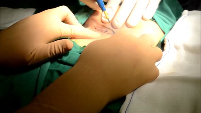
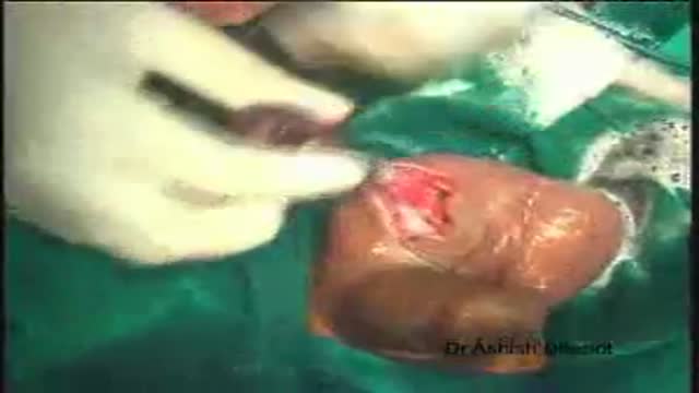
Excision of Pilonidal Cyst. Open method.

Guillain-Barre syndrome is a rare disorder in which your body's immune system attacks your nerves. Weakness and tingling in your extremities are usually the first symptoms. These sensations can quickly spread, eventually paralyzing your whole body. In its most severe form Guillain-Barre syndrome is a medical emergency. Most people with the condition must be hospitalized to receive treatment. The exact cause of Guillain-Barre syndrome is unknown. But it is often preceded by an infectious illness such as a respiratory infection or the stomach flu. There's no known cure for Guillain-Barre syndrome, but several treatments can ease symptoms and reduce the duration of the illness. Most people recover from Guillain-Barre syndrome, though some may experience lingering effects from it, such as weakness, numbness or fatigue.
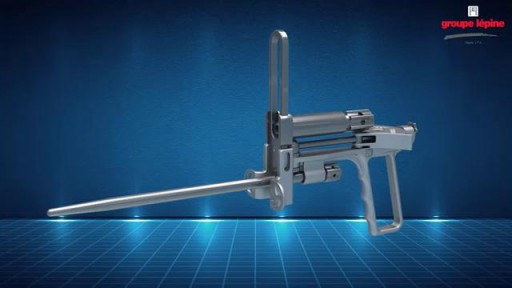
NEW WAVE Surgical Technique 3D Animation
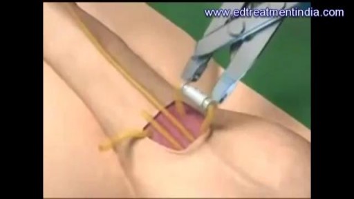
A penile prosthesis is another treatment option for men with erectile dysfunction. These devices are either malleable (bendable) or inflatable. The simplest type of prosthesis consists of a pair of malleable rods surgically implanted within the erection chambers of the penis. With this type of implant the penis is always semi-rigid and merely needs to be lifted or adjusted into the erect position to initiate sex. This type of implant is a good choice for men with spinal cord injuries and/or limited hand strength. Today, many men choose a hydraulic, inflatable prosthesis, which allows them to have an erection when they choose, and it's easier to conceal. It is also more natural. A penile implant is usually used when there is a clear medical cause for ED and when the problem is unlikely to resolve or improve naturally or with other medical treatments. Sometimes a penile prosthesis is implanted during surgery to reconstruct the penis when scarring has caused erections to curve (Peyronie's disease). Penile implant surgeries take about an hour and are typically done in an outpatient center. A man can resume sexual intercourse by 6 weeks after surgery.
Watch that video of The Real Human Body Decomposition Process

In this episode of Crash Course Anatomy & Physiology, Hank gives you a brief history of histology and introduces you to the different types and functions of your body's tissues.
Pssst... we made flashcards to help you review the content in this episode! Find them on the free Crash Course App!
Download it here for Apple Devices: https://apple.co/3d4eyZo
Download it here for Android Devices: https://bit.ly/2SrDulJ
Chapters:
Introduction 00:00
Nervous, Muscle, Epithelial & Connective Tissues 1:23
History of Histology 2:07
Nervous Tissue Forms the Nervous System 5:17
Muscle Tissue Facilitates All Your Movements 7:00
Identifying Samples 9:03
Review 9:48
Credits 10:22
Crash Course is on Patreon! You can support us directly by signing up at http://www.patreon.com/crashcourse
Want to find Crash Course elsewhere on the internet?
Facebook - http://www.facebook.com/YouTubeCrashCourse
Twitter - http://www.twitter.com/TheCrashCourse
Instagram - https://www.instagram.com/thecrashcourse/
CC Kids: http://www.youtube.com/crashcoursekids
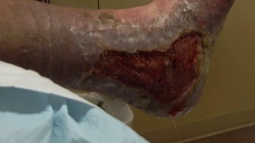
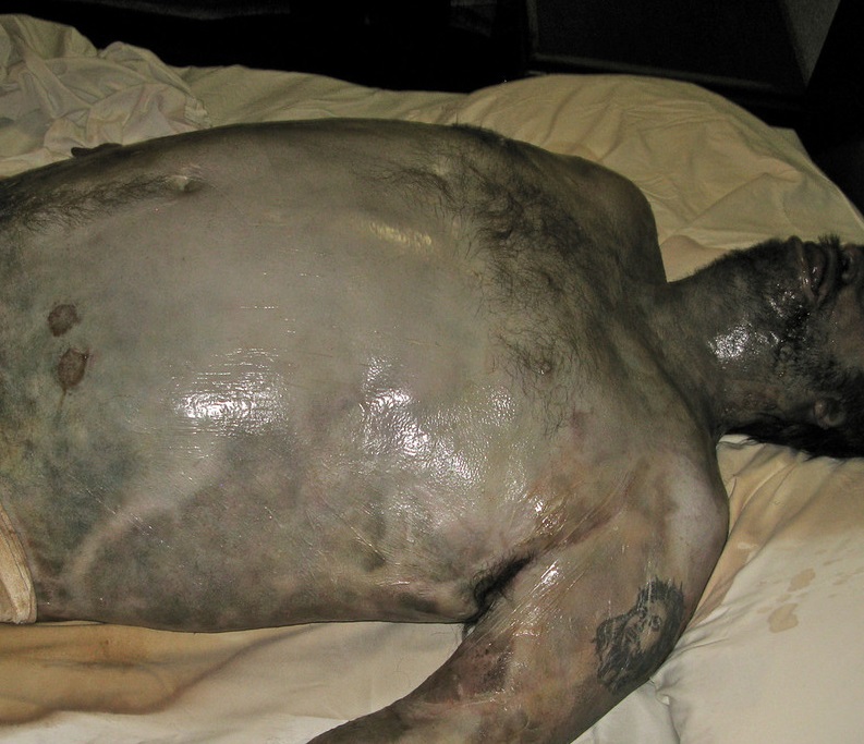
Debridement is the removal of necrotic tissue, foreign debris, bacterial growth, callus, wound edge, and wound bed tissue from chronic wounds in order to stimulate the wound healing process. Stimulation of wound healing mediated by debridement is thought to occur by the conversion of a chronic non-healing wound environment to an acute healing environment through the removal of cells that are not responsive to endogenous healing stimuli. Debridement is used commonly in standard wound treatment of diabetic foot ulcers (DFUs). Methods of debridement include surgery (sharp debridement), chemical debridement (antiseptics, polysaccharide beads, pastes), autolytic (hydrogels, hydrocolloids and transparent films), biosurgery (maggots), mechanical (hydrodebridement), and biochemical debridement (enzyme preparations). Callus is a buildup of keratinized skin formed under conditions of repeated pressure or friction and may contribute to ulcer formation by creating focal areas of high plantar pressure. The debridement of callus has been proposed to be relevant for both treatment and prevention of DFU. The purpose of this report is to retrieve and review existing evidence of comparative clinical effectiveness of different methods of debridement for the treatment of DFUs. Additionally examined in this report is the clinical effectiveness for treatment and prevention of DFU using callus debridement. Cost-effectiveness, and existing debridement guidelines for the treatment of DFUs will also be reviewed.
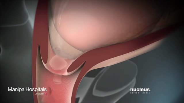
Orchidectomy and Orchidopexy in testis Torsion
