- Physical Examination
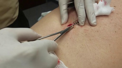
- Surgical Examination
- Ophthalmology
- Clinical Skills
- Orthopedics
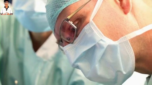
- Surgery Videos
- Laparoscopy
- Pediatrics
- Funny Videos
- Cardiothoracic Surgery
- Nursing Videos
- Plastic Surgery
- Otorhinolaryngology
- Histology and Histopathology
- Neurosurgery
- Dermatology
- Pediatric Surgery
- Urology
- Dentistry
- Oncology and Cancers
- Anatomy Videos
- Health and Fitness
- Radiology
- Anaesthesia
- Physical Therapy
- Pharmacology
- Interventional Radiology
- Cardiology
- Endocrinology
- Gynecology
- Emergency Medicine
- Psychiatry and Psychology
- Childbirth Videos
- General Medical Videos
- Nephrology
- Physiology
- Diet and Food Health
- Diabetes Mellitus
- Neurology
- Women Health
- Osteoporosis
- Gastroenterology
- Pulmonology
- Hematology
- Rheumatology
- Toxicology
- Nuclear Medicine
- Infectious Diseases
- Vascular Disease
- Reproductive Health
- Burns and Wound Healing
- Other
Top videos

Multiple sclerosis (MS) involves an immune-mediated process in which an abnormal response of the body’s immune system is directed against the central nervous system (CNS). The CNS is made up of the brain, spinal cord and optic nerves.


Multiple sclerosis (MS) is a disease of the central nervous system estimated to affect 2.3 million people worldwide. It is a chronic disease in which the immune system abnormally attacks the insulation and support around the nerve cells (myelin sheath) in the brain, spinal cord and optic nerves, causing inflammation and consequent damage. MS is a leading cause of non-traumatic disability in young people, usually striking between 20 and 40 years of age. There is no cure for MS, but research continues to better understand and treat the disease.

mply put, relapses, also known as flare ups, or (MS) attacks are new or worsening MS symptoms. But there is a concrete definition used by healthcare providers to identify MS attacks. To be considered an MS relapse: Old symptoms of MS must have become worse or new symptoms appeared.
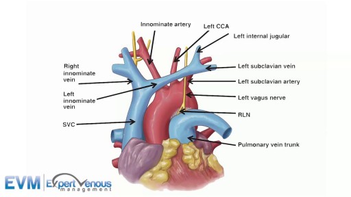
The superior vena cava (SVC, also known as the cava or cva) is a short, but large diameter vein located in the anterior right superior mediastinum.

G-Shot (G-Spot Amplification)
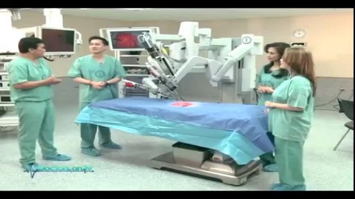
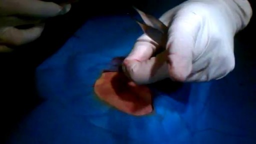
Understand how this world-class surgery platform operates a minimally invasive robotic surgery during a medical procedure for prostate cancer.
Possible causes are a blocked milk duct or bacteria entering the breast. It usually occurs within the first three months of breast-feeding. Symptoms include breast pain, swelling, warmth, fever, and chills. Antibiotics are required. Mild pain relievers can help with discomfort.

A bone-anchored hearing aid (BAHA) or bone-anchored hearing device,is a type of hearing aid based on bone conduction. It is primarily suited for people who have conductive hearing losses, unilateral hearing loss, single-sided deafness and people with mixed hearing losses who cannot otherwise wear 'in the ear' or 'behind the ear' hearing aids. They are more expensive than conventional hearing aids, and their placement involves invasive surgery which carries a risk of complications, although when complications do occur, they are usually minor. Two of the causes of hearing loss are lack of function in the inner ear(cochlea) and when the sound has problems in reaching the nerve cells of the inner ear. Example of the first include age-related hearing loss and hearing loss due to noise exposure. A patient born without external ear canals is an example of the latter for which a conventional hearing aid with a mould in the ear canal opening would not be effective. Some with this condition have normal inner ear function, as the external ear canal and the inner ear are developed at different stages during pregnancy. With normal inner anatomy, sound conducted by the skull bone improves hearing.
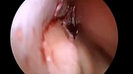
Repair Deviated Nasal Septum, Endoscopic Septoplasty, endoscopic surgery, Stapler repair of nasal septum, Dr B. Todd Schaeffer.
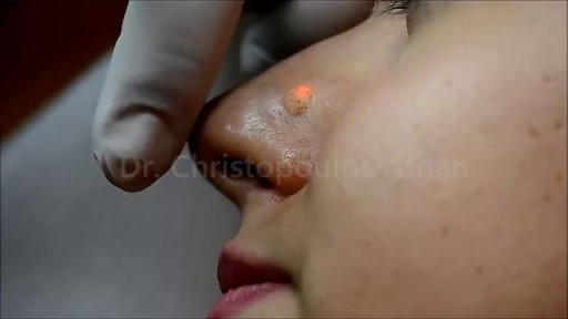
Most people develop several moles (nevi) throughout adulthood. Moles can be found anywhere on the body, usually in sun-exposed areas, and are usually brown, smooth, and slightly raised. In most cases, a nevus is benign and doesn't require treatment. Rarely, they turn into melanoma or other skin cancers. A nevus that changes shape, grows bigger, or darkens should be evaluated for removal.

Menorrhagia is the medical term for menstrual periods with abnormally heavy or prolonged bleeding. Although heavy menstrual bleeding is a common concern, most women don't experience blood loss severe enough to be defined as menorrhagia. With menorrhagia, you can't maintain your usual activities when you have your period because you have so much blood loss and cramping. If you dread your period because you have such heavy menstrual bleeding, talk with your doctor. There are many effective treatments for menorrhagia. Symptoms Signs and symptoms of menorrhagia may include: Soaking through one or more sanitary pads or tampons every hour for several consecutive hours Needing to use double sanitary protection to control your menstrual flow Needing to wake up to change sanitary protection during the night Bleeding for longer than a week Passing blood clots larger than a quarter Restricting daily activities due to heavy menstrual flow Symptoms of anemia, such as tiredness, fatigue or shortness of breath
