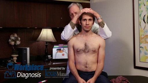
- Physical Examination
- Surgical Examination
- Ophthalmology
- Clinical Skills
- Orthopedics
- Surgery Videos
- Laparoscopy
- Pediatrics
- Funny Videos
- Cardiothoracic Surgery
- Nursing Videos
- Plastic Surgery
- Otorhinolaryngology
- Histology and Histopathology
- Neurosurgery
- Dermatology
- Pediatric Surgery
- Urology
- Dentistry
- Oncology and Cancers
- Anatomy Videos
- Health and Fitness
- Radiology
- Anaesthesia
- Physical Therapy
- Pharmacology
- Interventional Radiology
- Cardiology
- Endocrinology
- Gynecology
- Emergency Medicine
- Psychiatry and Psychology
- Childbirth Videos
- General Medical Videos
- Nephrology
- Physiology
- Diet and Food Health
- Diabetes Mellitus
- Neurology
- Women Health
- Osteoporosis
- Gastroenterology
- Pulmonology
- Hematology
- Rheumatology
- Toxicology
- Nuclear Medicine
- Infectious Diseases
- Vascular Disease
- Reproductive Health
- Burns and Wound Healing
- Other
Top videos
A breech birth occurs when a baby is born bottom first instead of head first. Around 3-5% of pregnant women at term (37–40 weeks pregnant) will have a breech baby. Most babies in the breech position are born by a caesarean section because it is seen as safer than being born vaginally.

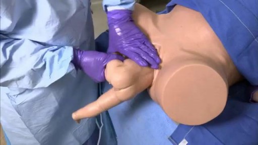
Breech Baby Position Exercise
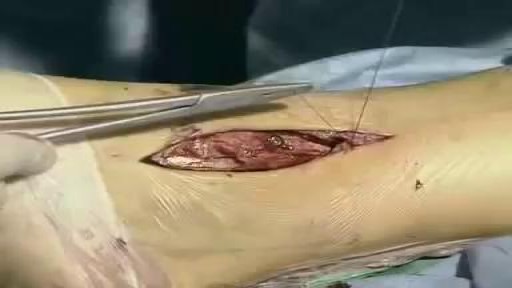
Unstable ankle joints after internal fixation of type B malleolar fractures exist. Residual instability most often occurs after trimalleolar fractures with initial joint dislocation. Treatment with an additional positioning screw generally produced a satisfactory result.
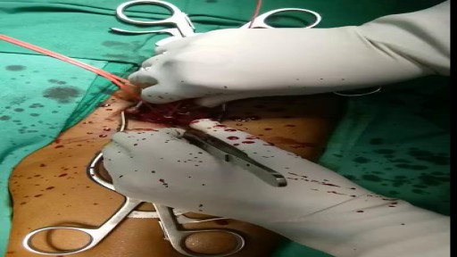
Femoral Embolectomy. Back. All emboli of the lower extremity, including a proximal saddle embolus at the aortic bifurcation, can be removed through the common femoral artery using Fogarty catheters. By passing these through the embolus, and by inflating the small balloon, the clot can be withdrawn and the flow restored

This baby was born with an adult sized tongue - and she just completed a surgery that will change her life.

Know About Cardiothoracic Surgery in 60 Seconds About what cardiothoracic surgery is, why it is done and what is the result of such surgery. A Major Session on #cardiothoracic #surgery at #Congress #2018HCC 2018 Healthcare and Cardiology Conference #BANGKOK http://cosmicseries.org/cardiology-conferences/

Skin cancer is the most common type of cancer. There are three major types of skin cancer — Basal Cell Carcinoma, Squamous Cell Carcinoma and melanoma. Out of these, Melanoma is the deadliest form of skin cancer. Melanoma appears on the skin as a new spot or growth or a change in an already existing mole. It is often fast growing and can spread to other parts of your body, including your bones, liver, and lungs to form a new cancer.

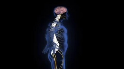
An estimated 12,500 spinal cord injuries occur in the U.S. every year, leaving the injured people, their friends, and their family, to cope with the aftermath of the catastrophe. For many, navigating the challenges of the health care system can feel a bit like going to medical school. Suddenly you're learning a veritable cornucopia of new terms, and may be spending endless hours Googling spinal cord anatomy to fill in the gaps in your knowledge. An educated patient is better equipped to advocate for his or her needs and interests. An education in spinal cord anatomy helps you understand what your doctor is saying, ask intelligent questions, and detect medical errors before they endanger your health.
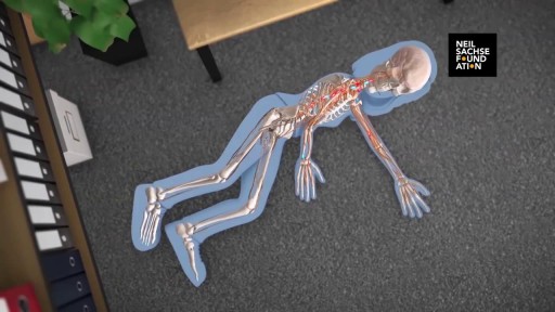
A detailed animation video explaining a spinal cord injury.
A spinal cord injury is not the sort of thing you have to wonder about having. If you've suffered a spinal cord injury, your life is in danger, and you'll know you're injured. You can't use symptoms to diagnose the sort of spinal cord injury you have, and every patient's prognosis is different. Some make a miraculous recovery within months; others need years of physical therapy and still make little to no progress.

Multiple sclerosis (MS) involves an immune-mediated process in which an abnormal response of the body’s immune system is directed against the central nervous system (CNS). The CNS is made up of the brain, spinal cord and optic nerves.

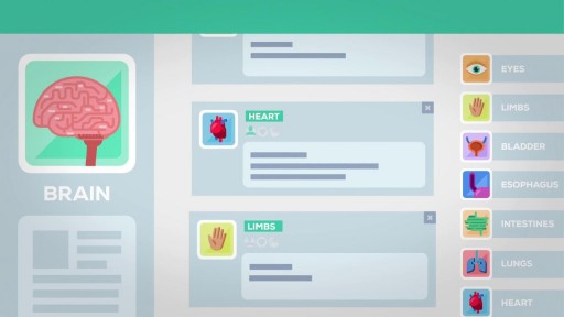
Multiple sclerosis (MS) is a disease of the central nervous system estimated to affect 2.3 million people worldwide. It is a chronic disease in which the immune system abnormally attacks the insulation and support around the nerve cells (myelin sheath) in the brain, spinal cord and optic nerves, causing inflammation and consequent damage. MS is a leading cause of non-traumatic disability in young people, usually striking between 20 and 40 years of age. There is no cure for MS, but research continues to better understand and treat the disease.

mply put, relapses, also known as flare ups, or (MS) attacks are new or worsening MS symptoms. But there is a concrete definition used by healthcare providers to identify MS attacks. To be considered an MS relapse: Old symptoms of MS must have become worse or new symptoms appeared.
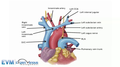
The superior vena cava (SVC, also known as the cava or cva) is a short, but large diameter vein located in the anterior right superior mediastinum.

G-Shot (G-Spot Amplification)
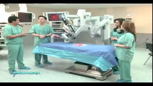
Understand how this world-class surgery platform operates a minimally invasive robotic surgery during a medical procedure for prostate cancer.