
- Physical Examination
- Surgical Examination
- Ophthalmology
- Clinical Skills
- Orthopedics
- Surgery Videos
- Laparoscopy
- Pediatrics
- Funny Videos
- Cardiothoracic Surgery
- Nursing Videos
- Plastic Surgery
- Otorhinolaryngology
- Histology and Histopathology
- Neurosurgery
- Dermatology
- Pediatric Surgery
- Urology
- Dentistry
- Oncology and Cancers
- Anatomy Videos
- Health and Fitness
- Radiology
- Anaesthesia
- Physical Therapy
- Pharmacology
- Interventional Radiology
- Cardiology
- Endocrinology
- Gynecology
- Emergency Medicine
- Psychiatry and Psychology
- Childbirth Videos
- General Medical Videos
- Nephrology
- Physiology
- Diet and Food Health
- Diabetes Mellitus
- Neurology
- Women Health
- Osteoporosis
- Gastroenterology
- Pulmonology
- Hematology
- Rheumatology
- Toxicology
- Nuclear Medicine
- Infectious Diseases
- Vascular Disease
- Reproductive Health
- Burns and Wound Healing
- Kidney Zone Podcast
- Other
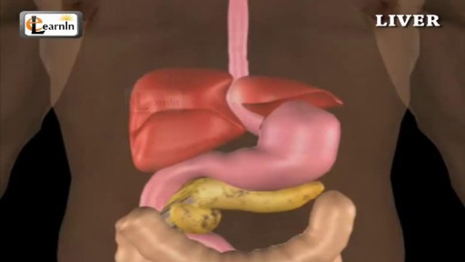
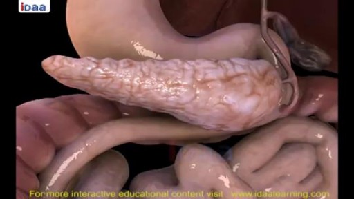
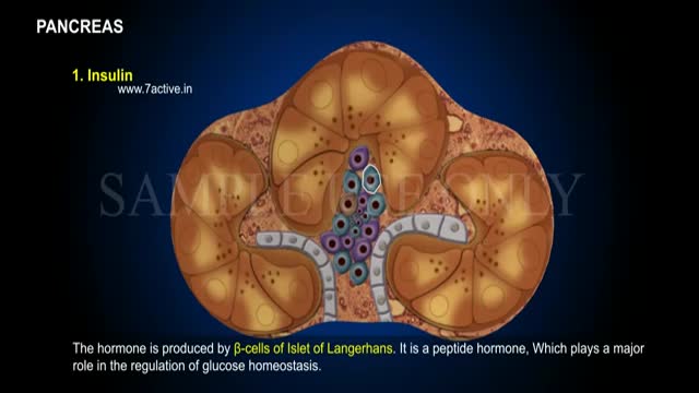

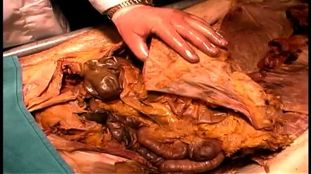
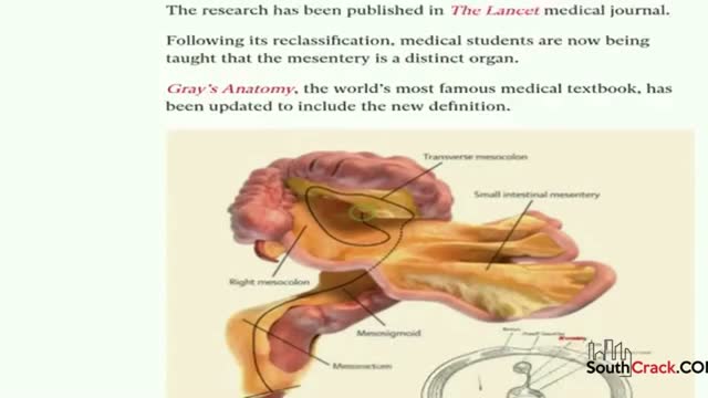
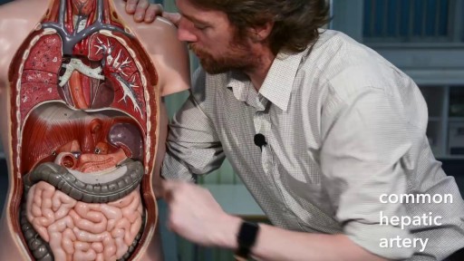

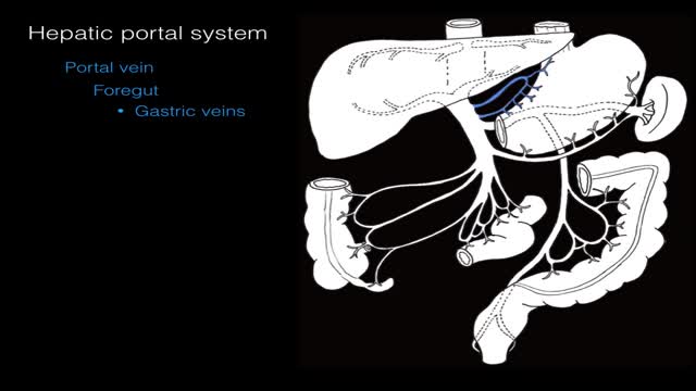



Liver Anatomy and Function

Educational only: videos on MedicalVideos.com are for general information and are not medical advice. Always consult your own doctor about your health.
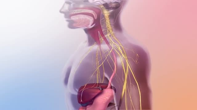
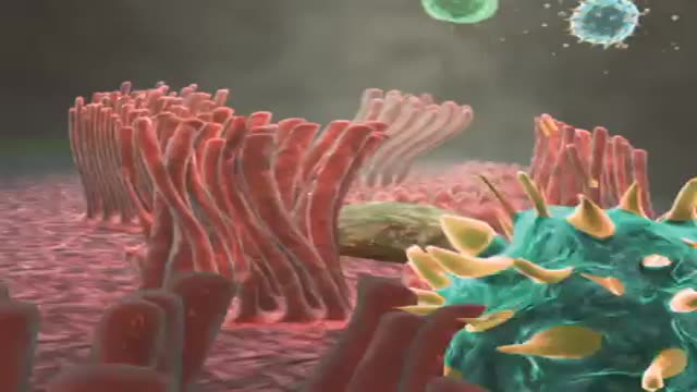
liver also detoxifies chemicals and metabolizes drugs. As it does so, the liver secretes bile that ends up back in the intestines. The liver also makes proteins important for blood clotting and other functions.
This video in 30 seconds
A histology lecture explains the structure and blood flow of the liver. It covers the dual blood supply from the portal vein and hepatic artery, bile production and storage, and the classic lobule model in which blood flows from portal triads toward a central vein while bile flows the opposite way. The lecture finishes by contrasting the anatomical lobule with the functional liver acinus and its zones.
Key points
- The liver receives nutrient-rich portal-vein blood and oxygenated hepatic-artery blood
- Bile is produced in the liver and stored in the gallbladder
- The classic lobule is hexagonal, with portal triads at its corners and a central vein in the middle
- Blood flows from the triads to the central vein while bile flows in the opposite direction
- The acinus model divides cells into three zones based on their blood supply
Chapters
- 0:04 — Liver functions and dual blood supply
- 1:11 — Bile production and storage
- 1:32 — Structure of the liver lobule
- 2:17 — Classic lobule model and blood flow
- 3:11 — The liver acinus and its zones
Video Transcript
A histology lecture on the liver. It covers the dual blood supply — nutrient-rich portal-vein blood and oxygenated hepatic-artery blood — bile production and storage, and the classic lobule model: blood percolating from the portal triads at the corners of the hexagonal lobule toward the central vein while bile flows the opposite way. It finishes by contrasting the anatomical lobule with the functional liver acinus.
Read the full transcript
The liver is an important as well as structurally and functionally complex organ. One of the important functions of the liver is to receive and take up nutrients and other molecules absorbed from the intestines before the blood is returned to the heart via the inferior vena cava. The liver has dual blood supplies, one venous and one arterial. The portal vein, which carries blood from the spleen, pancreas, and intestines, contributes about 75% of the blood volume to the liver. This blood is rich in nutrients and other absorbed molecules, but relatively poor in oxygen content.
The hepatic artery, a branch of the celiac trunk from the abdominal aorta, contributes about 25% of blood volume entering the liver. This blood is well oxygenated, but nutrient poor. Blood from the portal vein and hepatic artery mixes as it enters and passes through a liver labial, the classical structural unit of the liver. Blood then percolates through the labial and is collected by the hepatic or central vein. Central veins unite to form larger sub-lobular veins, which ultimately join hepatic veins. Blood flows to the heart via the inferior vena cava.
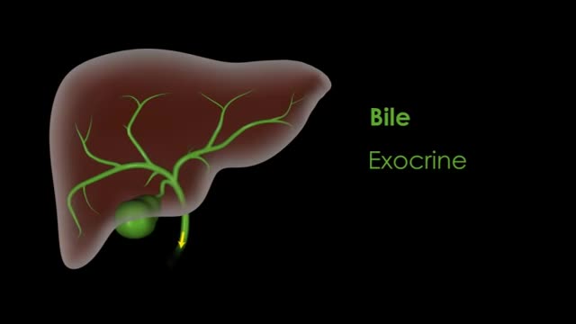
Bial comprises the exocrine secretory product of the liver. It is stored in the gallbladder. During digestion, bile is released from the gallbladder through the cystic duct and to the duodenum to aid in the digestion of fats. The filtration of blood and the production of bile takes place within the structural and functional unit of the liver. The lobule, where the terminal branches of the vessel's carrying incoming and outgoing blood meet. The liver lobule is roughly hexagonal in cross-section. In the center of the lobule lies the central vein, which collects blood from the lobule and carries it to the hepatic veins.
Located at the lobule periphery are branches of the vessel supplying incoming blood, the portal vein and the hepatic artery. Also located here is the bile duct. Collectively, the portal vein, hepatic artery and the bile duct together comprise the portal triad. Taking a horizontal cross-section of the lobule gives us an arrangement typically seen in histological preparations. In this view, we can identify the central veins and associated portal triads. There are two prevailing views of liver lobule organization, the classic liver lobule or anatomical model and the liver assonus.
The classic liver lobule is based on the structure of the lobule we just discussed. It is hexagonal in shape and is defined by portal triads surrounding a single central vein. To identify a lobule in a histological slide, first find a central vein. Next, locate the portal triad surrounding that vein, usually about six at the corners of the hexagonal prism. An imaginary line connecting the triads represents the outside border of the lobule. In the classic liver lobule, blood flows from the portal triads toward the central vein. Bile flows in the opposite direction, from the central position toward the portal triads of the lobule periphery.
Remember, the classic liver lobule model emphasizes the anatomical structure of the lobule. The liver assonus is another way to view liver structure. This model is important for understanding liver physiology and pathology. The structure of the assonus overlaps two classic liver lobules. The assonus is diamond or oval shaped. It involves approximately two portal triads and two central veins. To identify the assonus, find two adjacent portal triads. This makes the short axis. Next, find the closest central vein on either side of the triads. This is the long axis.
The resulting shape is a diamond or oval. Blood still flows from the portal triad to the central vein, but within the shape of the assonus, the flow is from the center to the periphery. Bile flows in the opposite direction, from the periphery towards the center. The assonus model is important because it emphasizes the metabolic differences and hepatocytes between the portal triads and the central vein. The cells in the assonus are divided into three zones and are numbered in the direction of blood flow, from one to three.
Cells in zone one lie along the short axis, between the portal triads. Thus, these cells are first to receive everything in blood, including oxygen, absorbed nutrients, and absorbed xenobiotics. Cells in zone two have characteristics intermediate between the cells of zones one and three. Cells in zone three are closest to the central vein, and therefore are the last to receive incoming blood. As blood moves towards the central vein, it becomes depleted of oxygen, nutrients, and xenobiotics. The liver lobule is the anatomical model. It is hexagonal in shape. It involves one anatomical liver lobule, and blood flows from the periphery to the center.
The liver assonus focuses on function. It is diamond or oval shaped. It involves two adjacent anatomical liver lobules. The blood flows from the middle, which we discuss as a short axis, to the periphery.
Transcript auto-generated from the video’s audio for accessibility and study. Educational content only — not medical advice.
Video Overview
From our Anatomy Videos collection: Anatomy videos: 3D human anatomy, dissections and body systems explained clearly for medical students and curious minds. Browse all Anatomy Videos videos →
Medical Videos
Free medical and surgery videos for patients, students and professionals — trusted since 2007.
Founded by Dr. Mohamed Ibrahim, MD, PhD — physician-scientist.
Follow us on Facebook · Instagram
SORT BY-
Top Comments
-
Latest comments