- Physical Examination
- Surgical Examination
- Ophthalmology
- Clinical Skills
- Orthopedics
- Surgery Videos
- Laparoscopy
- Pediatrics
- Funny Videos
- Cardiothoracic Surgery
- Nursing Videos
- Plastic Surgery
- Otorhinolaryngology
- Histology and Histopathology
- Neurosurgery
- Dermatology
- Pediatric Surgery
- Urology
- Dentistry
- Oncology and Cancers
- Anatomy Videos
- Health and Fitness
- Radiology
- Anaesthesia
- Physical Therapy
- Pharmacology
- Interventional Radiology
- Cardiology
- Endocrinology
- Gynecology
- Emergency Medicine
- Psychiatry and Psychology
- Childbirth Videos
- General Medical Videos
- Nephrology
- Physiology
- Diet and Food Health
- Diabetes Mellitus
- Neurology
- Women Health
- Osteoporosis
- Gastroenterology
- Pulmonology
- Hematology
- Rheumatology
- Toxicology
- Nuclear Medicine
- Infectious Diseases
- Vascular Disease
- Reproductive Health
- Burns and Wound Healing
- Other
Top videos
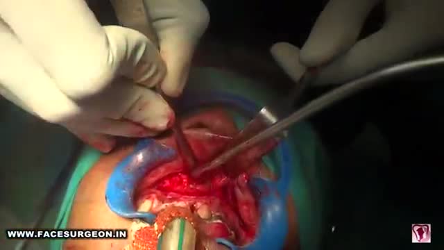
Anterior maxillary distraction for cleft retruded maxilla
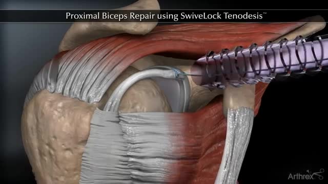
Proximal Biceps Repair using SwiveLock Tenodesis
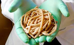
This video shows doctors removing two long ascaris worms from liver.

This 3D animation video explains airway clearance anatomy & physiology in the lungs.
Learn more about Baxter Respiratory Health products at www.hillrom.com/en/products-ca....tegory/non-invasive-
Rx Only. For safe and proper use of product mentioned herein, please refer to the Instructions for Use or Operator manual.
The information contained in these videos is provided for educational purposes only and is not intended nor implied to be a substitute for professional medical advice. You assume full responsibility for how you choose to use this information. Please speak with your healthcare provider about any questions you may have regarding a medical condition.
Baxter retains all right, title, and interest in and to the video, and retains the right to demand that you immediately cease use of the video and unembed the video. Baxter may discontinue or disable videos you have embedded at any time for any reason. You will not misrepresent the content contained in the video or use it in conjunction with price comparisons, in derogatory comparisons or in negative comparisons, with Baxter's competitor's products, or in derogatory or negative commentaries about Baxter's products - doing so may subject you to liability. Any and all claims made by you regarding the use, operation, quality, etc. of Baxter's products are your own, and you shall be responsible for ensuring that all such claims comply fully with all applicable federal, state and local laws.
US-FLC174-230024 v1
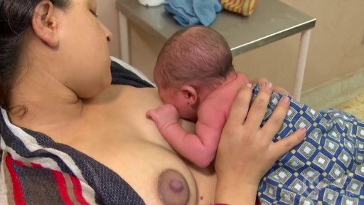
This video is intended primarily for mothers in the developing world, but may be helpful to breastfeeding mothers worldwide.
For education, Microsoft HoloLens will help make incredible leaps forward in productivity, collaboration, and innovation. See how Microsoft HoloLens transforms the way we teach anatomy and our understanding of the human body as we help to prepare the next generation of doctors.
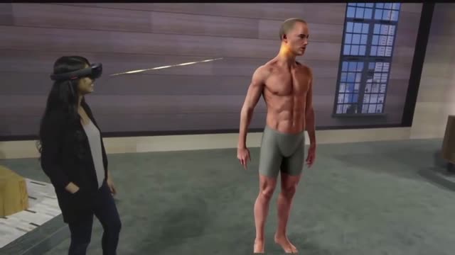
Microsoft HoloLens. Medical Education
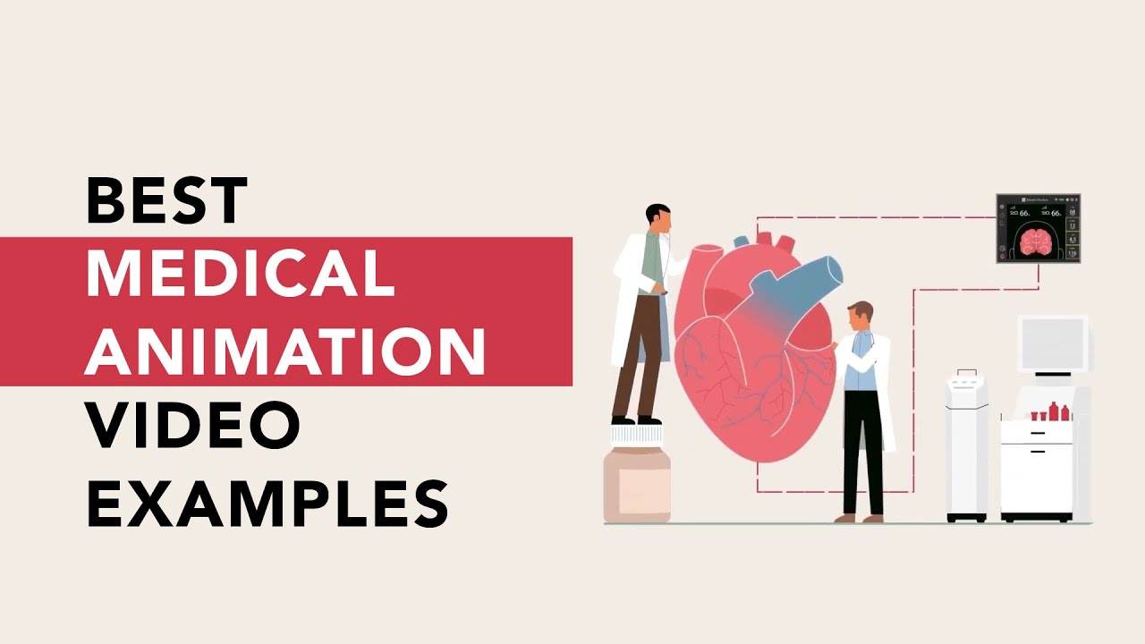
Get the best medical animation videos made at https://www.b2w.tv/healthcare-video-production
Check out more animated healthcare videos from out blog here https://www.b2w.tv/blog/health....care-marketing-video
Medical device manufacturers need to find new and innovative ways to explain their products to potential buyers.
It can be difficult for potential buyers to understand how a medical device works, and even more difficult to visualize how it would be used in a clinical setting.
Medical animation videos are the perfect way to showcase your medical devices.
They are engaging, easy to understand, and help potential buyers see how your product would fit into their workflow.
Jump to the video you like:
8. Pharming Healthcare 0:09
7. ThermoFisher Scientific 2:46
6. Fibrogen 5:49
5. OrthAlign 9:29
4. Edwards LifeSciences 11:34
3. Edwards LifeSciences 12:51
2. Edwards LifeSciences 13:43
1. Edwards LifeSciences 18:14
Check out more Healthcare Videos we have made for our clients:
1. Healthcare Explainer Video for WelbeHealth: https://on.b2w.tv/3OFRaWo
2. Healthcare Product Explainer Video for Edwards Lifesciences: https://on.b2w.tv/3OSdMDb
3. Healthcare Commercial Video for Coopervision: https://on.b2w.tv/45muvpf
4. Healthcare Marketing Video for OrthAlign: https://on.b2w.tv/3P8KBgD
5. Healthcare Video Marketing with The Video-First Approach: https://on.b2w.tv/3LiNDfW
6. 12 Best Brand Archetypes for Healthcare Videos: https://on.b2w.tv/3EIQ0Vu
Want to learn more about Healthcare Videos? Check out our blogs:
1. 10 Best Healthcare Marketing Videos: https://on.b2w.tv/47LxhpJ
2. 5 Animated Healthcare Commercial Videos: https://on.b2w.tv/47IgpAd
3. 11 Animated Healthcare Explainer Videos: https://on.b2w.tv/3Zd7fYM
4. How Long Does It Take To Make an Healthcare Explainer Video: https://on.b2w.tv/45nasak
5. Script for Healthcare Explainer Videos: https://on.b2w.tv/47IY1af
6. Guide to Making Your Own Healthcare Explainer Video: https://on.b2w.tv/3P6FKMR
#medicaldevice #medicalanimation #medicalanimations
Craziest Surgeries You'll Never Believe Occurred!
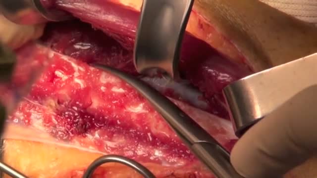
The peroneal artery is closely positioned to the fibula. The artery arises from the tibioperoneal trunk, distal to the takeoff of the anterior tibial artery (seen in the illustration below perforating the interosseous membrane). The peroneal artery sends perforators laterally to the skin of the lower leg, sometimes in a septocutaneous fashion via the lateral intermuscular septum, but often with muscular perforators. The length of the pedicle is usually short, but can be increased substantially by dissecting the peroneal artery and its venae from the fibula and using the distal bone for reconstruction.

NEET Motivational Video |Don't Give up 🔥|#neet2023#aiims
#neetmotivation
#aiims
#neet2023
#pw#dontgiveup
#dream
#mbbs
#neet2024
#doctor
#aiimsdelh#medical

8 months until the world’s first human head transplant
A brief demonstration of the different types of epileptic seizures based on the International Classification of Epileptic Seizures.
Treating Hernia with Laparscopic Inguinal Hernia Repair
Testing for the four features of Gerstmann Syndrome in this patient with two separate left sided strokes (left frontoparietal ischaemic stroke followed by left posterior parietal haemorrhagic stroke). He exhibits (i) acalculia, (ii) agraphia, (iii) left-right disorientation, and (iv) finger agnosia. Complicating the issue is his obvious nonfluent aphasia (expressive dysphasia) with paraphasic errors (replacing words with associated words (e.g. says 'fork' instead of 'spoon')) and some comprehension issues.
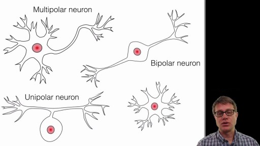
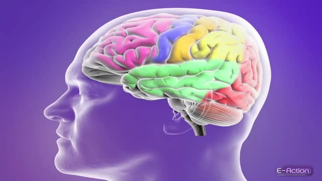
A neuron, also known as a neurone (British spelling) and nerve cell, is an electrically excitable cell that receives, processes, and transmits information through electrical and chemical signals. These signals between neurons occur via specialized connections called synapses.
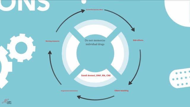
Remembering Medications & The Body Systems Affected

This video shows you how to conduct a clinical examination of the shoulder and to identify common causes of pain.
This video clip is part of the FIFA Diploma in Football Medicine and the FIFA Medical Network. To enrol or to find our more click on the following link http://www.fifamedicalnetwork.com
The Diploma is a free online course designed to help clinicians learn how to diagnose and manage common football-related injuries and illnesses. There are a total of 42 modules created by football medicine experts. Visit a single page, complete individual modules or finish the entire course.
The network provides the opportunity for clinicians around the world to meet and share ideas relating to football medicine. Ask about an interesting case, debate current practice and discuss treatment strategies. Create a profile and log on to interact with other health professionals from around the globe.
This is not medical advice. The content is intended as educational content for health care professionals and students. If you are a patient, seek care of a health care professional.
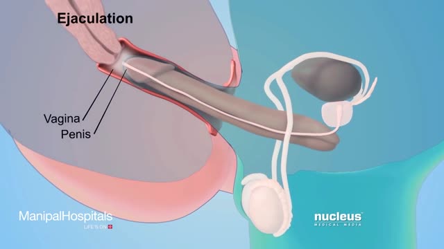
How To Use Male Condom Correctl
Diagnosis of HIV infection in infants is aided by HIV culture or DNA/RNA polymerase chain reaction (PCR); positive results are confirmed by repeating the test. In suspected cases, HIV testing should occur in the newborn period (ie, before the infant is 48 h old), at age 1-2 months, and again at age 3-6 months.
