- Physical Examination
- Surgical Examination
- Ophthalmology
- Clinical Skills
- Orthopedics
- Surgery Videos
- Laparoscopy
- Pediatrics
- Funny Videos
- Cardiothoracic Surgery
- Nursing Videos
- Plastic Surgery
- Otorhinolaryngology
- Histology and Histopathology
- Neurosurgery
- Dermatology
- Pediatric Surgery
- Urology
- Dentistry
- Oncology and Cancers
- Anatomy Videos
- Health and Fitness
- Radiology
- Anaesthesia
- Physical Therapy
- Pharmacology
- Interventional Radiology
- Cardiology
- Endocrinology
- Gynecology
- Emergency Medicine
- Psychiatry and Psychology
- Childbirth Videos
- General Medical Videos
- Nephrology
- Physiology
- Diet and Food Health
- Diabetes Mellitus
- Neurology
- Women Health
- Osteoporosis
- Gastroenterology
- Pulmonology
- Hematology
- Rheumatology
- Toxicology
- Nuclear Medicine
- Infectious Diseases
- Vascular Disease
- Reproductive Health
- Burns and Wound Healing
- Other
Physical Examination
A video from Loyola medical school, Chicago showing the cardiovascular medical and clinical examination
A video from Loyola medical school, Chicago showing the cardiovascular medical and clinical medical examination
Examination of the lower limbs from Loyola medical school, Chicago
Examination of the knee from Loyola medical school, Chicago
Examination of the upper limb by Loyola medical school, Chicago Part 2
Examination of the upper limb by Loyola medical school, Chicago

A video from Physical Exam Series of Loyola University Health System, Chicago showing the medical examination of the abdomen
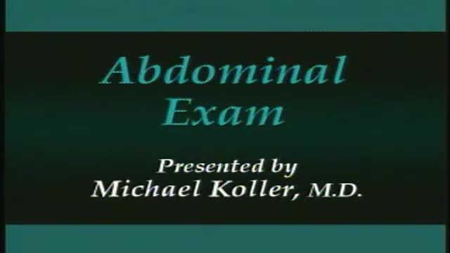
Medical examination of the abdomen from Loyola University, Chicago
Medical breast examination of a female from Loyola University,Chicago

Clinical case discussion for exams.
Useful for medical students and others.
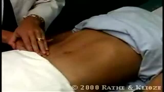
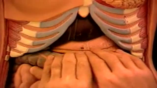
Spleen Palpation

Abdominal Aorta Palpation
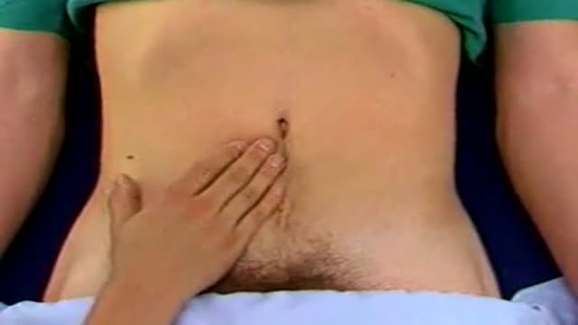
Deep Palpation of the Abdomen

Superficial Palpation of the Abdomen
Complete medical examination of the liver
Palpation for Abdominal Masses

Examination of neck veins and arteries - French Subtitled
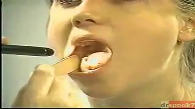
The exam should be performed in an orderly fashion as follows: 1. Have the patient stick out their tongue so that you can examine the posterior pharynx (i.e. the back of the throat). Ask the patient to say "Ah", which elevates the soft palate, giving you a better view. If you are still unable to see, place the tongue blade � way back on the tongue and press down while the patient again says "Ah," hopefully improving your view. This causes some people to gag, particularly when the blade is pushed onto the more proximal aspects of the tongue. It may occasionally be important to determine whether the gag reflex is functional (e.g. after a stroke that impairs CNs 9 or 10; or to determine if a patient with depressed level of consciousness is able to protect their airway from aspiration). This is done by touching a q-tip against the posterior pharynx, uvula or tongue. It is not necessary to do this during your routine exam as it can be quite noxious!
2. Note that the uvula hangs down from the roof of the mouth, directly in the mid-line. With an "Ah," the uvula rises up. Deviation to one side may be caused by CN 9 palsy (the uvula deviates away from the affected side), a tumor or an infection. CN9 Pasly Cranial Nerve 9 Dysfunction: Patient has suffered stroke, causing loss of function of left CN 9. As a result, uvula is pulled towards the normally functioning (ie right) side. 3. The normal pharynx has a dull red color. In the setting of infection, it can become quite red, frequently covered with a yellow or white exudate (e.g. with Strep. Throat or other types of pharyngitis).
4. The tonsils lie in an alcove created by arches on either side of the mouth. The apex of these arches are located lateral to and on a line with the uvula. Normal tonsils range from barely apparent to quite prominent. When infected, they become red, are frequently covered by whitish/yellow discharge. In the setting of a peritonsilar abscess, the tonsils appear asymmetric and the uvula may be pushed away from the affected side. When this occurs, the tonsil may actually compromise the size of the oral cavity, making breathing quite difficult.
5. Look carefully along the upper and lower gum lines and at the mucosa in general, which can appear quite dry if the patient is dehydrated.
6. Examine the teeth to get a sense of general dentition, particularly if the patient has a dental complaint. Pain produced by tapping on a tooth is commonly caused by a root abscess. Tooth Abscess: Tooth abscess involving left molar region. Associated inflammation of left face can clearly be seen. 7. Have the patient stick their tongue outside their mouth, which allows evaluation of CN 12. If there is nerve impairment, the tongue will deviate towards the affected side. Any obvious growths or abnormalities? Ask them to flip their tongue up so that you can look at the underside. If you see something abnormal, grasp the tongue with gauze so that you can get a better look. Left CN 12 Dysfunction: Stroke has resulted in L CN 12 Palsy. Tongue therefore deviates to the left.
8. Make note of any growths along the cheeks, hard palate (the roof of the mouth between the teeth), soft palate, or anywhere else. In particular, patients who smoke or chew tobacco are at risk for oral squamous cell cancer. Any areas which are painful or appear abnormal should also be palpated. Put on a pair of gloves to better explore these regions. What do they feel like? Are they hard? To what extent does a growth involve deeper structures? If the patient feels something that you cannot see, try to get someone else to hold the light source, freeing both your hands to explore the oral cavity with two tongue depressors.

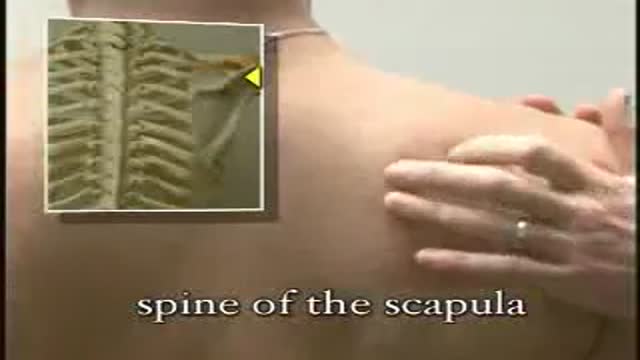
Shoulder Exam
I think that the most daunting aspect of the shoulder exam is appreciating the functional anatomy of this incredibly mobile joint. The primary benefit of the ball and socket arrangement is that it allows the hand to be positioned precisely in space, maximizing our ability to function. In terms of functionality, the shoulder might be best described as having a golf ball-on-a-tee design.
Location Of The Muscle Groups Is Approximated In The Pictures Above.
Start by looking at the normal (or more normal) side. Note any scars, obvious asymmetry, discoloration, swelling, or muscle asymmetry.
Palpation
Gently palpate around the shoulder, touching each of the landmarks noted above. Make note of pain.

Function and Anatomy:
Hinge type joint formed by the articulation of the Ulna and Radius (bones of the forearm), and Humerus (upper arm). Full extension is equal to 0 degrees, full flexion to ~ 150 degrees. Maximum supination (turning hand palm up so that it can hold a bowl of "soup") and pronation (palm down) are both 90 degrees
