
- Physical Examination
- Surgical Examination
- Ophthalmology
- Clinical Skills
- Orthopedics
- Surgery Videos
- Laparoscopy
- Pediatrics
- Funny Videos
- Cardiothoracic Surgery
- Nursing Videos
- Plastic Surgery
- Otorhinolaryngology
- Histology and Histopathology
- Neurosurgery
- Dermatology
- Pediatric Surgery
- Urology
- Dentistry
- Oncology and Cancers
- Anatomy Videos
- Health and Fitness
- Radiology
- Anaesthesia
- Physical Therapy
- Pharmacology
- Interventional Radiology
- Cardiology
- Endocrinology
- Gynecology
- Emergency Medicine
- Psychiatry and Psychology
- Childbirth Videos
- General Medical Videos
- Nephrology
- Physiology
- Diet and Food Health
- Diabetes Mellitus
- Neurology
- Women Health
- Osteoporosis
- Gastroenterology
- Pulmonology
- Hematology
- Rheumatology
- Toxicology
- Nuclear Medicine
- Infectious Diseases
- Vascular Disease
- Reproductive Health
- Burns and Wound Healing
- Kidney Zone Podcast
- Other








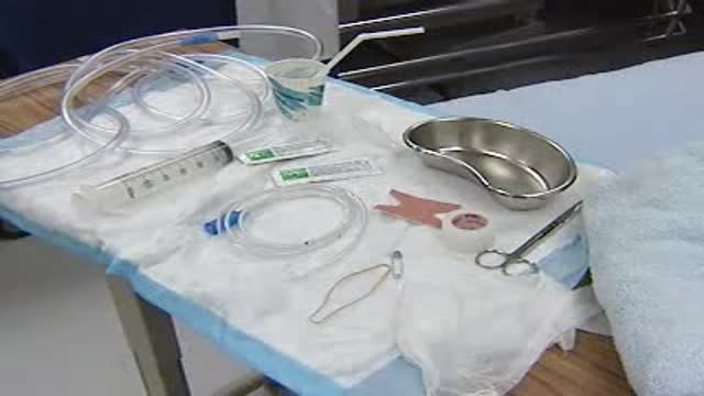

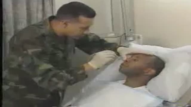


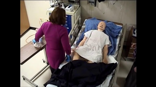

Loyola Abdomen exam

Educational only: videos on MedicalVideos.com are for general information and are not medical advice. Always consult your own doctor about your health.
A video from Physical Exam Series of Loyola University Health System, Chicago showing the medical examination of the abdomen
This video in 30 seconds
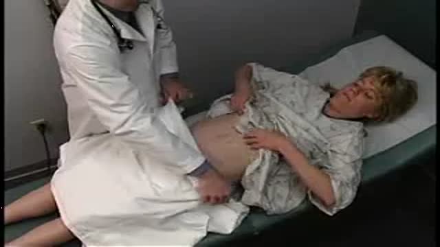
Dr. Michael Kohler teaches the abdominal examination using a standardized patient. The lesson covers positioning and draping, the surface anatomy of the abdominal wall and where the organs and vessels lie, then inspection of the abdomen's contour and listening for bowel sounds in all four quadrants.
Key points
- The patient is positioned supine with proper draping and exposure
- Bony landmarks and the four abdominal quadrants are identified
- The locations of major vessels and organs are described on the surface
- Inspection of abdominal contour and skin features is demonstrated
- Auscultation in all four quadrants is performed at the end
Chapters
- 0:19 — Introduction positioning and draping
- 1:56 — Surface anatomy of abdominal wall
- 5:04 — Four quadrants as reference points
- 9:00 — Major vessels of the abdomen
- 11:31 — Where the organs lie
- 16:58 — Inspection of the abdomen
- 18:44 — Auscultation in four quadrants
Video Transcript
Loyola's abdominal examination teaching video, with Dr. Michael Kohler examining a standardized patient, Thad. It begins with positioning and draping, works through the surface anatomy of the abdominal wall and where the organs lie, and goes on to inspection of the abdomen's contour and auscultation in all four quadrants.
Read the full transcript
Dr. Michael Kohler, and this is Thad, one of Loyola's standardized patients. The purpose of this video is to train first-year medical students on steps of the abdominal examination. And as a probability that will also include the surface anatomy that's relevant to the abdominal exam. And of course, before any physical examination, we always wash our hands, so I'm gonna first wash my hands. And with any examination, the patient needs to be in the proper position. And to do an abdominal examination, the patient should be flat or supine.
So Thad, if I can ask you to please lay back for me? So you as a physician need to make sure that the examining table is flat, and that you pull out the shelf for their legs so they can lie flat. The examiner needs to also make sure that they properly expose the abdomen. So you always need a sheet as well as a gown. It's always best to ask the patient to adjust the gown themselves. So Thad, if you could lift up your gown for me. Just a little bit higher, great.
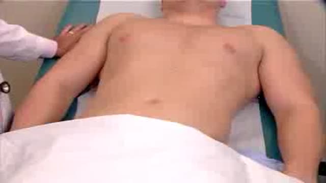
Now I've got good visualization of the abdomen here. We always ask patients to have their arms at their sides. They should not be above their head. The patient's hands are above their head that tenses up the skin and the muscles of the abdomen. And of course, the door should be shut in addition to draping. You as a physician should be on the patient's right side to do this examination. So let's begin by looking at some of the surface markings of the abdominal wall. The costal margin is the lower border of cartilage
of ribs seven to 10 anteriorly. While this is not the superior border of the abdomen, it is the bony prominence near the upper end of the abdomen. The xyfoid process is a finger-like projection from the body of the sternum in the midline. Also in the midline, there is a line known as the linea alba, which goes from the xyfoid process all the way down to the symphysis pubisis. You'll remember that line from your anatomy class. Then if you could lift your head off off the table, then notice there's contraction of the rectus abdominal muscles.
These muscles run longitudinally from the lower margins of the costal cartilage all the way down to the pubic bone and the symphysis pubis. They run approximately seven to 10 centimeters from the midline, and the lateral border of the rectus abdominal muscle is known as a linea semilunaris. I'll keep you in your head back, thanks. Also in the midline is the umbilicus, although it takes many different shapes and structures, whether it's an inni or an outie. The location of the umbilicus depends on the patient's body habitus. For most people, the umbilicus is located at the level of the fourth lumbar vertebrae,
however depending on the patient's weight, their size, and so forth, it can vary from L3 to L5. As we move inch virally, the bony prominence of the pubic bone is noted in the midline. The exact midline is a joint known as a symphysis pubis. It does not have a lot of movement, but there is an articulation there between the two pubic bones. As we move laterally, the bone is known as the pubic crest, and a little bit further later, about one inch out, there's a bony prominence known as the pubic tubercle.
That bony prominence is important because that is one of the points of reference for the inguinal ligament. As we move further laterally, you'll notice another bony prominence known as the anterior superior iliac spine. This is always easy to identify on any patient. Moving from the pubic tubercle to the anterior superior iliac spine is an inguinal ligament. As we move further from the anterior superior iliac spine, we can follow a bony prominence laterally, and that's known as the iliac crest. That iliac crest is a wing-like portion of the ilium bone,
which sort of flares out laterally, and it goes superiorly and posteriorly. And if we feel in back, it ends in what's known as the posterior superior iliac spine. There's four quadrants in the abdomen, and we use these four quadrants as reference points and as points of descriptions where we locate masses or we identify physical findings. It's sort of an arbitrary horizontal line, almost if you will. It's an imaginary horizontal line that runs through the umbilicus, and the vertical line also runs through the umbilicus. So, again, if we were to imagine those two lines over the abdomen,
we would have the right upper quadrant, the right lower quadrant, the left lower quadrant, and then the left upper quadrant. The patient's abdomen, of course, does extend superiorly above the costal margin and actually extends all the way up to the dome of the diaphragm. The location of the diaphragm does vary because as the muscle respiration, it elevates and lowers and elevates and lowers with each breath. With quiet respiration, there might only be approximately a one-centimeter movement up and down, but if a patient's exercising or taking deep breaths,
that varies as much as three to ten centimeters up and down. On the right side, you could locate the roughly horizontal plane of the dome of the diaphragm at about the fourth rib in the mid-clavicular line. Let's talk about that. I'm just going to lift your gait up a little bit higher. The clavicle is a bone that runs from the sternal notch all the way laterally to the shoulder. About midpoint of that clavicle, that would be the mid-clavicular line. For further reference to that anatomical landmark, I could refer you to the thorax training video where we go into much detail
all of the vertical lines around the axis of the chest. Let's move back down again to the inguinal ligament. As I said, that runs from the pubic tubercle laterally to the anterior superior iliac spine. Just above that, inguinal ligament lies the inguinal canal, which is approximately four centimeters long and runs above and parallel to the inguinal ligament. That is the site of inguinal hernias. Also in this general area are the inguinal lymph nodes. The superficial inguinal lymph nodes are divided into two groups. There's a horizontal group and a vertical group.
The horizontal inguinal lymph nodes lies approximately one centimeter below the area of the inguinal ligament and also parallel. When an examiner places their finger pads on the skin, they'll be feeling below the inguinal ligament and doing short circular motions such as this to feel the horizontal superficial inguinal lymph nodes. The vertical superficial inguinal lymph nodes again lie below that inguinal ligament and somewhat medial to the femoral artery. First, you need to identify the femoral artery pulse, which I think you can probably notice my fingertips moving back and forth.
The vertical superficial inguinal lymph nodes cluster near the end of the greater saftonous vein. So moving just medially to the femoral artery and going down is where I palpate for the vertical superficial inguinal lymph nodes. It's important to make that distinction of the horizontal and the vertical, almost forming a T because they do drain separate areas of the body. Next, let's review the surface markings of some of the major arteries that go through the abdomen. The abdominal aorta begins as a direct continuation of the thoracic aorta at the level of T12 or L1
where the thoracic aorta passes through the diaphragm. It's what's known as the aorta caitus, which is approximately the midline of the diaphragm. The abdominal aorta will then descend in the midline but slightly to the left just in front of the first four lumbar vertebrae. The total length of the abdominal aorta would be approximately 10 centimeters at most. And usually its internal diameter is approximately 2 centimeters. In a thin person, pulsations are often seen between the costal margins, especially when the person exhales and holds their breath. This pulsation is completely normal and is not indicative of any pathology.
At about the level of L4 or about the level of the umbilicus, the abdominal aorta divides into the common iliac arteries, one for the right and one for the left. At about four centimeters from the midline, they then divide into internal and external branches of the iliac artery. The external branch continues laterally and goes deep to the inguinal ligament. When it runs underneath the inguinal ligament as it emerges going down towards the thigh, it's known as the femoral artery. So the femoral artery is just a continuation of the external iliac artery.
It's about in the midpoint between the anterior superior iliac spine and the pubic tubercle. So it's about the midpoint of the inguinal ligament. If you place your fingers down, you'll feel the femoral pulses I'm feeling right now. The renal arteries take off somewhat higher in the right upper and lower quadrant from the abdominal aorta and they take off at approximately right angles at about the level of L1 or L2. Now let's talk about some of the surface markings of the major abdominal organs. Of course, the position and shape of the viscera depend on the patient's posture
versus if they're supine or standing. It also depends on respiration as many organs are pushed down by the diaphragm with each respiratory cycle. It also depends on the physiologic state of the organ. For example, if you've just eaten and your stomach is full, your stomach will be distended and will be further down in the abdomen. The pancreas, let's talk about that first. The pancreas is not normally palpable organ. It's a deep structure that's in the retroperitoneum. It sort of lies in both upper quadrants. The head of the pancreas conforms to the C-shaped curve of the duodenum just to the right of the midline.
As the head of the pancreas crosses over the midline, it's known as the neck and the body of the pancreas. From this point, it takes a superior and leftward approach towards the higher surface of the spleen. The spleen is just inferior to the diaphragm. It's also found at the levels of the 9th, 10th, and 11th ribs. If we identify a line in the mid-axilla, the spleen is actually posterior to that mid-axillary line as it runs down the 9th, 10th, and 11th ribs. The spleen lies just above the kidney and behind the stomach.
And again, as I said before, oftentimes the tail of the pancreas comes up to the spleen's hylum. Okay, turn back. The liver is the largest organ in the abdomen. In fact, it takes up at approximately 1⁄40 of the body weight of a normal adult. The liver is in the right upper quadrant but also crosses into the epigastric area. The superior border of the liver conforms to the shape of the dome of the diaphragm. And because the liver is right underneath the diaphragm, the liver does move with each respiration.
At rest, the upper border of the liver is approximately in the 5th costal cartilage in the mid-clavicular line. The lower border is somewhat on a oblique surface and as the patient takes a breath, that comes down. And we'll be talking about palpating for the liver edge later in this video. The stomach lies primarily in the left upper quadrant and in the epigastric area. It terminates just across the midline into the duodenum, the first part of the duodenum. Again, the shape and structure and location of the stomach varies widely depending on the body position
as well as when the patient lasts 8. In a thin patient, the stomach oftentimes takes sort of a J shape and can drop as low as about the level of the umbilicus before it curves up to the duodenum. In more broadly built patients, the stomach tends to lie more transversely rather than a J shape. Again, the stomach is not normally a palpable organ. Also not palpable usually are the kidneys because they are very posterior retroparous neal organs. The upper poles of the kidneys are protected by the 11th and 12th ribs posteriorly,
although the inferior poles might be approximately 4 centimeters above the iliac crest, which is all the way laterally over here. The normal kidneys are about 11 centimeters long, 5 to 6 centimeters wide, and 3 to 5 centimeters thick, and they're approximately 4 or 5 centimeters from the midline. They're usually located between the 12th thoracic vertebrae to the first 3 lumbar vertebrae. The bladder when empty is posterior to the pubic bones and not palpable. However, as the bladder distends with urine, it becomes dome shaped and can rise up above the level of the pubic bone towards the umbilicus.
Normally it does not get above a halfway point between the simpusous pubic and the umbilicus, but in pathologic conditions that can extend all the way up to the level of the umbilicus. The colon, the large intestine, is approximately 5 feet long. It begins in the right lower quadrant as he seek them in ascending colon and traverses upwards to what's known as the hepatic flexure. At that point, we have the transverse colon, which is the area between the hepatic flexure and the splenic flexure. This is the longest and most mobile part of the colon, and it may actually sort of cross down the midline
at the level of the umbilicus before it goes superiorly in towards the left at the splenic flexure. Then we have the descending colon, which comes down from the left upper quadrant to the left lower quadrant. At the brim of the pelvis, the descending colon turns into the sigmoid colon, which then changes to rectum and finally anus. Now we're ready to examine the abdomen. The four steps of the examination begin of course with inspection, but then we move right to oscultation, and this is different than in any other organ system.
And the reason why we oscultate right after inspection is that anytime we would percussor, palpate the abdomen that can stimulate the bowel and falsely increase the bowel sounds. So we always first inspect and then we move to oscultation right away. And again, the exam is always done on skin and not over an article of clothings or gown. Always warn the patient before you touch their abdomen. Many people are jitterish or ticklish, so always let the patient know before you actually do maneuver what you are going to do and examine from the right side of course.
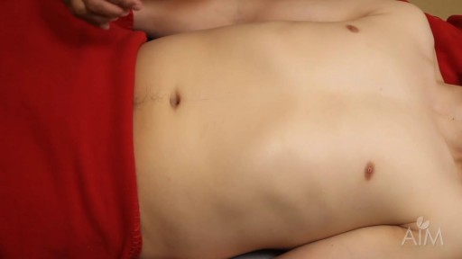
The first thing we're going to do is just inspect the abdomen. We're going to look at the abdominal skin, see if there's any skin lesions. We're going to look for scars. We're going to look for striae, which might be pink or purple and colored, and be indicative of Cushing syndrome. We'll look for dilated veins on the abdominal wall, which could be a sign of liver cirrhosis. We also are going to look for skin rashes. Oftentimes people will have a rash where their clothing rubs against their skin,
against the belt line or else in the inguinal area, you might notice a rash. And looking at the umbilicus, you're going to be looking to see if there's any sign of herniation. And also look for signs of pulsation on the skin. After you've done a general inspection, then you're going to note the contour of the abdomen. And to do that, you might need to actually look down a little bit tangentially at the abdomen instead of standing straight up. Basically what you're looking for is this contour here.
In fact, the contour is basically flat. The contour can also be concave down or what we call scaphoid, in which it's sort of depressed or hollowed out. Conversely, it could also be rounded or pertubrant or distended, where the actual abdomen is shaped as pertubrant. That could be because the patient is overweight, from gas, from pregnancy, from asides, from a tumor. A pertubrant or distended abdomen isn't a specific diagnosis. It's again just something you notice in part of your inspection. The next part of the examination is off-scotation.
So with that, I'm going to place my stethoscope on your skin and listen. And we do ask that you listen in all four quadrants. You listen with the diaphragm of your stethoscope and not with the bell. And gently place the diaphragm on the patient's abdomen. Let just the weight of the stethoscope hold it. You don't need to push down hard on the abdomen. Now I'm moving to the right lower quadrant, lower quadrant, and the left upper quadrant. You need to listen until you hear bowel sounds. If you don't hear bowel sounds in a quadrant, you need to listen for a full two minutes
before you can say that the bowel sounds are absent in any given quadrant. But normally bowel sounds are present at a rate of 3 to 34 a minute, so they're usually very easily heard. Also while you're listening in the upper quadrants, you might notice breweries. This could be a sound of blockage, either the aorta or the renal arteries. But for a brewery to be significant, you should have both the systolic as well as the diastolic.
Transcript auto-generated from the video’s audio for accessibility and study. Educational content only — not medical advice.
Video Overview
From our Physical Examination collection: Watch physical examination videos step by step — cardiovascular, respiratory, abdominal and neurological exams demonstrated for medical students and clinicians. Browse all Physical Examination videos →
Medical Videos
Free medical and surgery videos for patients, students and professionals — trusted since 2007.
Founded by Dr. Mohamed Ibrahim, MD, PhD — physician-scientist.
Follow us on Facebook · Instagram
SORT BY-
Top Comments
-
Latest comments