
- Physical Examination
- Surgical Examination
- Ophthalmology
- Clinical Skills
- Orthopedics
- Surgery Videos
- Laparoscopy
- Pediatrics
- Funny Videos
- Cardiothoracic Surgery
- Nursing Videos
- Plastic Surgery
- Otorhinolaryngology
- Histology and Histopathology
- Neurosurgery
- Dermatology
- Pediatric Surgery
- Urology
- Dentistry
- Oncology and Cancers
- Anatomy Videos
- Health and Fitness
- Radiology
- Anaesthesia
- Physical Therapy
- Pharmacology
- Interventional Radiology
- Cardiology
- Endocrinology
- Gynecology
- Emergency Medicine
- Psychiatry and Psychology
- Childbirth Videos
- General Medical Videos
- Nephrology
- Physiology
- Diet and Food Health
- Diabetes Mellitus
- Neurology
- Women Health
- Osteoporosis
- Gastroenterology
- Pulmonology
- Hematology
- Rheumatology
- Toxicology
- Nuclear Medicine
- Infectious Diseases
- Vascular Disease
- Reproductive Health
- Burns and Wound Healing
- Kidney Zone Podcast
- Other
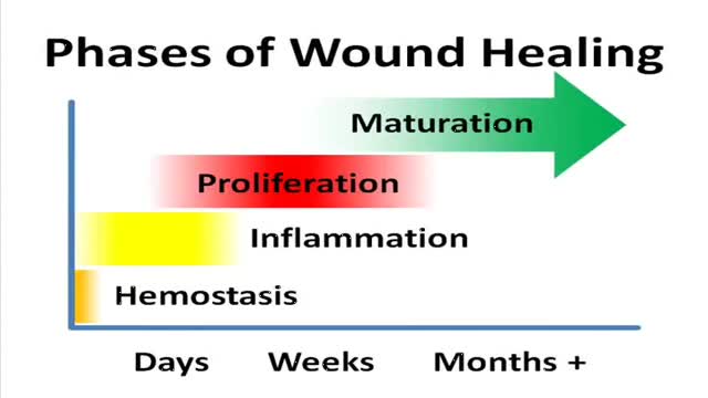







Phases of Wound Healing

Educational only: videos on MedicalVideos.com are for general information and are not medical advice. Always consult your own doctor about your health.
Wound healing, or wound repair, is the body's natural process of regenerating dermal and epidermal tissue. When an individual is wounded, a set of complex biochemical events takes place in a closely orchestrated cascade to repair the damage. These events overlap in time and may be artificially categorized into separate steps: the inflammatory, proliferative, and remodeling phases (Some authors consider healing to take place in four or more stages, by splitting different parts of inflammation or proliferation into separate steps.). In the inflammatory phase, bacteria and debris are phagocytized and removed, and factors are released that cause the migration and division of cells involved in the proliferative phase.
This video in 30 seconds
This video explains the phases of wound healing, the body's natural process of regenerating dermal and epidermal tissue. It describes the inflammatory, proliferative and remodeling phases, including phagocytosis of bacteria and debris and the release of factors that drive cell migration and division in the proliferative phase.
Key points
- Explains wound healing as regeneration of dermal and epidermal tissue
- Covers the inflammatory phase
- Covers the proliferative phase
- Covers the remodeling phase
Video Transcript
This teaching module explains the phases of wound healing. It describes haemostasis, in which platelets form a clot and release signals that recruit other cells; the inflammatory phase, in which leukocytes and macrophages clear bacteria and debris and release growth factors; the proliferative phase of granulation tissue, contraction and epithelialisation; and the maturation phase, in which collagen is reorganised and remodelled and the scar gains tensile strength while remaining weaker than uninjured tissue and at risk of breakdown.
Read the full transcript
Hemostasis occurs right after initial injury. At the time of injury, epinephrine, a chemical that constricts peripheral blood vessels, is released in an attempt to minimize bleeding into the soft tissues. The key cell responsible for this function is the platelet, which causes the body to form a clot to prevent further bleeding. There is an increased aggregation of platelets to enable the wounded vessel to complete the clotting process. Platelets also release key cytokines, such as platelet-derived growth factor, that call in cells to participate in later phases of healing.
The objective of the hemostasis phase of wound healing is to control bleeding. In hemostasis, the inflammatory phase begins. The local signs and symptoms that occur during the inflammatory phase are swelling, fission of blood, red histamine, heat, histamine response, and pain. The inflammatory phase is characterized by a host of cells, leukocytes and macrophages, infiltrating the wound sites. Bleeding is controlled by hemostasis. Any bacteria that is present is destroyed by leukocyte-destroyed bacteria, cleansing the wound of cellular debris. Macrophages replace the leukocytes and produce a host of cytokines and growth factors
and tractants to other cells needed for tissue repair. Macrophages also convert macromolecules into the amino acids and sugars necessary for wound healing. It is thought that the macrophage also attracts contractual cells to the wound to encourage wound contraction. Vasodilation, with resultant edema, warmth, and ruber, are the result of factors secreted from the macrophage and other in response to the inflammatory process. The objectives of the inflammatory phase of wound healing are to clean debris and bacteria, and prevent infection. Scar tissue formation is characterized by three distinct phases.
Granulation, contraction, epithelialization. Click on one of the phases to learn more. She was generated, producing red fibroblasts. It gives tissue its tensile granulation tissue, its margins contract or pull together, decreasing the size of the wound, tissue, realization, divide, and tissue. Epithelial cells will not migrate across a dry surface or necrotic tissue. Curation phase, the collagen fibers reorganize, remodel, and mature, gaining tensile strength. Collagen fibers, proteoblacens, and fibromectin, distributed. The scar becomes less cellular and gains tensile strength. This tissue will always be at risk for breakdown, because its tensile strength is less than that of fibroblasts,
with collagen fibers is a complicated process, key growth factors in the development of procollagen. Procollagen fibers mature into collagen fiber, connected within a single collagen fiber. Research is currently underway to determine the chemical details associated with collagen synthesis.
Transcript auto-generated from the video’s audio for accessibility and study. Educational content only — not medical advice.
Video Overview
From our Health and Fitness collection: Exercise science, injury prevention, rehabilitation and healthy-living guidance from medical professionals. Browse all Health and Fitness videos →
Medical Videos
Free medical and surgery videos for patients, students and professionals — trusted since 2007.
Founded by Dr. Mohamed Ibrahim, MD, PhD — physician-scientist.
Follow us on Facebook · Instagram
SORT BY-
Top Comments
-
Latest comments