- Physical Examination
- Surgical Examination
- Ophthalmology
- Clinical Skills
- Orthopedics
- Surgery Videos
- Laparoscopy
- Pediatrics
- Funny Videos
- Cardiothoracic Surgery
- Nursing Videos
- Plastic Surgery
- Otorhinolaryngology
- Histology and Histopathology
- Neurosurgery
- Dermatology
- Pediatric Surgery
- Urology
- Dentistry
- Oncology and Cancers
- Anatomy Videos
- Health and Fitness
- Radiology
- Anaesthesia
- Physical Therapy
- Pharmacology
- Interventional Radiology
- Cardiology
- Endocrinology
- Gynecology
- Emergency Medicine
- Psychiatry and Psychology
- Childbirth Videos
- General Medical Videos
- Nephrology
- Physiology
- Diet and Food Health
- Diabetes Mellitus
- Neurology
- Women Health
- Osteoporosis
- Gastroenterology
- Pulmonology
- Hematology
- Rheumatology
- Toxicology
- Nuclear Medicine
- Infectious Diseases
- Vascular Disease
- Reproductive Health
- Burns and Wound Healing
- Other
Top videos
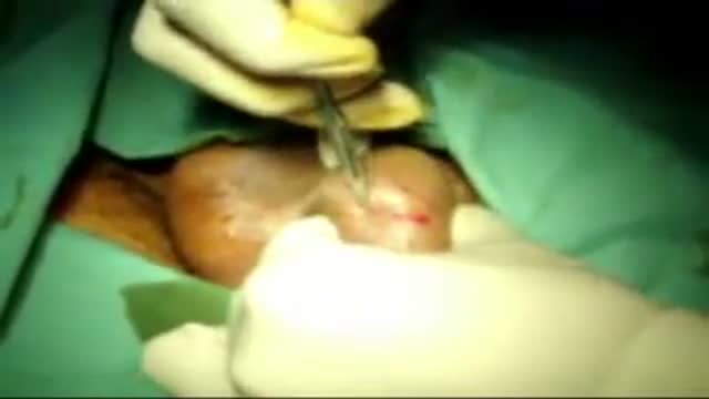
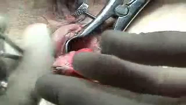
open multi puncture testicular biopsy to retrieve sperm for ICSI (IntaCytoplasmic Sperm Injection)
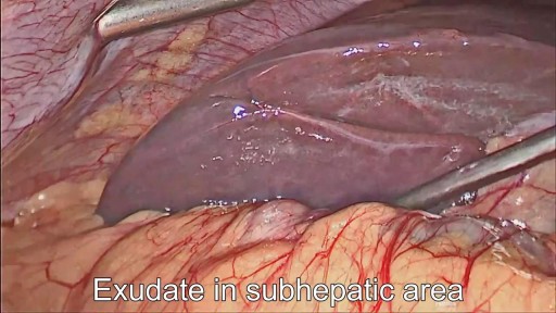
28 years old gentleman presented with huge liver abscess in the right lobe, with repeated attempts of percutaneous aspirations in the past. He was evaluated and subjected to Laparoscopic drainage. This video depicts feasibility of laparoscopy in deep seated liver abscesses. Video created by: Dr. Juneed M. Lanker Fellow Minimal Access Surgery Apollo Hospitals Chennai.
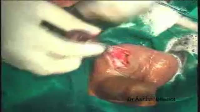
Orchidectomy and Orchidopexy in Testicular Torsion
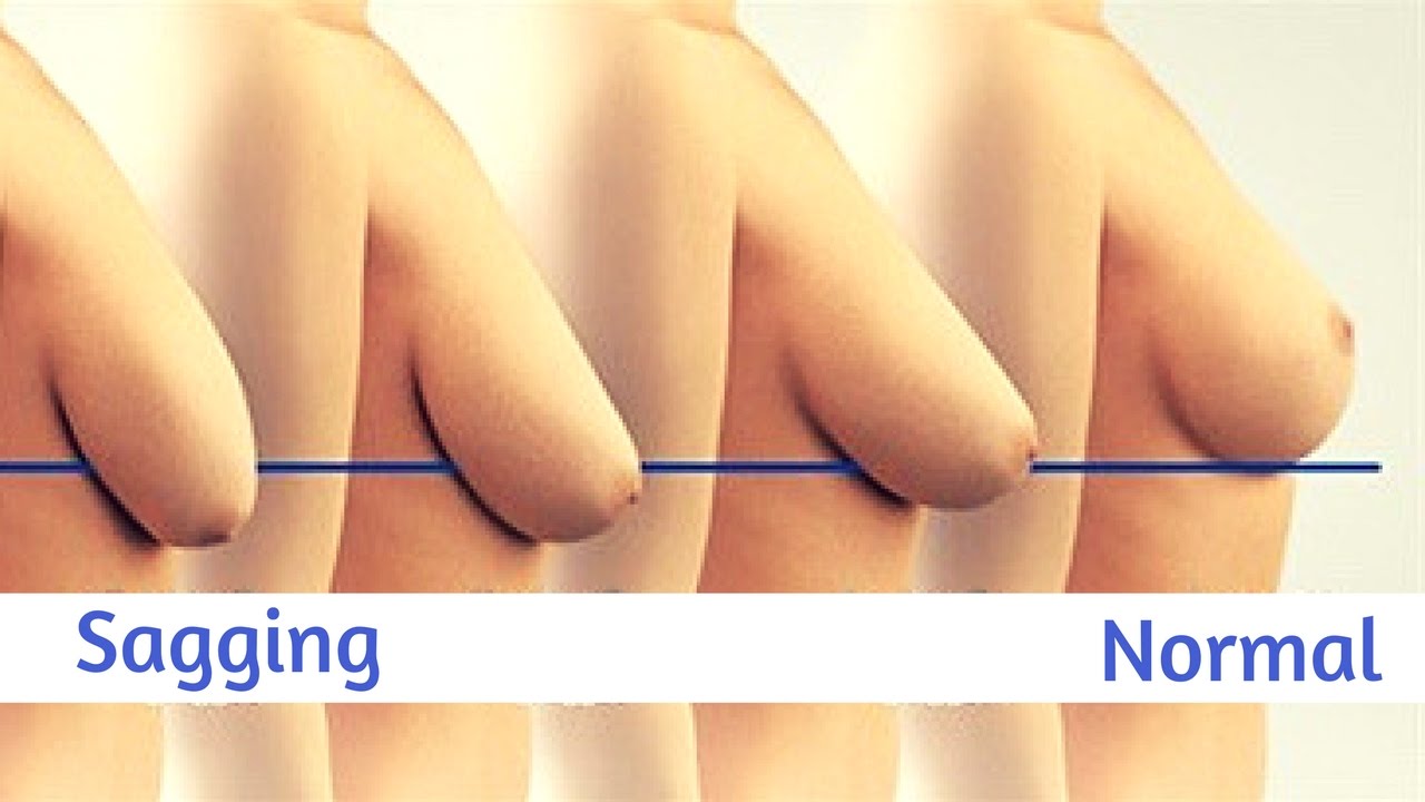
Watch that video to know How To Firm And Lift Your Sagging Breasts Naturally

Christopher J. Rapuano, MD, Director of the Cornea Service at Wills Eye Institute describes his surgical approach of a Combined Penetrating Keratoplasty (PK) and Cataract Surgery
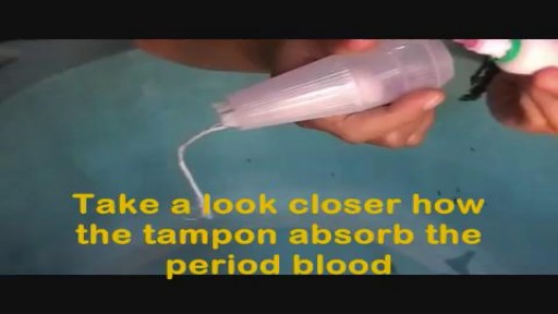
How to Use Tampons for The First Time Demo Video
The Epley maneuver or repositioning maneuver is a maneuver used to treat benign paroxysmal positional vertigo of the posterior or anterior canals

In order to be able to look at tissues under a microscope, we need to first stain them with the right technique. Learn the main staining techniques used in histology today on our full video: https://khub.me/aux9w
Oh, are you struggling with learning anatomy? We created the ★ Ultimate Anatomy Study Guide ★ to help you kick some gluteus maximus in any topic. Completely free. Download yours today: https://khub.me/e0th1
As you probably know, histology is the study of the microscopic anatomy of cells and tissues. So we use staining methods to visualize and distinguish the different parts of cells and tissues since cells and their structures are usually transparent or colorless. The types of dyes used to color cells and their components can either be specific to particular structures, chemical groups or even molecules, and it can also be non-specific in which case most of the cell is stained in the same way.
When staining tissue samples, dyes that are used are either acidic or basic or a combination of the two. And why is that, you might be asking. Well, cellular structures such as nucleic acids or proteins have charged groups which are known as phosphate groups or carboxyl groups, just to name a couple. The dyes used in histology are colored organic compounds which also have a charge. Acidic dyes carry a negative charge and so they bind to positively-charged cell structures.
In the full version of this tutorial, we will cover some of the most common types of dyes used in histological staining of cells and their structures:
- basic dyes vs acidic dyes vs neutral dyes;
- hematoxylin and eosin;
- PAS - staining;
- Golgi method;
- Toluidine blue;
- Masson's trichrome;
- Osmium tetroxide;
To master this topic, click on the link and carry on watching the full video (available to Premium members): https://khub.me/aux9w !
Want to test your knowledge on the different types of cells and tissues? Take this quiz: https://khub.me/3g19f
Read more on how to interpret different histological sections on this complete article which goes through the different stains used in histology https://khub.me/saimh
For more engaging video tutorials, interactive quizzes, articles and an atlas of Human anatomy and histology, go to https://khub.me/pkvz2
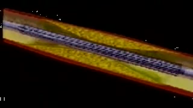
This video depicts how a stent is placed in the coronary artieries. We first place a guiding wire in the heart artery through a catheter, usually from the groin. Then the stent is inflated by a balloon in the artery, which is then removed. The stent remains permanently. Blood thinners, aspirin and plavix, are both required after a stent is placed in your heart artery.
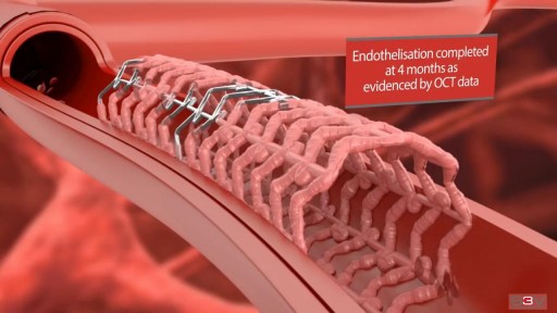
Angioplasty is a procedure to restore blood flow through the artery. You have angioplasty in a hospital. The doctor threads a thin tube through a blood vessel in the arm or groin up to the involved site in the artery. The tube has a tiny balloon on the end.
Medical Examination of the Lower Limbs
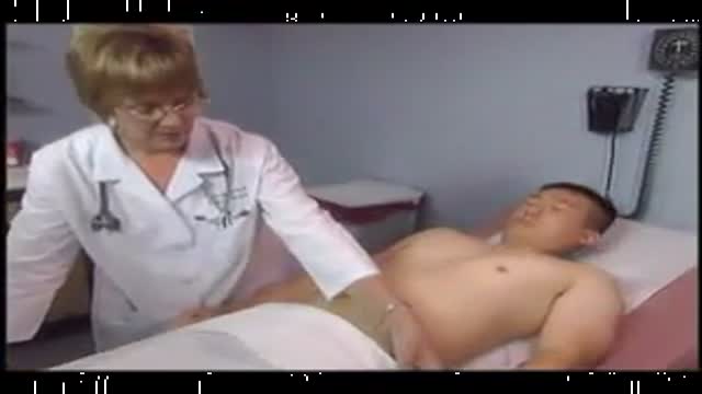
Physical Examination of the abdomen
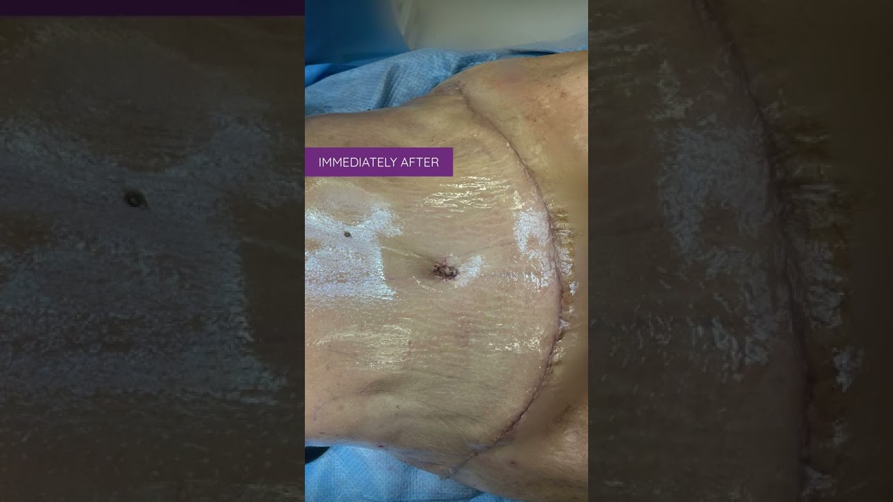
Liposuction in tummy tuck requires special planning and technique. I need to ensure that the blood circulation is well maintained for good healing. Yet proper liposuction is important to have a nice flat and contoured tummy.
#hdliposuction #tummytuck #lipoabdominoplasty #surgicalplanning #skinremovalsurgery #imeediatelyafter #plasticsurgeondubai #cocoonaclinic #drsanjayparashar #dubai
For more information visit www.drsanjayparashar.com
For more content, follow me on my social media
Instagram : https://www.instagram.com/drsanjayparashar/
Facebook : https://www.facebook.com/drsanjayparashar

: Frederick Lang, M.D., and Jeffrey Weinberg, M.D., neurosurgeons at MD Anderson Cancer Center, answer frequently asked questions about what to expect when you’re having brain tumor surgery.
Learn more about the MD Anderson Brain and Spine Center: www.mdanderson.org/brainandspine
Request an appointment at MD Anderson by calling 1-877-632-6789 or online: https://my.mdanderson.org/RequestAppointment

Please remember that this video is to be used for educational purposes. You must follow your facility or colleges' policies and procedure checklists to ensure you are completing the skill satisfactorily. Thanks for watching!
Music from #Uppbeat (free for Creators!):
https://uppbeat.io/t/swoop/blue-sea
License code: W9DFUQ4II7YVHA59
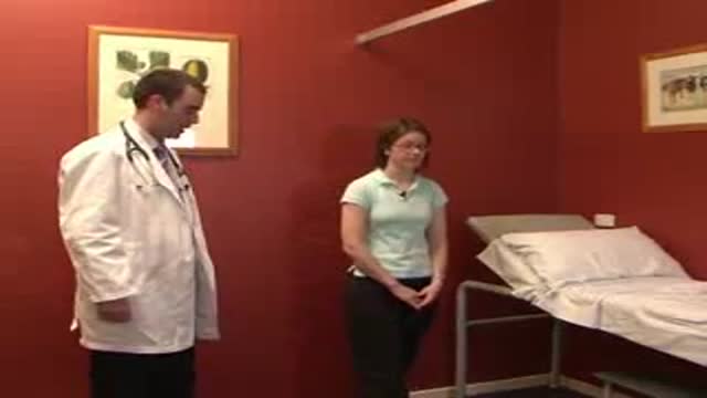
Loyola Full Male Exam Part 1 A video from Loyola medical school, Chicago showing the full examination of the male
Hemorrhoidectomy Operation Video
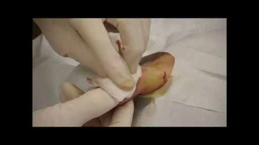
Finger Abscess Incision and Drainage. Digital block with drainage.
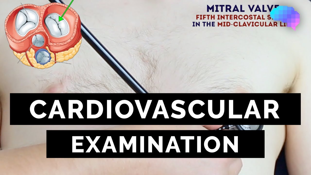
This video demonstrates how to perform a cardiovascular examination in an OSCE station.
You can access our step-by-step OSCE guide to accompany this video here: https://geekymedics.com/cardio....vascular-examination
Check out our other awesome clinical skills resources including:
• 🔥 Geeky Medics Bundles (discounted products): https://app.geekymedics.com/purchase/bundles/
• ✨ 1000+ OSCE Stations: https://app.geekymedics.com/pu....rchase/osce-stations
• 🏥 Geeky Medics OSCE Revision Book: https://app.geekymedics.com/purchase/book/
• 📝 150+ PDF OSCE Checklists: https://geekymedics.com/pdf-osce-checklists/
• 🗂️ 3000+ OSCE Flashcards: https://app.geekymedics.com/pu....rchase/flashcard-col
• 📱 Geeky Medics OSCE App: https://geekymedics.com/geeky-medics-app/
• 🩺 Medical Finals SBA Question Pack: https://app.geekymedics.com/pu....rchase/medical-stude
• 💊 PSA Question Pack: https://app.geekymedics.com/pu....rchase/prescribing-s
Chapters:
- Introduction 00:00
- General inspection 00:35
- Hands 00:46
- Schamroth's window test 01:07
- Capillary refill 01:27
- Pulses 01:35
- Carotid auscultation 02:21
- Carotid pulse 02:43
- Jugular venous pressure 02:55
- Hepatojugular reflux 03:09
- Inspection of the face 03:21
- Inspection of the chest 03:49
- Apex beat 04:12
- Heaves and thrills 04:28
- Heart valve ausculation 04:49
- Accentuation manoeuvres 05:45
- Lung base auscultation 06:23
- Sacral and pedal oedema 06:43
- Summary 07:10
Subscribe to our newsletter to be the first to know about our latest content: https://geekymedics.com/newsletter/ ✉️
Join the Geeky Medics community: 👩👩👧👧
Twitter: http://www.twitter.com/geekymedics
Instagram: https://instagram.com/geekymedics
Facebook: http://www.facebook.com/geekymedics
Always adhere to your medical school/local hospital guidelines when performing examinations or clinical procedures. DO NOT perform any examination or procedure on patients based purely upon the content of these videos. Geeky Medics accepts no liability for loss of any kind incurred as a result of reliance upon the information provided in this video.
Normal heart sounds and aortic regurgitation/stenosis sounds
Recorded on a Thinklabs Digital Stethoscope (https://www.thinklabs.com)
Some people have found this video useful for ASMR purposes.

Surgical site infections (SSIs) remain a prevalent threat to patient safety. Proper surgical hand scrub or rub techniques are essential to decreasing the incidence of SSIs. This video provides instructions on the anatomical surgical hand scrub procedure using the brushstroke method. Learn more from the Department of Hospital Epidemiology and Infection Control (HEIC) at The Johns Hopkins Hospital: http://www.hopkinsmedicine.org/heic
