
- Physical Examination
- Surgical Examination
- Ophthalmology
- Clinical Skills
- Orthopedics
- Surgery Videos
- Laparoscopy
- Pediatrics
- Funny Videos
- Cardiothoracic Surgery
- Nursing Videos
- Plastic Surgery
- Otorhinolaryngology
- Histology and Histopathology
- Neurosurgery
- Dermatology
- Pediatric Surgery
- Urology
- Dentistry
- Oncology and Cancers
- Anatomy Videos
- Health and Fitness
- Radiology
- Anaesthesia
- Physical Therapy
- Pharmacology
- Interventional Radiology
- Cardiology
- Endocrinology
- Gynecology
- Emergency Medicine
- Psychiatry and Psychology
- Childbirth Videos
- General Medical Videos
- Nephrology
- Physiology
- Diet and Food Health
- Diabetes Mellitus
- Neurology
- Women Health
- Osteoporosis
- Gastroenterology
- Pulmonology
- Hematology
- Rheumatology
- Toxicology
- Nuclear Medicine
- Infectious Diseases
- Vascular Disease
- Reproductive Health
- Burns and Wound Healing
- Kidney Zone Podcast
- Other





Abdomen Examination Video

Educational only: videos on MedicalVideos.com are for general information and are not medical advice. Always consult your own doctor about your health.
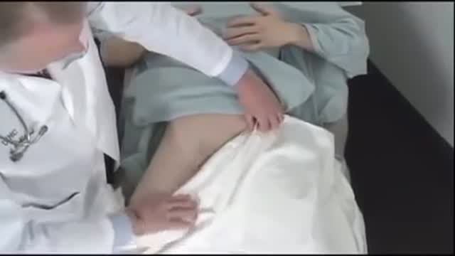
Complete examination of the abdomen including all the items: inspection, palpation, percussion and auscultation Video
This video in 30 seconds
A step-by-step teaching demonstration of the adult abdominal examination. The examiner works through inspection and hernia checks with coughing, auscultation before palpation, light and deep palpation while watching the patient's face, liver and spleen techniques including percussion of the liver span, and then moves on to leg pulses.
Key points
- Inspection of contour and skin and hernia checks with coughing open the exam
- Auscultation is performed before palpation
- Light and deep palpation are demonstrated while watching the patient's face
- Liver and spleen palpation techniques are demonstrated
- Percussion of the liver span and leg pulses follow
Chapters
- 0:00 — Inspection and hernia checks
- 1:30 — Auscultation of the abdomen
- 2:10 — Light and deep palpation
- 4:17 — Liver and spleen palpation
- 5:22 — Percussion of liver span
- 7:02 — Femoral pulses and leg pressures
Video Transcript
A step-by-step teaching demonstration of the adult abdominal examination, performed on a patient addressed as Mr. Johnson. The examiner works through inspection and hernia checks with coughing, auscultation before palpation so bowel sounds are not stimulated, light and deep palpation while watching the patient's face, liver and spleen techniques including percussion of the liver span, and then moves on to the pulses of the legs.
Read the full transcript
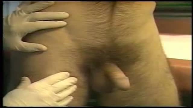
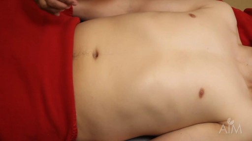
We want to begin the examination of the abdomen and the first part of that examination is to inspect the contour of the abdomen as well as inspecting the skin of the abdomen. The next part of the examination is to inspect for hernias. The patient should be disrobed and you should have visibility of the person's abdomen from approximately this area down to the syphusis pubis. Are you comfortable? Would you like to have your gown on, Mr. Johnson? Comfortable? Okay. We want to have exposure of the patient's femoral area and we can do that by placing the drape in this fashion.
And we will then ask the patient to cough. Mr. Johnson, would you turn your head to the other side and cough for me? And we want to observe, cough, we want to observe this part of the abdomen as well as this part of the abdomen. This part of the abdomen for umbilical or incisional hernias. And then of course we want to look in this area, in the inguinal area and in the femoral area to see if there are any pulsations when the person coughs. Cough again. Again.
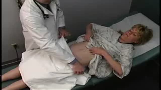
We can then place our hands in the inguinal area and cough. And in the femoral area and cough again. Okay. At this point we can return the sheet to preserve the modesty of the patient. And the first part of the examination of the abdomen is auscultation. We auscultate before we palpate the abdomen because if bowel sounds were absent, we might artificially stimulate bowel sounds by palpation. So one places the stethoscope anywhere in the abdomen and listens for 50 to 20 seconds to determine whether or not bowel sounds are present.
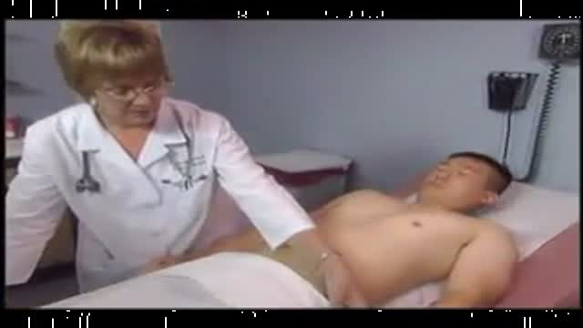
We can then place the stethoscope about one inch above the umbilicus and about one inch lateral to the umbilicus, evaluating for renal artery breweries. There are two types of palpation. One is light palpation, the other is deep palpation. Light palpation is used to ascertain whether or not the patient is having any tenderness in the abdomen. If a person is having any tenderness in the abdomen, you evaluate that area last. Are you having any tenderness in your abdomen, Mr. Johnson? No. Okay. The technique for palpation is basically to place your hand firmly on the abdomen and with your fingers in this position, press down very lightly while you're observing the patient's face.
It's critically important to evaluate the patient while you're watching his or her face to see if the patient winces even though you are asking them whether there's any tenderness. So by placing my hand gently on the abdomen, I'm going to look at the patient's face and ask, any tenderness here? No. What about over here? No. Any tenderness here? No. Over here? No. Here? No. Any tenderness here? No. And over here? No. And finally here? No. You then proceed with deep palpation. Deep palpation uses one hand applied to the skin and the other hand firmly presses down and provides the pressure.
The hand at the bottom is the hand which is palpating for masses. The hand at the top is the hand that is pressing down and using the pressure to push the lower hand down. And you use a rolling motion, again, looking at the patient's face. You've already ascertained that there is no tenderness present in Mr. Johnson's case. But you then use a rolling motion as you roll over the descending colon. And you can ask, are you having any tenderness? Coming over here across the transverse colon? Any tenderness?
No. What about over here? I know this may feel a little uncomfortable, but if there's any pain, please let me know. No. Any tenderness here? No. Now the ascending colon and finally down toward where the cecum exists. After you've done that, we want to go on to special palpation. Special palpation is done of the liver and of the spleen. And there are several techniques which one can use. We can start low in the abdomen and start in this fashion going up toward the costal margin as the patient takes a deep breath.
And out. Again. Out. Again. We could also place our left hand under the right costal margin and do the same technique as was just demonstrated. We could also do something called the hooking technique which is placing your hands in this part of the abdomen, standing in this position and actually trying to hook the liver edge as the patient takes a deep breath. Take a big breath.
And out. Again. And out. All of these techniques are useful to determine the lower edge of the liver. The upper edge of the liver is done by percussion of the chest from where the percussion note goes from resonant over lung tissue to dull over the liver perencoma. Resonant.
Resonant. Resonant. Dull.
So this is the upper border of the liver. This was the lower. One usually measures the span of the liver in the mid-clavicular line and normally about 10 centimeters or 12 centimeters is the normal span for a liver. It's normal. Okay. We can go on to evaluation of the spleen and again we start low in the abdomen starting here and moving up and take a big breath. And out. Again. And out. Again. And out. We might place our hand over the left costal margin to support it and to palpate the spleen in this fashion.
Normally the spleen is not palpable whereas the lower edge of the liver may very well be palpable in many individuals. All four quadrants of the abdomen are also evaluated by percussion. If a person is very well developed and has a very muscular abdominal wall, if the person has a cites which is fluid in the abdomen or if the person is just frankly obese all of these exams obviously a lot more difficult to evaluate. We're now going to move on to evaluation of the pulses of the lower extremity.
First the femoral pulses.
Evaluating also the timing of the femoral pulse with the radial pulse. These two pulses peak at the same time. And in the patient who has co-arctation of the aorta one will have a delay in the femoral pulses. Remember that co-arctation of the aorta is a very important cause of a systemic hypertension of the upper extremity and actually hypotension in the lower extremity. Any patient who especially if the person is young who has hypertension should be evaluated for blood pressure in the upper extremities and as we talked earlier should be evaluated for blood pressure in the lower extremities.
And lower pressure is always abnormal. Lower pressure in the lower extremity compared to the upper extremity in association with a delay in femoral pulses is very strongly suggestive of co-arctation of the aorta. We're now going to proceed with examination of the peripheral pulses before we go on to examination of the genitalia.
Transcript auto-generated from the video’s audio for accessibility and study. Educational content only — not medical advice.
Video Overview
From our Physical Examination collection: Watch physical examination videos step by step — cardiovascular, respiratory, abdominal and neurological exams demonstrated for medical students and clinicians. Browse all Physical Examination videos →
Medical Videos
Free medical and surgery videos for patients, students and professionals — trusted since 2007.
Founded by Dr. Mohamed Ibrahim, MD, PhD — physician-scientist.
Follow us on Facebook · Instagram
SORT BY-
Top Comments
-
Latest comments