
- Procedures & Tests
- Symptoms A-Z
- Symptom Checker
- Kidney Diet
- First Aid
- Anatomy Atlas
- Ask MedicalVideos
- Drug Guide
- Lab Values
- Vital Signs
- Muscles A–Z
- Clinical Signs
- Medical Abbreviations
- Medical Terminology
- Physical Examination
- Surgical Examination
- Ophthalmology
- Clinical Skills
- Orthopedics
- Surgery Videos
- Laparoscopy
- Pediatrics
- Funny Videos
- Cardiothoracic Surgery
- Nursing Videos
- Plastic Surgery
- Otorhinolaryngology
- Histology and Histopathology
- Neurosurgery
- Dermatology
- Pediatric Surgery
- Urology
- Dentistry
- Oncology and Cancers
- Anatomy Videos
- Health and Fitness
- Radiology
- Anaesthesia
- Physical Therapy
- Pharmacology
- Interventional Radiology
- Cardiology
- Endocrinology
- Gynecology
- Emergency Medicine
- Psychiatry and Psychology
- Childbirth Videos
- General Medical Videos
- Nephrology
- Physiology
- Diet and Food Health
- Diabetes Mellitus
- Neurology
- Women Health
- Osteoporosis
- Gastroenterology
- Pulmonology
- Hematology
- Rheumatology
- Toxicology
- Nuclear Medicine
- Infectious Diseases
- Vascular Disease
- Reproductive Health
- Burns and Wound Healing
- Kidney Zone Podcast
- Other
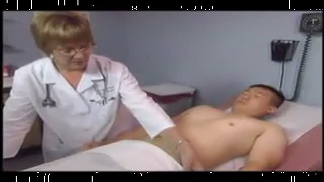
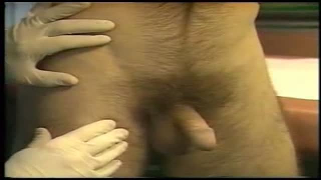
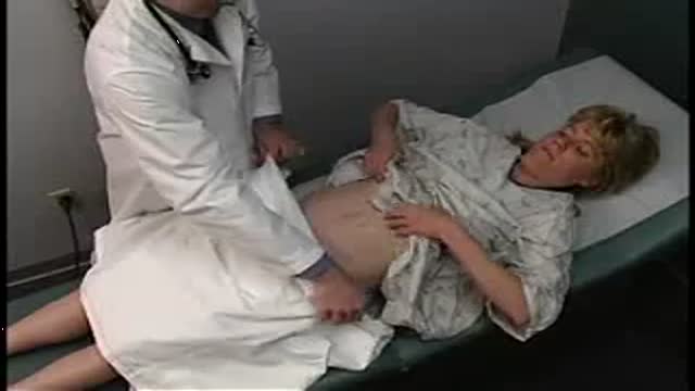

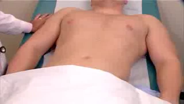

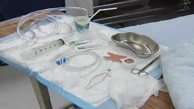
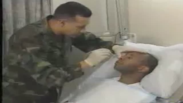

abdomen physical examination

Educational only: videos on MedicalVideos.com are for general information and are not medical advice. Always consult your own doctor about your health.
a video of abdominal physical examination including all the required items:
-Inspection
-Palpation
-Percussion
-Auscultation
This video in 30 seconds
A narrated teaching video demonstrates the complete abdominal physical examination on a healthy patient. It works through preparation, inspection, auscultation, percussion and palpation of the abdomen, and examination of the liver, spleen and right kidney, noting that normal findings and variations differ between patients.
Key points
- Preparation includes the patient emptying the bladder and lying relaxed and comfortable
- Inspection covers skin, symmetry, contour, peristalsis and the umbilicus
- Auscultation listens for bowel sounds and for bruits over arteries when indicated
- Percussion assesses tympany and dullness and the liver and gastric areas
- Palpation proceeds from light to deep across all quadrants, checking tenderness and masses
Chapters
- 0:01 — Examination overview and preparation
- 0:49 — Inspection of the abdomen
- 1:09 — Auscultation for bowel sounds
- 2:38 — Percussion of abdomen and organs
- 3:19 — Palpation technique across quadrants
- 4:15 — Tenderness and peritoneal signs
Video Transcript
A narrated teaching video of the complete abdominal physical examination on a healthy patient. It covers inspection, auscultation, percussion and palpation of the abdominal wall, then percussion and palpation of the liver and spleen and palpation of the right kidney — beginning with the preparation: bladder emptied, patient lying down, relaxed and comfortable, with the examiner noting that normal findings and variations differ between patients.
Read the full transcript
This video presents examination of the abdomen, including inspection, auscultation, percussion, and palpation of the abdominal wall, percussion and palpation of the liver, percussion and palpation of the spleen, and palpation of the right kidney and a organ. In the video, the examiner will assess a healthy patient. Keep in mind that other patients may have the same normal findings, or may exhibit normal variations or abnormal findings. Before you examine the abdomen, make sure that the patient has emptied her bladder. Ask her to lie down and relax and make her as comfortable as possible.
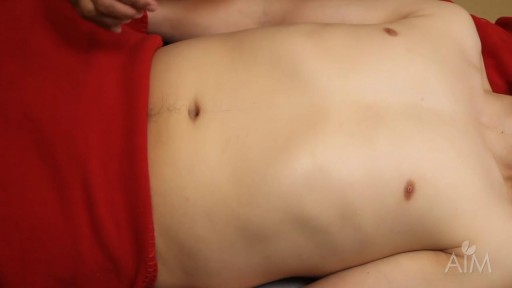
Expose her lower chest and abdomen, but keep her genitalia covered. Inspect the skin for scars, stri-e, dilated veins, or rashes. Inspect the symmetry and contour of the abdomen. And note any peristalsis, pulsations, or masses. Also observe the contour of the umbilicus and look for signs of inflammation, or listen for bowel sounds by placing the diaphragm of the stethoscope gently on the right lower quadrant. Listen to their pitch, quality, and frequency. If the patient has hypertension, listen for bruise over the right renal artery,
aorta, and left renal artery. If present, a bruise would sound like this. In a patient with hypertension, a bruise raises suspicion of renal artery stenosis, but most bruise have other causes. If you suspect arterial insufficiency in the legs, listen for bruise over the aorta, right iliac artery, and left iliac artery. Then identify and listen over the right femoral artery and left femoral artery. Lightly percuss the abdomen to assess the distribution of tympani and dullness. Tympani indicates gas in the stomach or intestine. Dullness suggests fluid or feces.
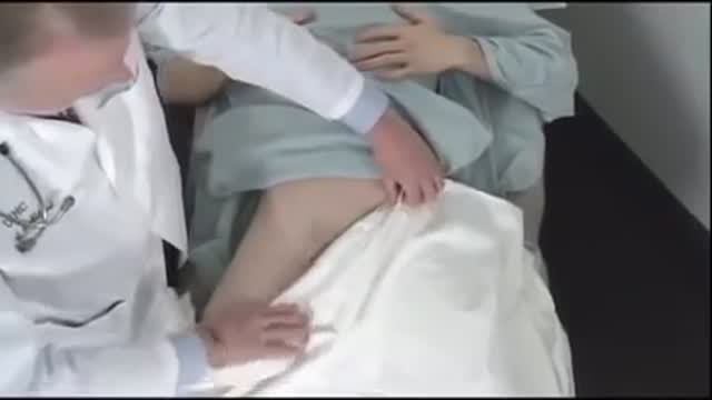
Note any large area of dullness that might suggest a mass or enlarged organ. Briefly percuss the lower anterior chest. On the right, liver dullness is usually present. On the left, you may hear the tympani of the gastric air bubble. Help aid the abdomen, beginning gently and saving painful areas for last. With your fingers together, place your hand flat on the abdomen and press, using a light dipping motion. Moving smoothly, feel in all quadrants identifying any tenderness or increased resistance to your hand. When resistance is present, try to relax the patient and palpate gently again.
Just let me know if anything's uncomfortable. Then palpate more deeply in all four quadrants as you feel for any masses or tenderness. One hand on top of the other may make it easier to feel deeply. If you feel a structure that suggests a segment of colon, roll it under your fingers in one direction, then another, and try to assess its shape. Here the sigmoid colon is palpable. Abdominal pain, tenderness, and involuntary muscular rigidity suggests peritoneal irritation. To localize it, ask the patient to cough and then show you where it hurts.
Then try to localize the tenderness with one finger. If necessary, feel for rebound tenderness. Now I'm going to push down here and I'm going to let go very quickly. And I want you to tell me whether it hurts more when I push down or when I let go. Press your fingers in firmly and slowly, then quickly withdraw them. Watch and listen for signs of pain. It hurts more when you push down. Unlike in this patient, pain induced or worsened by withdrawal is rebound tenderness and suggests peritoneal inflammation.
Transcript auto-generated from the video’s audio for accessibility and study. Educational content only — not medical advice.
Medical Videos
Free medical and surgery videos for patients, students and professionals — trusted since 2007.
Founded by Dr. Mohamed Ibrahim, MD, PhD — physician-scientist.
Follow us on Facebook · Instagram
SORT BY-
Top Comments
-
Latest comments