
- Physical Examination
- Surgical Examination
- Ophthalmology
- Clinical Skills
- Orthopedics
- Surgery Videos
- Laparoscopy
- Pediatrics
- Funny Videos
- Cardiothoracic Surgery
- Nursing Videos
- Plastic Surgery
- Otorhinolaryngology
- Histology and Histopathology
- Neurosurgery
- Dermatology
- Pediatric Surgery
- Urology
- Dentistry
- Oncology and Cancers
- Anatomy Videos
- Health and Fitness
- Radiology
- Anaesthesia
- Physical Therapy
- Pharmacology
- Interventional Radiology
- Cardiology
- Endocrinology
- Gynecology
- Emergency Medicine
- Psychiatry and Psychology
- Childbirth Videos
- General Medical Videos
- Nephrology
- Physiology
- Diet and Food Health
- Diabetes Mellitus
- Neurology
- Women Health
- Osteoporosis
- Gastroenterology
- Pulmonology
- Hematology
- Rheumatology
- Toxicology
- Nuclear Medicine
- Infectious Diseases
- Vascular Disease
- Reproductive Health
- Burns and Wound Healing
- Kidney Zone Podcast
- Other


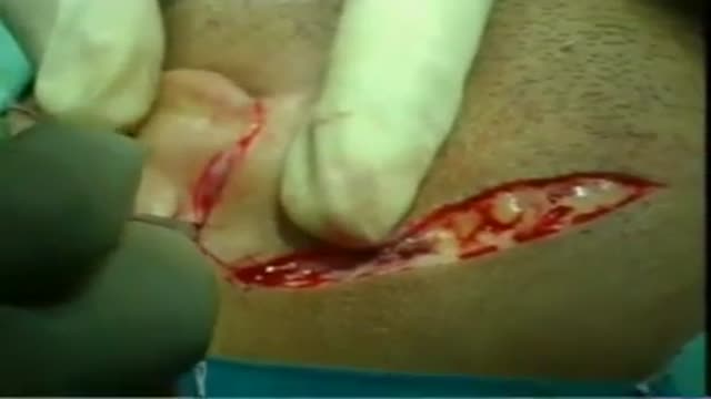
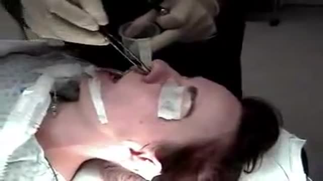
Endoscopic Nasal Septoplasty

Educational only: videos on MedicalVideos.com are for general information and are not medical advice. Always consult your own doctor about your health.
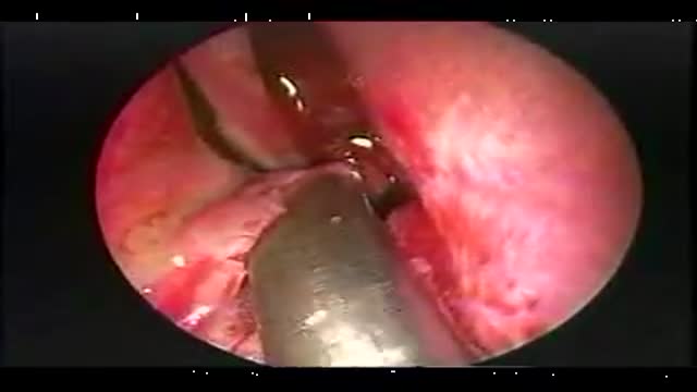
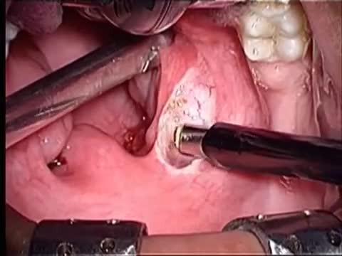
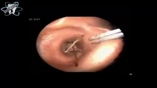
The endoscopic resection of a sharp bony nasal septal spur
This video in 30 seconds
A narrated endoscopic septoplasty shows removal of a sharp septal spur beneath the left middle turbinate. The surgeon describes raising the lining tissue from both surfaces of the spur and freeing it from the vomer with a chisel. The spur is then removed with cup forceps and the flaps are laid back with temporary packing.
Key points
- An incision is made over the end of the septal spur
- The lining is elevated from the upper and lower surfaces of the spur
- A chisel worked with a two-tap technique frees the spur from the vomer
- The freed spur is grasped and removed with cup forceps
- The flaps are repositioned and packing is placed at the end
Chapters
- 0:01 — The septal spur presentation
- 0:10 — Raising the mucosal flaps
- 1:10 — Freeing the spur with a chisel
- 1:56 — Removing the spur with forceps
- 2:19 — Repositioning flaps and packing
Video Transcript
A narrated endoscopic septoplasty in which a sharp septal spur beneath the left middle turbinate is removed. The surgeon describes raising the mucoperichondrium and mucoperiosteum from both surfaces of the spur, freeing it from the vomer with a chisel worked in a two-tap technique, removing it with cup forceps, and laying the flaps back with temporary packing.
Read the full transcript
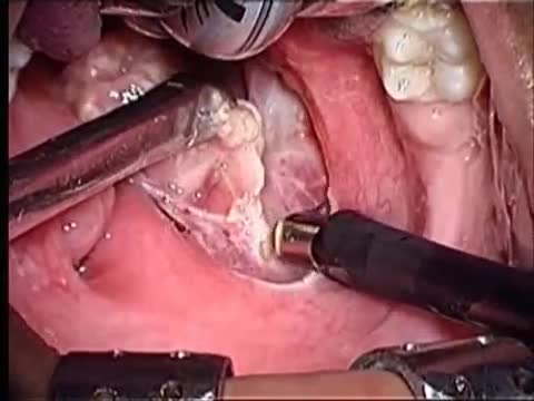
This patient presented with a sharp septal spur on the left side protruding underneath the left middle terminate.
After an incision over the end of the spur was made with a sickle knife, a fur elevator was used to gently raise the mucopericondrium and mucoperiosteum.
Notice how the mucosa and the pericondrium is being elevated on the superior surface. And then the lower surface of the sharp edge of the spur has its mucopericondrium elevated by working underneath the septal spur.
This spur presented with a very sharp keel-like edge and was being removed both for access to the nasal cavity and to relieve obstruction. The sub-pericondrial dissection is continued superiorly and inferiorly until enough of the spur is exposed to enable a straight chisel to be inserted.
The surgeon guides the chisel along the base of the spur while the assistant gently taps using a two-tap light heavy light heavy technique. You can see after a few taps the spur is now become freed
anteriorly, but there is still a superior and posterior attachment to the vulmar which must be divided. The chisel is angled slightly and
continued to be used to free the spur from its attachment to the vulmar. Once the spur has been freed it was grabbed with cup foresups and gently removed from the nasal cavity.
Notice the spur extending above and the flat thin plate of vulmar.
You can see how this spur would cause nasal obstruction as well as prevent access to the middle mucus. At the end of the procedure the flaps are simply placed back in position and held there with temporary packing.
Transcript auto-generated from the video’s audio for accessibility and study. Educational content only — not medical advice.
Video Overview
From our Plastic Surgery collection: Rhinoplasty, liposuction, breast augmentation, facelifts and reconstructive procedures performed step by step. Browse all Plastic Surgery videos →
Medical Videos
Free medical and surgery videos for patients, students and professionals — trusted since 2007.
Founded by Dr. Mohamed Ibrahim, MD, PhD — physician-scientist.
Follow us on Facebook · Instagram
SORT BY-
Top Comments
-
Latest comments