
- Physical Examination
- Surgical Examination
- Ophthalmology
- Clinical Skills
- Orthopedics
- Surgery Videos
- Laparoscopy
- Pediatrics
- Funny Videos
- Cardiothoracic Surgery
- Nursing Videos
- Plastic Surgery
- Otorhinolaryngology
- Histology and Histopathology
- Neurosurgery
- Dermatology
- Pediatric Surgery
- Urology
- Dentistry
- Oncology and Cancers
- Anatomy Videos
- Health and Fitness
- Radiology
- Anaesthesia
- Physical Therapy
- Pharmacology
- Interventional Radiology
- Cardiology
- Endocrinology
- Gynecology
- Emergency Medicine
- Psychiatry and Psychology
- Childbirth Videos
- General Medical Videos
- Nephrology
- Physiology
- Diet and Food Health
- Diabetes Mellitus
- Neurology
- Women Health
- Osteoporosis
- Gastroenterology
- Pulmonology
- Hematology
- Rheumatology
- Toxicology
- Nuclear Medicine
- Infectious Diseases
- Vascular Disease
- Reproductive Health
- Burns and Wound Healing
- Kidney Zone Podcast
- Other



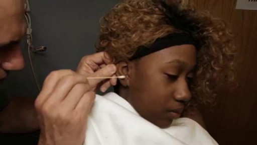
Ear Examination

Educational only: videos on MedicalVideos.com are for general information and are not medical advice. Always consult your own doctor about your health.
Complete clinical examination of the ears with all the associated tests
This video in 30 seconds
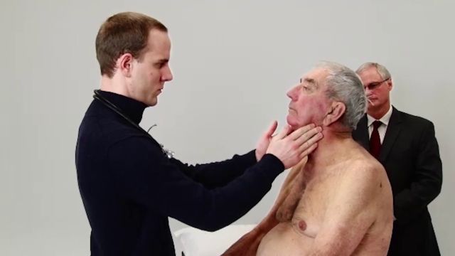
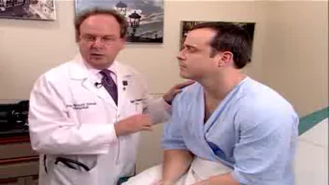
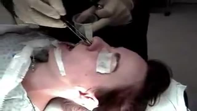
A bedside teaching examination of the ears is performed on a patient in this examination series. The examiner inspects and palpates the external ear, tests hearing first by whisper and then with tuning-fork tests, and demonstrates the otoscopic examination. The session finishes with an inspection of the nose and sinuses.
Key points
- The external ear is inspected and palpated for tenderness
- Hearing is first tested with a whispered voice
- Tuning-fork tests compare air and bone conduction in both ears
- The otoscope is used to view the canal and tympanic membrane
- The nose and sinuses are inspected at the end
Chapters
- 0:00 — Inspecting the external ear
- 0:42 — Whisper test for hearing
- 1:07 — Tuning-fork tests: Rinne and Weber
- 2:56 — Otoscopic examination demonstrated
- 4:39 — Inspecting nose and sinuses
Video Transcript
A bedside teaching examination of the ears, performed on the 'Mr. Johnson' patient of this examination series. The examiner inspects and palpates the external ear, tests hearing first by whisper and then with the tuning-fork tests (Rinne, then Weber), explaining the technique as he goes, and finishes with an inspection of the nose and sinuses.
Read the full transcript
The first part of the examination of the ears is inspection of the external ear. One looks at the external ear and notices the helix, the antihelix, the tragus, the antitragus, and the external auditory canal. We want a palpate also. Mr. Johnson, has this heard at all? Okay. Tenderness by pulling on the lobule or the antitragus will provide pain if the person is having an acute otitis external. We're now going to evaluate auditory acuity. And there are basically two ways of doing that. One by whispering something in the patient's ear and the second is a 244 test.
Mr. Johnson, would you take your hand and place it in your external canal and tell me what I've said? Train. Okay. And if you now include the other canal? Possibly. Okay. We're now going to proceed with the evaluation of auditory acuity by a tuning fork test. Typically one uses a 512 Hertz tuning fork. This happens to be a 128, which is certainly acceptable, but a 512 is generally the one which is used. There are two tuning fork tests, the Weber and the RINY test. I'm going to demonstrate the RINY test first.
Strike the tuning fork by placing it into oscillation, by striking the heel of your hand, and then placing the vibrating tuning fork on the mastoid process of the patient. Mr. Johnson, do you hear or feel anything? Yes. Okay. Tell me when you stop hearing it or feeling it. It stops. Okay. What about now? I can hear it. Okay. So air conduction is greater than bone conduction, and that's normal. AC greater than BC. The examination is conducted in the other ear as well. And do you hear or feel anything?
Mm-hmm. Tell me when you stop hearing it. Okay. What about now? I can hear it. Okay. So air conduction is greater than bone conduction, bilaterally. The second test is the Weber test. And one strikes the tuning fork and places it midline on the patient's forehead. And the patient is asked, do you hear or feel anything? Yes. Okay. Tell me what you hear or feel. Vibration. Okay. Where do you feel it? Both sides of my head. Does it feel equal, or is it in one place louder than another?
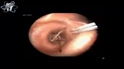
No. It feels equal. We call that no lateralization, and that is the normal Weber test. I'm now going to demonstrate the otoscopic examination. The otoscopic head is put onto the handle, and the speculum, the largest diameter speculum, is used. In order to visualize the patient's tympanic membrane and external auditory canal in the right ear, the examiner places his left hand on the mastoid process and pulls up, out, and back, thereby straightening the external canal. The instrument may either be held this way to enter the canal, or this way.
Either way is acceptable. The most important part, though, is pulling up, out, and back to straighten the canal. In doing such, you will straighten the canal, place the speculum into the external auditory canal, and visualize the external canal and the tympanic membrane. You'll notice the malleus, the umbo, the light reflex pointing anteriorly, and perhaps other ossicles in the middle ear. If the examiner is on the left ear, the examiner uses his or her right hand to pull on the tragus and ear lobe up, out, and back,
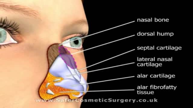
and the otoscope can be entered into the external canal either as was demonstrated on the other ear or held in this manner, and the speculum is inserted as I'm demonstrating now. We're next going to inspect the nose and the nasal skeleton as well as the sinuses. Any tenderness here? No. Mr. Johnson? Mm-hmm. Any tenderness here? No. Tenderness here? No. So what about here? Any tenderness here? No. This is evaluation of the frontal, maxillary, and ethmoid sinuses. Evaluation of the nose is done by having the person extend the neck and using a light source,
and elevating the tip of the nose, looking in the narrowings to see the position of the septum, and if there's any discharge or separation of the septum. To examine the left ear, the examiner uses his or her right hand to pull on the tragus and ear lobe up, out, and back, and the otoscope can be entered into the external canal either as was demonstrated on the other ear or held in this manner, and the speculum is inserted as I'm demonstrating now. We're next going to inspect the nose and the nasal skeleton as well as the sinuses.
Any tenderness here? No. Mr. Johnson? Mm-hmm. Any tenderness here? No. Tenderness here? No. So what about here? Any tenderness here? No. This is evaluation of the frontal, maxillary, and ethmoid sinuses. So the evaluation of the nose is done by having the person extend the neck and using a light source and elevating the tip of the nose, looking in the narrowings to see the position of the septum, and if there's any discharge or separation of the septum.
Transcript auto-generated from the video’s audio for accessibility and study. Educational content only — not medical advice.
Video Overview
From our Physical Examination collection: Watch physical examination videos step by step — cardiovascular, respiratory, abdominal and neurological exams demonstrated for medical students and clinicians. Browse all Physical Examination videos →
Medical Videos
Free medical and surgery videos for patients, students and professionals — trusted since 2007.
Founded by Dr. Mohamed Ibrahim, MD, PhD — physician-scientist.
Follow us on Facebook · Instagram
SORT BY-
Top Comments
-
Latest comments