
- Physical Examination
- Surgical Examination
- Ophthalmology
- Clinical Skills
- Orthopedics
- Surgery Videos
- Laparoscopy
- Pediatrics
- Funny Videos
- Cardiothoracic Surgery
- Nursing Videos
- Plastic Surgery
- Otorhinolaryngology
- Histology and Histopathology
- Neurosurgery
- Dermatology
- Pediatric Surgery
- Urology
- Dentistry
- Oncology and Cancers
- Anatomy Videos
- Health and Fitness
- Radiology
- Anaesthesia
- Physical Therapy
- Pharmacology
- Interventional Radiology
- Cardiology
- Endocrinology
- Gynecology
- Emergency Medicine
- Psychiatry and Psychology
- Childbirth Videos
- General Medical Videos
- Nephrology
- Physiology
- Diet and Food Health
- Diabetes Mellitus
- Neurology
- Women Health
- Osteoporosis
- Gastroenterology
- Pulmonology
- Hematology
- Rheumatology
- Toxicology
- Nuclear Medicine
- Infectious Diseases
- Vascular Disease
- Reproductive Health
- Burns and Wound Healing
- Other
Top videos
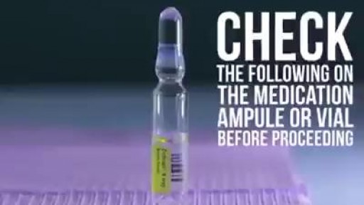

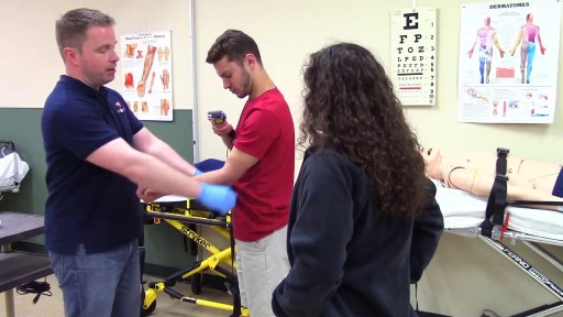
Learn how to give an intramuscular injection

The eyes A close up of a young person's eyes. The eyes are responsible for four-fifths of all the information our brain receives. Here you can find out a bit more about how they work, common problems that affect vision and the work Sightsavers does to treat and prevent avoidable blindness. You can also find out more about the people whose lives have been changed thanks to donations from people like you. How do eyes work? (click image to see enlarged version or click here for text alternative) Graphic of an eye with information about its different parts The images we see are made up of light reflected from the objects we look at. This light enters the eye through the cornea. Because this part of the eye is curved, it bends the light, creating an upside down image on the retina (this is eventually put the right way up by the brain). The retina is a complex part of the eye, but only the very back of it is light sensitive. This part of the retina has roughly the area of a 10p coin, and is packed with photosensitive cells called rods and cones. Cones are the cells responsible for daylight vision. There are three kinds – each responding to a different wavelength of light: red, green and blue. The cones allow us to see images in colour and detail. Rods are responsible for night vision. They are sensitive to light but not to colour. In darkness, the cones do not function at all. How do we see an image? The lens focuses the image. It can do this because it is adjustable – using muscles to change shape and help us focus on objects at different distances. The automatic focusing of the lens is a reflex response and is not controlled by the brain. Once the image is clearly focused on the sensitive part of the retina, energy in the light that makes up that image creates an electrical signal. Nerve impulses can then carry information about that image to the brain through the optic nerve.
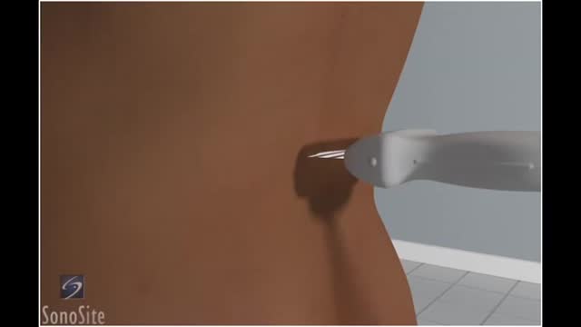
Lumbar puncture is a common emergency department procedure used to obtain information about the cerebrospinal fluid (CSF) for diagnostic and, less commonly, therapeutic reasons. Please refer to the full article on Lumbar Puncture for more details on the lumbar puncture procedure. Lumbar puncture is typically performed via “blind” surface landmark guidance. The surface landmark technique is reported to be successful in a high percentage of attempted lumbar punctures; however, surface landmark identification of underlying structures has been shown to be accurate only 30% of the time. [1] Unsuccessful identification of proper landmarks often leads to increased difficulty in obtaining CSF, if the procedure is performed, and a higher rate of complications. Few alternatives are available in these cases. If available, fluoroscopic-guided lumbar puncture may be performed. If not, treatment is sometimes initiated empirically without obtaining CSF. Disadvantages of using fluoroscopy include limited availability or necessary transport of the patient outside of the emergency department, inability to directly visualize the spinal canal, and inherent radiation exposure
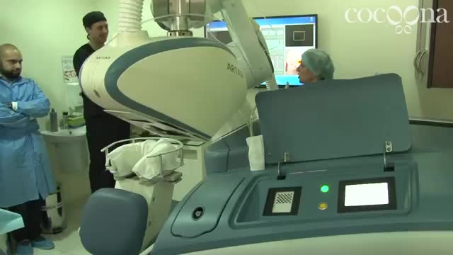
Introducing ARTAS in Cocoona Delhi & Dubai by Dr Sanjay Parashar - Hair Transplant Robot
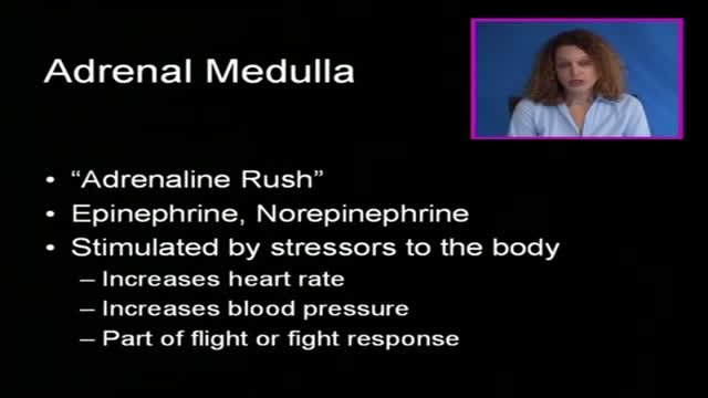
A pheochromocytoma (fee-o-kroe-moe-sy-TOE-muh) is a rare, usually noncancerous (benign) tumor that develops in cells in the center of an adrenal gland. You have two adrenal glands, one above each kidney. Your adrenal glands produce hormones that give instructions to virtually every organ and tissue in your body. If you have a pheochromocytoma, an adrenal gland releases hormones that cause persistent or episodic high blood pressure. If left untreated, a pheochromocytoma can result in severe or life-threatening damage to other body systems, especially the cardiovascular system. Most people with a pheochromocytoma are between the ages of 20 and 50, but the tumor can develop at any age. Surgical treatment to remove a pheochromocytoma usually returns blood pressure to normal.
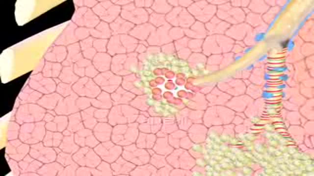
Emphysema gradually damages the air sacs (alveoli) in your lungs, making you progressively more short of breath. Emphysema is one of several diseases known collectively as chronic obstructive pulmonary disease (COPD). Smoking is the leading cause of emphysema. Your lungs' alveoli are clustered like bunches of grapes. In emphysema, the inner walls of the air sacs weaken and eventually rupture — creating one larger air space instead of many small ones. This reduces the surface area of the lungs and, in turn, the amount of oxygen that reaches your bloodstream. When you exhale, the damaged alveoli don't work properly and old air becomes trapped, leaving no room for fresh, oxygen-rich air to enter. Treatment may slow the progression of emphysema, but it can't reverse the damage.

Mommy Makeover Plastic Surgery Boca Raton FL
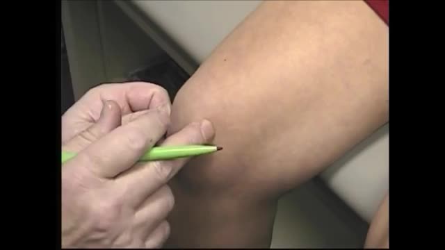
When oral medications do not relieve knee pain, but you're not to the point of pursuing knee surgery, one of the following injections or procedures may help. Hyaluronic acid supplements – Although not technically medications, these substances are injected into knee joints to supplement naturally occurring hyaluronic acid. In healthy joints hyaluronic acid acts as a shock absorber and lubricant, allowing joints to move smoothly over each other. However, the acid appears to break down in people with osteoarthritis. Injecting it into a joint may lessen pain and inflammation. The injections are given weekly for three or five weeks, depending on the product (examples are Synvisc and Hyalgan). A small amount of joint fluid is removed first to make room for the hyaluronic acid. Corticosteroid Injections – Doctors sometimes inject corticosteroids directly into the knee joint for quick relief of pain and inflammation. Their benefits may last anywhere from a few days to more than six months. While the injections bring targeted relief to the joint and lack many of the side effects of oral corticosteroid medications, they are not without risks. Repeated knee injections may actually contribute to cartilage breakdown. For that reason your doctor will likely put a limit on the number of injections you can receive. Read a report from the British Medical Journal on corticosteroid injections for knee osteoarthritis. Arthrocentesis – Also called joint fluid aspiration, arthrocentesis is removal of joint fluid through a hollow needle inserted into the joint space of the knee. Although the purpose of removing joint fluid from the knee is usually so that it can be tested in the lab, removing excess fluid can also quickly ease pain and swelling. Often after withdrawing fluid, doctors use the same puncture site where the fluid was removed to inject a corticosteroid preparation and/or anesthetic into the knee joint to further relieve pain and inflammation.
When you’re depressed, it can feel like you’ll never get out from under a dark shadow. However, even the most severe depression is treatable. So, if your depression is keeping you from living the life you want to, don’t hesitate to seek help. Learning about your depression treatment options will help you decide what approach is right for you. From therapy to medication to healthy lifestyle changes, there are many effective treatments that can help you overcome depression and reclaim your life.

Every day, specialists deliver high-quality care in 68 disciplines in health centres across Canada. Yet many Canadians know very little about what many specialists actually do, and the important role these disciplines play in Canada’s health care system.
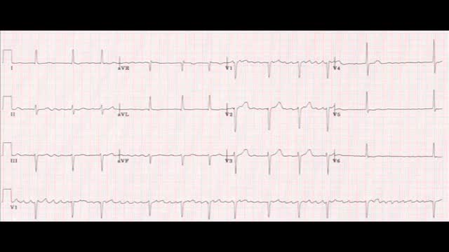
AFib is caused by abnormal electrical impulses in the atria, which are the upper chambers of the heart. The result is a rapid and irregular pumping of blood through the atria. These chambers fibrillate, or quiver, rapidly.

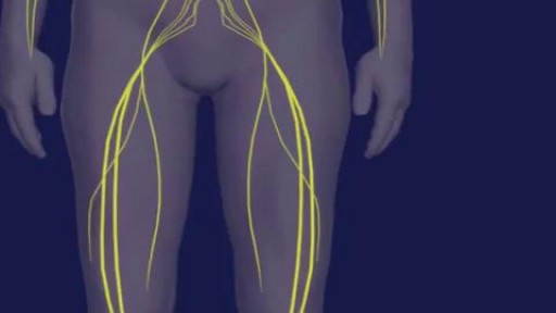
Chemokuren geven neuropathische pijnen. Behandeling met het supplement palmitoylethanolamide en topicale analgetische creme geven het volgende resultaat
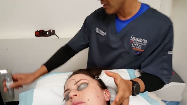
What is a mole? Many people refer to a mole as any dark spot or irregularity in the skin. Doctors use different terms. But the following types of skin marks such as these are not treated the same way moles are and are not discussed here: Birthmarks Abnormal formations of blood vessels (hemangiomas) Keratoses (benign or precancerous spots, which appear after about age 30 years)

Woman suffers allergic reaction to nut protein in boyfriend's sperm.A British woman has become the first recorded case of someone suffering an allergic...
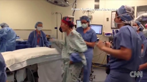
Cancer Patient Dances in OR before her Surgery
If a patient comes to you with a painful, throbbing, swollen, red face (a ''fat face'), perhaps with fever, trismus and lymphadenitis, he is probably suffering from an acute dental or oral infection, most probably an alveolar abscess. He may have: (1) An alveolar abscess begins as an infection in the bone around a non-vital infected tooth. He has severe pain, which becomes less as pus is released into more superficial tissues and his face starts to swell. After 36 hours of cellulitis he usually has a fluctuant abscess which needs draining. If drainage is delayed, the pus in his abscess discharges spontaneously through a sinus (26-8) in his gum or face, which may become chronic. First, control infection with antibiotics, and then drain the abscess, either by incising it where it is pointing, or by removing the infected tooth, which acts as a cork to prevent the pus escaping, or by doing both these things. If you remove a tooth before you have controlled the infection with antibiotics, and while his face is still severely swollen, you may spread the infection; your task will also be more difficult. (2) A periodontal abscess at the side of a tooth, caused by spread from an infected gum. (3) A pericoronal abscess caused by infection of the gum over the crown of an unerupted and impacted tooth, usually a lower third molar (''an infected wisdom tooth'). Often, an abscess does not form, and the gum round the tooth is merely inflamed.

Sliced Fingertip Makeup Tutorial
