
- Physical Examination
- Surgical Examination
- Ophthalmology
- Clinical Skills
- Orthopedics
- Surgery Videos
- Laparoscopy
- Pediatrics
- Funny Videos
- Cardiothoracic Surgery
- Nursing Videos
- Plastic Surgery
- Otorhinolaryngology
- Histology and Histopathology
- Neurosurgery
- Dermatology
- Pediatric Surgery
- Urology
- Dentistry
- Oncology and Cancers
- Anatomy Videos
- Health and Fitness
- Radiology
- Anaesthesia
- Physical Therapy
- Pharmacology
- Interventional Radiology
- Cardiology
- Endocrinology
- Gynecology
- Emergency Medicine
- Psychiatry and Psychology
- Childbirth Videos
- General Medical Videos
- Nephrology
- Physiology
- Diet and Food Health
- Diabetes Mellitus
- Neurology
- Women Health
- Osteoporosis
- Gastroenterology
- Pulmonology
- Hematology
- Rheumatology
- Toxicology
- Nuclear Medicine
- Infectious Diseases
- Vascular Disease
- Reproductive Health
- Burns and Wound Healing
- Kidney Zone Podcast
- Other

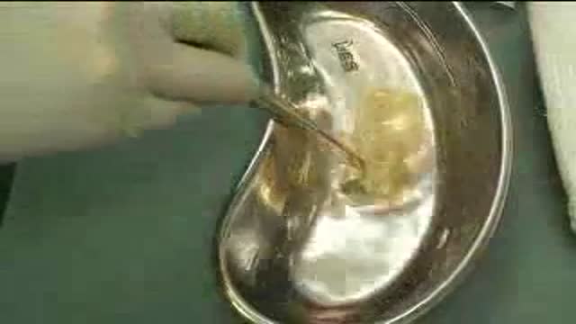



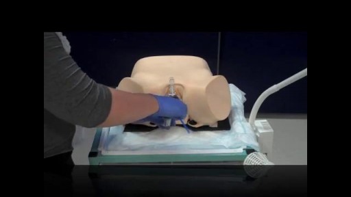
Anterior Elevate Mesh Repair performed by Dr. Robert Moore and Dr. John Miklos

Educational only: videos on MedicalVideos.com are for general information and are not medical advice. Always consult your own doctor about your health.
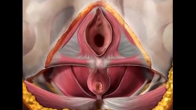
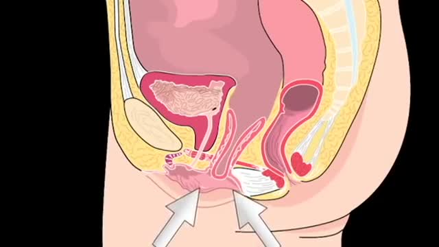
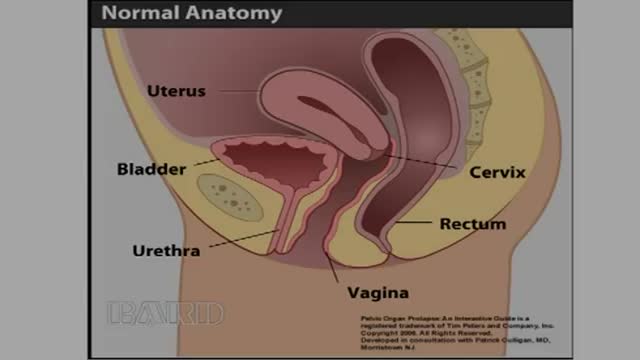
Anterior vaginal wall relaxation (cystocele) is one of the most commonly diagnosed forms of pelvic organ prolapse in women. More than 200,000 cystocele repairs are completed yearly, however to date the procedures that are completed do not provide very high cure rates and/or poor anatomic outcomes. Successful treatment of anterior vaginal wall prolapse remains one of the most challenging aspects of pelvic reconstructive surgery we face. We have developed very good procedures that provide excellent support for the posterior wall (ie rectoceles) and the apex of the vagina (ie vaginal vault prolapse) and reproduce normal anatomy. We were one of the first centers in the country to utilize grafts in rectocele repairs and have seen improved cure rates to over 90% with minimal complications. It has been known for many years that abdominal sacralcolpopexy with placement of a mesh graft at the top of the vagina for vaginal vault prolapse is the most successful procedure in the literature. We have made advancements with this procedure as well in being able to offer our patients a laparoscopic minimally invasive approach for sacralcolpopexy, with the same excellent cure rates (>92%) and with hospital stays typically less than 24 hours and reduced complications. However the anterior wall has been one of the most difficult compartments in the vagina to get good anatomic results and high cure rates with traditional repairs and at the same time not cause sexual dysfunction, pain with intercourse, voiding dysfunction (ie incontinence or urgency/frequency syndrome), or a shortened or scarred down vagina. The transobturator approach was developed as a less invasive way to place an anterior wall graft (see below) however this still involved blind needle passes and the graft did not support the apex of the vagina, therefore the search for improvements in these procedures is ongoing.
Video Transcript
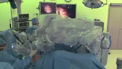
Drs. Robert Moore and John Miklos present their anterior Elevate mesh repair for cystocele (anterior vaginal wall prolapse), recorded at a surgical meeting in Lake Como, Italy. The narrated presentation explains the minimally invasive vaginal approach and where it fits among prolapse repair options.
Read the full transcript
Hi, I'm Dr. Robert Moore of the Atlanta Center of Leproscopic Urogyne Ecology in Atlanta, Georgia. My partner, Dr. John Miklos, and myself have one of the busiest practices in the United States in laparoscopic and minimally invasive vaginal reconstructive surgery. We're actually here in Lake Como, Italy to present our latest surgical technique in the advancements and the minimally invasive treatments of vaginal prolapse utilizing a mesh procedure vaginally. I actually was just traveling in Poland and we were the first to do this interior elevate procedure in the country of Poland. We actually helped bring the technology to the United States.
We actually helped develop the technology and the procedure in the pursuit of finding a less invasive procedure to treat cysticeal and vaginal prolapse with a minimally invasive vaginal procedure. The procedure itself involves a one small incision vaginally and the placement of an anterior wall vaginal mesh procedure. We've actually been invited by the International Urogyne Ecology Association meeting that's taking place in Lake Como, Italy to present this new surgical technique. The following video will demonstrate this technique. Cysticeals or anterior wall prolapse occurs in many women concomitantly with uterine prolapse or vaginal vault prolapse.
In a recent Cochrane review, mesh has been shown to be beneficial in the treatment of cysticeals. However, procedures such as the transoperator perigee system, although giving excellent level 2 support, do not truly support level 1 or the apex. The elevate procedure has been developed as a less invasive method to place an anterior wall graft with no transoperator needle passes and also offering true level 1 support with a safe and strong attachment to the sacrospondous ligaments. The procedure has begun with infiltration of the anterior vaginal wall with a local anesthetic solution,
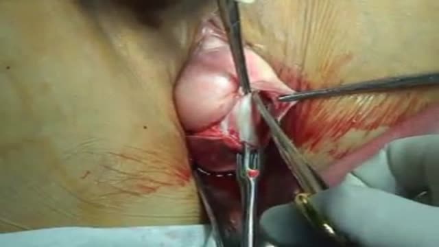
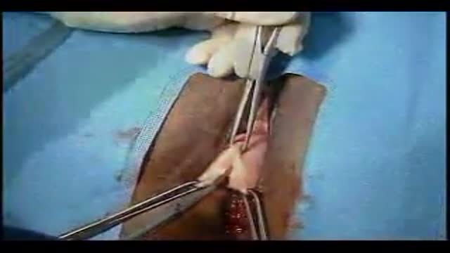
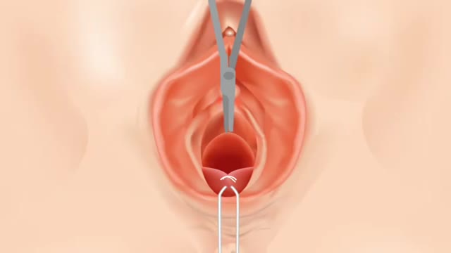
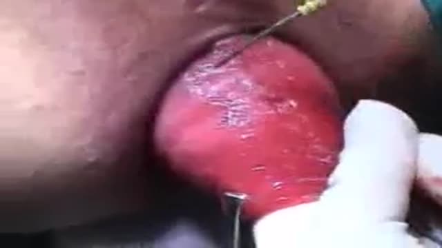
with epinephrine or another vasoconstrictor to not only help minimize bleeding during the dissection, but also for hydro dissection to allow us to get into a deeper dissection plane. This allows for a thicker vaginal epithelial flap to be created, which ultimately we believe helps minimize risk of mesh extrusion. The bladder is dissected off the vaginal epithelium out to the lateral sidewall, and then blunt dissection can be utilized to take the bladder off of the sidewall all the way up to the level of the eschial spine. The dissection is completed in an identical fashion on the patient's opposite side.
Again, the bladder is dissected away from the vaginal epithelium and out laterally just beyond the isopubic ramus at the level of the bladder neck, and again, the bladder is dissected away from the sidewall all the way up to the level of the eschial spine. The eschial spine is palpated and the sacrospondous ligament can then be isolated by taking the index finger and sweeping it medially from the eschial spine over to the sacrum, isolating and palpating the ligament. It's also very important to dissect the bladder off of the apex of the vagina or the cervix if the uterus is still in place.
The ligament can then be identified on the opposite side again by sweeping medially from the eschial spine to the sacrum. The body of the graft is made up of a type 1 macroporous monofilament soft polypropylene mesh that's actually 50% less dense and lighter than the original perigee mesh. The bladder neck arms of the mesh have a self-fixating tip on it that the anterior needle inserts into. The needle is then taken into the dissection and inserted into the pelvic sidewall into the obturator and ternus muscle just beyond the disengeting ischupubic ramus.
The arm is pushed into the midline of the graft is lined up with the urethra. The opposite side is completed in identical fashion. Again, the needle is inserted into the dissection and then pushed into the pelvic sidewall just beyond the disengeting ischupubic ramus into the obturator and ternus. It's taken in deep enough so as to not place any tension at the bladder neck. The distal portion of the graft is then held in place with a suture at the bladder neck. This suture helps prevent the graft from sliding out of position.
Sutures are then placed through the apex of the vagina or the peri cervical ring if the uterus is still in place. These sutures should either be permanent or delayed absorbable such as PDS. The length of the graft is then trimmed according to the patient's vaginal length. The mesh arm that will fixate to the sacrospinus ligament is then inserted onto the apical needle. The protective sheath is then slid over the needle and locked into place. This protects the tip from catching on tissue as it's passed into the vagina.
The needle is then designed in a shape for easy and safe access to the sacrospinus ligament. When the sheath retracts it allows the tip of the needle to be exposed for fixation into the sacrospinus ligament. The ligament is isolated and the needle is then brought up into the vagina. It is then placed approximately 2 centimeters, medial to the ischial spine, the trigger is pulled, the sheath retracts, and this allows the tip to be fixated down into the sacrospinus ligament, a safe distance away from the pedendal complex.
The other apical arm is then placed in an identical fashion. The arm is placed on the needle itself and the protective sheath then covers over the needle and the fixating tip. It's locked into place and then the needle is brought up into the vaginal dissection, the ischial spine isolated, and the needle placed approximately 2 centimeters medial from the ischial spine. The trigger on the handle is pulled, the sheath retracts, which allows the tip to penetrate into the sacrospinus ligament approximately 12 millimeters. These arms are then passed through the loosening eyelets at the top of the graft.
The previously placed apical sutures are then fed through the apex of the graft and these are tied into place. This attaches the top of the graft to the apex of the vaginal vault. The adjustment tool is then utilized to push the graft up into position onto the apical arms. Each side is done sequentially, elevating the vaginal vault up into position. If any tension is felt at the apex, it can be loosened prior to locking the graft into place. The graft is locked into place with locking eyelets that are slid down over the arms.
The adjustment tool is then utilized to push the eyelets up onto the mesh arms, locking the graft into place, and fixing the position of the apex of the vagina with a bilateral sacrospinus ligament suspension. The excess mesh and the mesh arm is then removed. Care should be taken to leave approximately 1 centimeter of mesh above the locking eyelet to ensure fixation. Little to no vaginal epithelium should be excised and then the vaginal incision can be closed with a running, delayed, absorbable suture. The anterior elevate system gives us a new minimally invasive technique to treat cystocelinal vault prolapse through a single incision
and a safe minimally traumatic fixation to the sacrospinus ligaments without any needle passes through the groins or buttocks.
Transcript auto-generated from the video’s audio for accessibility and study. Educational content only — not medical advice.
Video Overview
From our Gynecology collection: Pelvic exams, Pap smears, IUD insertion, hysteroscopy and women's health procedures explained step by step. Browse all Gynecology videos →
Medical Videos
Free medical and surgery videos for patients, students and professionals — trusted since 2007.
Founded by Dr. Mohamed Ibrahim, MD, PhD — physician-scientist.
Follow us on Facebook · Instagram
SORT BY-
Top Comments
-
Latest comments