
- Physical Examination
- Surgical Examination
- Ophthalmology
- Clinical Skills
- Orthopedics
- Surgery Videos
- Laparoscopy
- Pediatrics
- Funny Videos
- Cardiothoracic Surgery
- Nursing Videos
- Plastic Surgery
- Otorhinolaryngology
- Histology and Histopathology
- Neurosurgery
- Dermatology
- Pediatric Surgery
- Urology
- Dentistry
- Oncology and Cancers
- Anatomy Videos
- Health and Fitness
- Radiology
- Anaesthesia
- Physical Therapy
- Pharmacology
- Interventional Radiology
- Cardiology
- Endocrinology
- Gynecology
- Emergency Medicine
- Psychiatry and Psychology
- Childbirth Videos
- General Medical Videos
- Nephrology
- Physiology
- Diet and Food Health
- Diabetes Mellitus
- Neurology
- Women Health
- Osteoporosis
- Gastroenterology
- Pulmonology
- Hematology
- Rheumatology
- Toxicology
- Nuclear Medicine
- Infectious Diseases
- Vascular Disease
- Reproductive Health
- Burns and Wound Healing
- Kidney Zone Podcast
- Other
Chest drain insertion: What to Expect & Recovery
A chest drain removes air or fluid from around the lung through a tube between the ribs, letting the lung re-expand. It is usually inserted on the ward or in emergency care under local anaesthetic, and recovery is about the underlying cause — the drain itself typically stays for days while air leaks seal or fluid clears.
Before surgery: how to prepare
- Chest drains are often inserted urgently — the team will explain while preparing. Tell them about blood thinners, allergies and previous chest surgery or radiation, which change the approach.
- Imaging (X-ray, ultrasound or CT) guides placement; ultrasound guidance is standard for fluid drains.
- You will be positioned sitting up or on your side, and the area numbed; ask for pain relief before positioning if moving hurts.
- Understand the bottle system: the drain connects to an underwater-seal bottle that must stay below chest level and upright — this governs how you move around.
The day of surgery
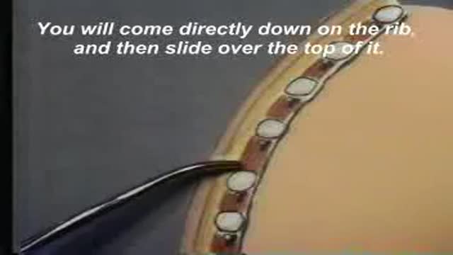
Under local anaesthetic (with sedation sometimes), a tube is inserted between the ribs into the pleural space and connected to an underwater-seal drain. A chest X-ray confirms position. Air bubbling or fluid draining shows it is working. Pain at the site is managed actively — good pain control lets you breathe deeply and cough, which is the real therapy for the lung.
In hospital
Recovery timeline
Every recovery is individual, but most follow this general arc:
Warning signs: when to get help
- Increasing breathlessness at any point, especially after drain removal (possible recurrence)
- New surgical emphysema — crackly swelling of the neck or chest skin — that is spreading
- The drain falling out, disconnecting, or the bottle being knocked over (cover the hole with a dressing and call staff immediately if in hospital; seek urgent help at home — drains are rarely managed at home)
- Fever, or drain fluid becoming cloudy with worsening chest pain (infection)
- Coughing up blood
Related topics
Watch: real operation videos
From the MedicalVideos library. Some surgical videos are graphic and intended for education.
Frequently asked questions
Why does the bottle have to stay below my chest?
The underwater seal is a one-way valve: air and fluid can leave your chest but cannot be sucked back in. Raising the bottle above chest level can siphon fluid back toward the lung. Keep it upright and below you, including in bed and when walking.
Will the pneumothorax come back?
Recurrence after a first spontaneous pneumothorax is a real possibility — which is why repeat episodes, or a persistent air leak, prompt discussion of a surgical procedure (pleurodesis or bullectomy) to fix the lung to the chest wall and prevent recurrence.
Medically reviewed by Dr. Mohamed Ibrahim, Assistant Professor of Medicine, Transplant Nephrology, University of Maryland School of Medicine. Last reviewed: 12 November 2026.
← More General & Other Surgery · All surgery recovery guides