
- Physical Examination
- Surgical Examination
- Ophthalmology
- Clinical Skills
- Orthopedics
- Surgery Videos
- Laparoscopy
- Pediatrics
- Funny Videos
- Cardiothoracic Surgery
- Nursing Videos
- Plastic Surgery
- Otorhinolaryngology
- Histology and Histopathology
- Neurosurgery
- Dermatology
- Pediatric Surgery
- Urology
- Dentistry
- Oncology and Cancers
- Anatomy Videos
- Health and Fitness
- Radiology
- Anaesthesia
- Physical Therapy
- Pharmacology
- Interventional Radiology
- Cardiology
- Endocrinology
- Gynecology
- Emergency Medicine
- Psychiatry and Psychology
- Childbirth Videos
- General Medical Videos
- Nephrology
- Physiology
- Diet and Food Health
- Diabetes Mellitus
- Neurology
- Women Health
- Osteoporosis
- Gastroenterology
- Pulmonology
- Hematology
- Rheumatology
- Toxicology
- Nuclear Medicine
- Infectious Diseases
- Vascular Disease
- Reproductive Health
- Burns and Wound Healing
- Kidney Zone Podcast
- Other
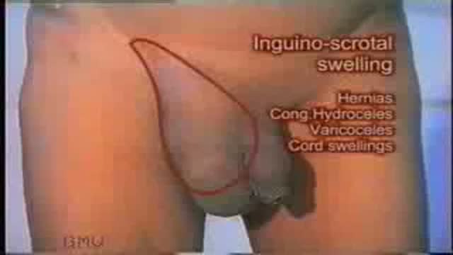
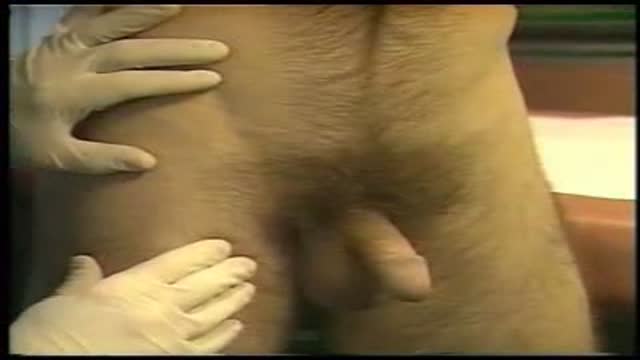
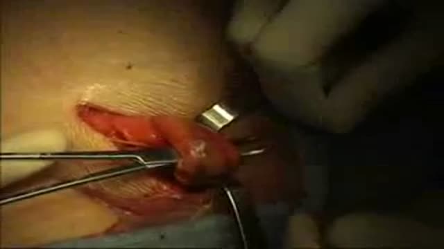
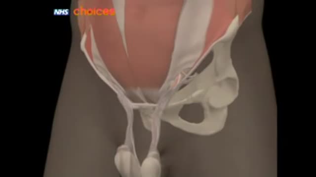



Scrotal swelling exam

Educational only: videos on MedicalVideos.com are for general information and are not medical advice. Always consult your own doctor about your health.
A testicular examination is mainly performed on male patients who present with testicular pathology e.g. pain, swelling, a lump. Although titled testicular examination it involves the examination of the penis, scrotum and testes. As this is an intimate examination it is pertinent to gain a good rapport with your patient, maintain good communication and ensure the patient’s dignity at all times. Remember to offer a chaperone for this skill. For the purposes of your exam, you will most likely be examining a mannequin.
This video in 30 seconds
This surgical-teaching narration demonstrates the clinical examination of a scrotal swelling. It works through inspection with the patient standing, palpation of the swelling, cord, and opposite side, and tests for fluctuation, transillumination, reducibility, and cough impulse. It finishes with examination of the abdomen and lymph nodes.
Key points
- Inspection in the standing position notes whether the swelling is unilateral, scrotal, or inguino-scrotal
- The shape, surface, and overlying skin of the swelling are inspected
- Palpation checks temperature, tenderness, consistency, and the opposite cord
- Fluctuation and transillumination are tested for cystic swellings
- Reducibility, cough impulse, and examination of abdomen and lymph nodes complete the assessment
Chapters
- 0:03 — Inspection of the swelling
- 4:23 — Palpation of the swelling
- 6:15 — Fluctuation and transillumination tests
- 10:29 — Reducibility and cough impulse
- 11:19 — Palpating testis and cord
- 13:36 — Abdomen and lymph node examination
Video Transcript
A classic surgical-teaching narration on the clinical examination of a scrotal swelling. It works through inspection with the patient standing, palpation of the swelling, cord, and opposite side, the tests for fluctuation, transillumination, reducibility, and cough impulse, and finishes with examination of the abdomen and lymph nodes.
Read the full transcript
Examination of a scrotal swelling. Let us begin the examination of a scrotal swelling with inspection. Let the patient stand with the scrotum and abdomen fully exposed. Inspection of a scrotal or invoino-scrotal swelling should always be done in standing position because the direct hernias and the varicoseal may be totally missed in lying down position and if hernia be present its full extent can be seen only in standing position. On inspection observe the swelling and note first whether it is on one side of the scrotum that is unilateral or both sides that is bilateral.
Second whether it is only in the scrotum that is scrotal or does it extend to the inguinal region that is invoino-scrotal. Third whether the testis is seen separate from the swelling as seen in this insisted hydrosil of the cord or is the testis incorporated within the swelling as in this testicular tumor or in a hydrosil. And lastly observe the penis whether it is pushed to the opposite side and whether it is partly buried in the swelling. Note how the penis is buried in the scrotum in this case of a large bilateral hydrosil
and in this unilateral hydrosil it is pushed to the opposite side. Now note the shape and surface of the swelling. Is it globular, retort shaped or irregular? Scrotal swellings that is testicular and epididymal swellings and hydrosils are usually globular and a hydrosil often shows a characteristic constriction around its central portion as shown by the arrows in these cases which is diagnostic of a tunica vaginalis hydrosil. Next measure the size of the swelling in centimeters in all three dimensions using a measuring tape. Now inspect the overlying skin.
Normal scrotal skin has rugositis which may be diminished in a hydrosil. Thick edymata skin which on palpation will be firm and non-tender is a feature of filarial elephantiasis. You can also see a ramshorn penis in this case due to filarial lymphedema of the penis. Redness and edema are a feature of scrotal wall cellulitis or as seen in this patient acute epididymal architis. Skin excoriation and vesicles are seen in urinary extroversation. Blackening and sloughing of the scrotal skin is a feature of four nearest gangrene which is an idiopathic gangrene of the scrotal skin secondary to infection and cellulitis.
Very large hydrosil may show friction ulcers on the lateral and posterior aspects due to friction with the thighs. Also look for ulcers, sinuses and scars as seen in tuberculous epididymitis. It is extremely important to lift the scrotum and inspect its posterior surface. Otherwise a posterior sinus or ulcer arising from the epididymus may be missed. Note the tuberculous sinus on the posterior wall seen on lifting the scrotum. Lastly ask the patient to cuff and look for visible expansile impulse in the swelling and over both the inguinal canals.
So on inspection we observe the swelling to note whether it is unilateral or bilateral, scrotal or inguino scrotal and its relation to the testis and penis. Then we noted its shape and surface noting in particular the central constriction of a hydrosil and measured the size of the swelling. Then we inspected the overlying skin for rugosities, redness, ulcers and sinuses and lastly we looked for a cuff impulse. Now let us proceed to palpation. As always start the palpation with temperature and tenderness. First look for any local rise of temperature over the swelling.
Pulpate the swelling with the back of the fingers first on the normal side and then over the swelling for comparison. Then pulpate over the swelling first very gently and then with slight firmness to look for tenderness. In a scrotal swelling the first point to be noted is whether the top of the swelling can be reached. Hold the root of the scrotum between the thumb in front and other fingers behind and pulpate the cord structures above the swelling by moving the thumb from medial to lateral side and feeling the cord as it slips medially.
Normal cord structures are felt as a whip cord like vas and multiple string like fibres of the cremaster muscle. In a unilateral scrotal swelling always pulpate the opposite normal cord and compare the thickness and feel. If only the cord structures are felt above the swelling it means that the top of the swelling can be reached. That is the swelling is entirely scrotal not inguino scrotal. Now pulpate the surface of the swelling and note whether it is smooth or nodular. Note the nodular uneven surface of a testicular tumor.
Then note the consistency of the swelling whether solid or cystic and lift the swelling to note its weight. Note the weight of the normal test is for comparison. Malignant testicular tumors feel very heavy as compared to cystic swellings like hydrocele. If the swelling has a cystic consistency then test for fluctuation, transillumination, reducibility and cuff impulse. Fluctuation is the presence of transmitted impulse in two planes at right angles to each other. Since the scrotum is very mobile fix the swelling between the ring and little fingers of both hands stretching the skin slightly.
Then keep the thumb and index fingers of the left and right hands at the upper and lower poles respectively. Apply intermittent pressure at the lower pole and note that the thumb and index fingers at the upper pole move apart from each other showing a positive fluctuation test and indication of presence of fluid in the swelling. This is another case of a large hydrocele. Observe the separation of the fingers at the upper pole. Transillumination. Transillumination is the demonstration of transmission of light through the swelling. If a dark room is available, transillumination should be tested in darkness.
Place a pencil torch on the lateral or medial aspect of the scrotal swelling and the whole swelling will be illuminated and show a red glow indicating positive transillumination and presence of clear fluid in the swelling. The walls of the swelling should also be thin and translucent. Thick and opaque walls may interfere with transillumination. If a dark room is not available, observe through a roll of black paper placed on the anterior medial or lateral side of the swelling holding the torch diagonally opposite. The roll of the paper will create a dark zone where the transillumination will be appreciated.
Note the positive transillumination as seen through the roll of paper whereas the same is not appreciated on the surrounding skin due to broad daylight. Two precautions should be observed while testing transillumination in scrotum. Firstly, do not place the torch posteriorly where the testists will block the light and the light will not reach the fluid. Note the positive transillumination with the torch applied laterally and negative transillumination when it is applied posteriorly. And secondly, if the skin is thin or if the swelling is small, first place the pencil torch on the normal side of the scrotum
to note the normal skin illumination in the patient. Then test over the swelling so that normal skin illumination is not mistaken for transillumination. Congenital hydrocelles in children are brightly transilluminant due to thin skin and thin transparent covering membranes. Epidermal cysts are also brilliantly transilluminant. Note this cyst of epididymis which is seen separate from the testists and is related to the head of the epididymis at the upper pole of the testists. It is brilliantly transilluminant. A multi-loculated cyst of epididymis gives a characteristic Chinese lantern type of transillumination.
The septae between the cysts cast shadows akin to the shadows cast by the wooden frame of the lantern and this is the typical Chinese lantern type of transillumination. This is another case of multi-loculated cyst of epididymis. Note the shadows of the septae and the Chinese lantern pattern. Sometimes a scrotal swelling contains fluid. It is fluctuent but shows negative transillumination. This is seen if the fluid is opaque for example, hematosil, pyosil or chylosil and if the wall is thick and opaque as in long standing hydrosil. Reducibility. Now raise the scrotum and compress the swelling gently towards the external inguinal ring.
Does it reduce in size? A hydrosil cannot be reduced. A varicoseal and a congenital hydrosil are scrotal swellings but are reducible. Now hold the swelling gently in the hand and ask the patient to cuff. Look and feel for an expansile impulse in the swelling. Then keep the hand over the inguinal canal and again ask the patient to cuff looking for an expansile cuff impulse of an associated hernia. Ask the patient to look away from you while cuffing so that the blast of air and sputum particles is not directed to you.
Having completed the palpation of the swelling, palpate the testis, epididymis and spermatic cord if they are separately palpable. In this case of hydrosil, the testis is not palpable but in a small hydrosil, testis may be felt within the swelling in the posterior portion. If the swelling is separate from the testis, palpate the head and tail of the epididymis to ascertain whether the swelling is arising from the epididymis or from the cord. If it appears to be in the cord above the testis, apply downward traction to the testis and see whether the swelling gets pulled down and becomes fixed.
This is the positive traction test. This swelling is an insisted hydrosil of the cord within the cord. Note that it becomes fixed when I pull the testis down. This is a positive traction test. Now palpate the cord. Normally you feel the firm whip cord like wasps and fibres of cremaster muscle felt as a number of strings. A nodular wasp is seen in tuberculosis. Thickened cord with a bag of worms feel is typical of a varicoseal. The dilated tortuous veins which become turgid on standing have the characteristic roundworm like feel, diagnostic of a varicoseal.
Next, palpate the opposite testis, epididymis and cord to confirm that they are normal. This completes the palpation of the scrotal swelling. To revise on palpation, first note the temperature and tenderness of the swelling. Then palpate the cord structures above the swelling to note whether the top of the swelling can be reached. Then note the surface weight and consistency of the swelling and its relation to the testis, epididymis and spermatic cord. If the swelling is cystic, test for fluctuation, transillumination, reducibility and cuff impulse. And lastly, palpate the opposite side also.
Now let us proceed to the focal and general examination. In supine lying down position, palpate the inguinal and external iliac lymph nodes on both sides. Examine the abdomen carefully for presence of any lump and organomegaly. In particular, in case of a varicoseal, look for a left renal mass. And in a case of malignant testis, look for parietic lymph nodes and also the left supraclavicular lymph nodes. In this case of a malignant testicular lump, note the heavy testis, the large parietic lymph nodes and the left supraclavicular lymph nodes.
If the patient has epididymorchitis, do a perectal examination for prostratitis and enlarged seminal vesicles. This concludes the clinical examination of a case of scrotal swelling.
Transcript auto-generated from the video’s audio for accessibility and study. Educational content only — not medical advice.
Video Overview
From our Surgical Examination collection: Pre-op assessments, wound and hernia exams, and clinical surgical skills demonstrated step by step by surgeons. Browse all Surgical Examination videos →
Medical Videos
Free medical and surgery videos for patients, students and professionals — trusted since 2007.
Founded by Dr. Mohamed Ibrahim, MD, PhD — physician-scientist.
Follow us on Facebook · Instagram
SORT BY-
Top Comments
-
Latest comments