
- Procedures & Tests
- Symptoms A-Z
- Symptom Checker
- Kidney Diet
- First Aid
- Anatomy Atlas
- Ask MedicalVideos
- Drug Guide
- Lab Values
- Vital Signs
- Muscles A–Z
- Clinical Signs
- Medical Abbreviations
- Medical Terminology
- Physical Examination
- Surgical Examination
- Ophthalmology
- Clinical Skills
- Orthopedics
- Surgery Videos
- Laparoscopy
- Pediatrics
- Funny Videos
- Cardiothoracic Surgery
- Nursing Videos
- Plastic Surgery
- Otorhinolaryngology
- Histology and Histopathology
- Neurosurgery
- Dermatology
- Pediatric Surgery
- Urology
- Dentistry
- Oncology and Cancers
- Anatomy Videos
- Health and Fitness
- Radiology
- Anaesthesia
- Physical Therapy
- Pharmacology
- Interventional Radiology
- Cardiology
- Endocrinology
- Gynecology
- Emergency Medicine
- Psychiatry and Psychology
- Childbirth Videos
- General Medical Videos
- Nephrology
- Physiology
- Diet and Food Health
- Diabetes Mellitus
- Neurology
- Women Health
- Osteoporosis
- Gastroenterology
- Pulmonology
- Hematology
- Rheumatology
- Toxicology
- Nuclear Medicine
- Infectious Diseases
- Vascular Disease
- Reproductive Health
- Burns and Wound Healing
- Kidney Zone Podcast
- Other
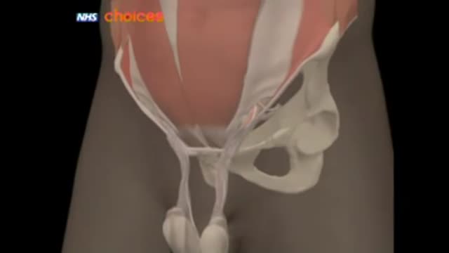



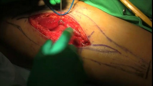
Inguino-Scrotal swelling

Educational only: videos on MedicalVideos.com are for general information and are not medical advice. Always consult your own doctor about your health.
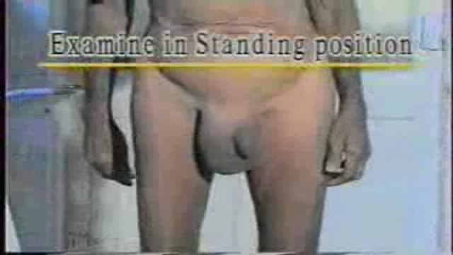
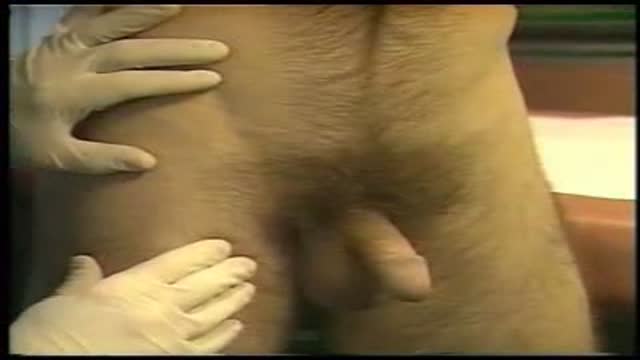
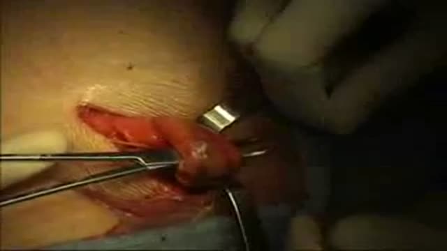
examination of a case of inguino-scrotal swelling as in cases of oblique inguinal hernia..etc
This video in 30 seconds
A clinical-examination lecture examines inguinoscrotal swelling, covering hernias, hydrocele, varicocele, and cord swellings. It works through inspection with the patient standing, palpation and reducibility, ring and canal tests, and related abdominal, prostate, and respiratory checks that complete the assessment.
Key points
- Inspection in the standing position covers shape, size, skin, and cough impulse
- Palpation covers cord structures, the pubic tubercle, consistency, and tenderness
- Reducibility is tested for different hernia types, including obstructed and strangulated presentations
- Internal and superficial ring tests and canal palpation are demonstrated
- Related checks include the abdominal wall, prostate examination, and respiratory system
Chapters
- 0:02 — Introduction to inguinoscrotal swellings
- 0:22 — Inspection with patient standing
- 4:15 — Palpation of cord and swelling
- 8:01 — Testing reducibility of hernia
- 10:35 — Internal ring occlusion test
- 14:36 — Percussion and auscultation findings
- 15:30 — Abdominal wall and related examinations
- 17:21 — Respiratory system examination
Video Transcript
A classic clinical-examination lecture on inguinoscrotal swelling, delivered in full over about eighteen minutes. It works through inspection with the patient standing (shape, size, skin), palpation and reducibility of hernias, and the related checks — abdominal scars, the urethral meatus, prostate examination in older patients, and the respiratory system — that complete the assessment.
Read the full transcript
Examination of an Inguyanoscrotal Swelling Inguyanoscrotal Swelling extends from the scrotum to the Inguyanal region and includes Inguyanal hernias, congenital and insisted hydrosil, varicoseal and chord swellings. Let us begin their study with inspection. To inspect an Inguyanoscrotal Swelling, the patient should be in a standing position stripped below the waist because the full extent of hernia can be seen only in standing position and direct hernias and varicoseals may not be seen at all in lying down position. So with the patient in standing position, first observe the shape and location of the
swelling and note whether it is unilateral or bilateral. This swelling is pyreformin shape, broad in the scrotum and narrow over the medial half of the Inguyanal ligament. This is the shape of an indirect hernia. Compare it with this direct hernia which is globular in shape over the medial half of the Inguyanal ligament, usually not entering the scrotum. This is a bilateral hernia. The left hernia is large and is standing to descend into the scrotum while the right is globular only over the medial half of the Inguyanal ligament.
Now measure its exact size in centimeters. In case of a hernia, the size should be measured when the hernia is fully distended by asking the patient to cuff. Next, inspect the skin overlying the swelling which would be normal in an uncomplicated hernia. Look for inflammation or redness, scars and discoloration. Redness and edema of skin suggest acute inflammation and in case of a hernia, acute strangulation. Note the red, tense and shiny skin over this strangulated hernia. This patient has a scar of previous hernia surgery indicating a recurrent hernia.
This is a thin scar but if the scar is irregular and ragged, think of postoperative wound infection as the probable cause of recurrence. And irregular pigmentation and discoloration may be seen in a patient wearing hernia truss for a long time. Now observe the swelling closely for visible peristalsis. An enterosil, that is hernia containing bowel loops, may show peristalsis especially when the covering layers are thin. Then ask the patient to cuff and look for a visible expansile impulse in the swelling. Note how the swelling expands during cuffing.
This is a positive cuff impulse diagnostic of an inguinal hernia. Of course, if the expansile impulse is not seen, that does not rule out hernia. An omentosil with adhesions as seen here or an obstructed or strangulated hernia will not show expansile cuff impulse. Lastly, observe the position of the penis. A hernia will push the penis to the opposite side. And a very large hernia as seen in this patient will bury the penis in the scrotal skin. To revise, on inspection, we first observed the shape and location of the swelling, noted
whether it is unilateral or bilateral and measured the exact size in centimeters. Then we observed the skin over the swelling for redness, scars and discoloration and looked for visible peristalsis in the swelling. Then we asked the patient to cuff and looked for an expansile impulse. And lastly, we noted the displacement or burial of the penis. Now let us proceed to the palpation. With the patient still in standing position, feel with the back of the fingers for temperature. First on the normal side to note the normal skin temperature and then over the swelling.
Then press over the swelling gently for tenderness before proceeding further with palpation. First, pulpit the cord structures between the thumb in front and other fingers behind the root of the scrotum. It is not possible to reach the top of the swelling in scrotum indicating that this is an inguino-scrotal swelling. Next, pulpit the pubic tubercle and confirm that the swelling is medial to it and above the inguinal ligament. A femoral hernia as seen in this female patient is lateral to the pubic tubercle and below the inguinal ligament.
Pulpit the testis and check whether the swelling stops above the testis or incorporates the testis in it. An acquired hernia will always stop above the testis. A congenital type of hernia will include the testis within its sac and the testis may also get reduced into the abdomen along with the intestines when the hernia is reduced. If the testis is absent and the scrotum is empty as seen here, then it is a case of undecended testis with hernia. Now feel the consistency of the swelling. A hernia feels soft and elastic when it contains intestines.
It is a little firm and doughy if it contains momentum and if the hernia is strangulated it is tense and tender. A bag of worms feel is characteristic of a varicose. Now note the soft and elastic enterosil, firm and doughy omentosil, tense and tender strangulated hernia and a bag full of worms feel in a varicose. The dilated tortuous veins seen in the cord above the testis look and feel like entangled round worms. Now ask the patient to turn his head to the opposite side and cuff forcefully.
Keep your hand over the swelling and feel for an expansile impulse on cuffing, that is the swelling increases in size and feels more tense during the act of cuffing. If the swelling is not visible, keep the hand over the medial half of the inguinal canal from superficial to deep inguinal rings and ask the patient to cuff. In a small hernia only a cuff impulse may be felt over the medial half of the inguinal canal. To differentiate the impulse of direct, indirect and femoral hernias, Ziemann's technique of
palpation is applied. Stand on the side of the hernia, reduce the hernia and place three fingers, index finger over the internal ring, middle finger over the external ring and ring finger over the femoral ring which is 4 cm below and lateral to the pubic tubercle. Now ask the patient to close the nose and mouth and blow. An impulse is felt by the index finger in indirect hernia, by the middle finger in direct hernia and by the ring finger in femoral hernia. Now test for reducibility. Ask the patient to lie down.
A direct hernia will usually reduce by itself on lying down. While an indirect hernia usually needs to be manually reduced. And a congenital hydrocele may not get reduced manually but it reduces slowly over several hours during sleep. To reduce the swelling, lift the scrotum in the left hand and apply even pressure with the right hand starting at the apex of the swelling and squeezing it towards the superficial in vinyl ring. If the contents are difficult to reduce, ask the patient to flex the thigh, adduct it and
rotate it internally to relax the pillars of the superficial in vinyl ring and the obliques. This is an obstructed hernia and it could not be reduced even when the patient was relaxed under anesthesia. Now observe how the hernia reduces. This hernia reduces with difficulty in the initial part and the last portion just slips in. This is typical of an enterosil containing coils of small intestine. Also note the gurgling sound heard and felt as the intestines in the enterosil get reduced. In this second case which is an omentosil, the first part reduces with ease but the
last part reduces with difficulty due to omental adhesions in the sac. If the hernia can be reduced only partly, then probably it is an omentosil with adhesions in the sac preventing the contents from returning to the abdomen. If the hernia cannot be reduced at all, then look for signs of obstruction and strangulation. This is a strangulated hernia. The skin is tense and shiny and it is tender. It contains gangrenous loops of bobble. This is an obstructed hernia in a child. Note the distended coils of intestines in the abdomen featuring a step-lager pattern.
After the swelling is reduced, perform the internal ring occlusion test. Place the hernia, place the thumb 1.25 cm above the mid-inguinal point to occlude the deep-inguinal ring completely. Mid-inguinal point is the midpoint between anterior superior iliac spine and the symphysis pubis. After occluding the deep-inguinal ring, ask the patient to cuff. If the hernia is indirect, it will not bulge out on occluding the deep-inguinal ring. This is the positive occlusion test. Note that the hernia does not come out when the internal ring is occluded, but when the
finger is removed and the patient cuffs, it comes out immediately. But if the hernia is direct, as you will see in this case, it will bulge out medial to the occluding finger on cuffing. This is the negative internal ring occlusion test. This is because the direct sag does not come out through the internal ring, but medial to it. After reducing the hernia with the patient in recumbent position, invaginate the scrotal skin over the index finger, starting near the bottom of the scrotum. Assess by the pubic tubercle.
Assess the size of the superficial-inguinal ring. Normally, it just admits one finger. It is dilated if the hernia is coming out of it. Now advance the finger through the superficial-inguinal ring into the inguinal canal. In an indirect-inguinal hernia, it goes along the inguinal canal, that is upwards, backwards and outwards. In a direct hernia, the finger will go directly backwards through the defect in the posterior wall of the inguinal canal. Then try to pulpit the superior remmers of ospeobis, which is felt there in a direct hernia, while it is difficult to feel in an indirect hernia where the transversal
is spacia is intact. Now keep the finger along the length of the inguinal canal and ask the patient to cuff. In an indirect hernia, the tip of the finger facing the internal ring feels the expansile impulse. While in a direct hernia, the expansile impulse is felt by the pulp of the finger, which is facing the posterior wall. As the patient cuffs, the inguinal canal is felt to close around the finger. Judge the sphincteric strength of the conjoined tendon from the force of the contracting muscle around the finger.
Thus the invagination test gives us a lot of information. The size of the superficial or external inguinal ring, direction of the hernia tract, direction of the expansile impulse and the sphincteric strength of the conjoined tendon. If the external ring is small, don't try to force your finger through it. Use the little finger or just abandon the test. Also this test is not performed in children. To revise on palpation, first look for a local rise of temperature and tenderness. Then palpate the core structures above the testes to confirm that the
swelling is inguino scrotal and note the relation of the swelling to the pubic tubercle and to the testes. Now palpate the swelling to note its consistency and compress it to the test reducibility. Ask the patient to cuff and look for a cuff impulse. And lastly perform the internal ring occlusion test and the invagination test. Now we proceed to percussion. Ask the patient to cuff to distend the swelling. Then percuss gently. A resonant note indicates presence of intestines that is enterocle. All other swellings give a dull note.
On auscultation, peristalsis may be heard over an enterocle. Otherwise, auscultation is of no diagnostic value in the examination of inguino scrotal swellings. Having completed the examination of hernia, palpate the testes, epididymis and spheromatic cord of the opposite side to confirm that they are normal. Ask the patient to cuff and inspect and palpate very carefully for an expansion impulse over the opposite inguinal canal and superficial inguinal ring. In this case of a left inguinal hernia, note the positive cuff impulse over the right inguinal canal. Now looking for a possible cause for the hernia.
Test the tone of the abdominal wall muscles. Examine the abdomen for scars and acitis. Examine the urethra for a stricture. Do a perectal examination for enlarged prostate. And examine the respiratory system for evidence of chronic bronchitis or tuberculosis with chronic cuff as the straining factor. In standing position, observe the lower abdomen. If the muscle tone is poor, there is undue protrusion of the lower abdomen. Then in lying down position, ask the patient to raise his neck and shoulders. If the muscle tone is poor, you will see longitudinal bulgings
over the lateral parts of both inguinal ligaments and iliac crests. They are termed as malhygnes bulgings. And they indicate poor tone of the oblique muscles. In a person with good abdominal muscle tone, the abdominal wall is flat and retracted over the flanks. Next, examine the abdomen. A scar of appendicitomy or urethral lithotomy may suggest injury to the iliohypogastric nerve as the cause for weakness of the conjoined tendon. Note the scar of appendicitomy in this patient with right inguinal hernia. Also, look for evidence of free fluid in the abdomen.
Now wear gloves and inspect the urethral meiasis for narrowing and pulpit along the length of the urethra for a stretcher as a straining factor. In an elderly patient, do a perectal examination for enlarged prostate, which is the commonest cause for direct inguinal hernias in the elderly. Lastly, examine the respiratory system in details for chronic bronchitis or tuberculosis. This completes the clinical examination of an inguinal sprotal swelling.
Transcript auto-generated from the video’s audio for accessibility and study. Educational content only — not medical advice.
Medical Videos
Free medical and surgery videos for patients, students and professionals — trusted since 2007.
Founded by Dr. Mohamed Ibrahim, MD, PhD — physician-scientist.
Follow us on Facebook · Instagram
SORT BY-
Top Comments
-
Latest comments