
- Physical Examination
- Surgical Examination
- Ophthalmology
- Clinical Skills
- Orthopedics
- Surgery Videos
- Laparoscopy
- Pediatrics
- Funny Videos
- Cardiothoracic Surgery
- Nursing Videos
- Plastic Surgery
- Otorhinolaryngology
- Histology and Histopathology
- Neurosurgery
- Dermatology
- Pediatric Surgery
- Urology
- Dentistry
- Oncology and Cancers
- Anatomy Videos
- Health and Fitness
- Radiology
- Anaesthesia
- Physical Therapy
- Pharmacology
- Interventional Radiology
- Cardiology
- Endocrinology
- Gynecology
- Emergency Medicine
- Psychiatry and Psychology
- Childbirth Videos
- General Medical Videos
- Nephrology
- Physiology
- Diet and Food Health
- Diabetes Mellitus
- Neurology
- Women Health
- Osteoporosis
- Gastroenterology
- Pulmonology
- Hematology
- Rheumatology
- Toxicology
- Nuclear Medicine
- Infectious Diseases
- Vascular Disease
- Reproductive Health
- Burns and Wound Healing
- Kidney Zone Podcast
- Other
Audiometry (hearing test)
Category: Brain, Nerve & Sleep Tests · All brain, nerve & sleep tests
What it is
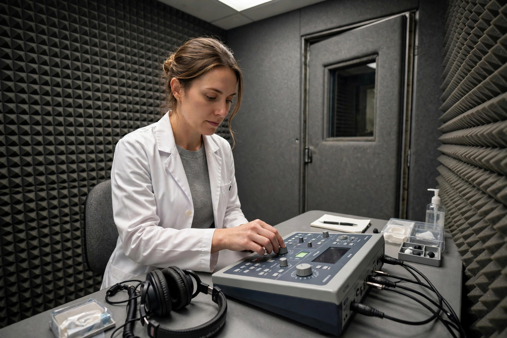
Audiometry measures hearing thresholds: you listen to tones of different pitches and volumes through headphones (and a bone-conduction vibrator behind the ear) and signal when you just hear them. The results plot an audiogram that distinguishes conductive hearing loss (outer/middle ear) from sensorineural loss (inner ear/nerve).
Why it is done
It assesses hearing loss of any cause, monitors noise exposure and ototoxic medicines, investigates one-sided loss (which may need imaging), and determines hearing aid needs.
How to prepare
Wax blocking the canal distorts results, so ears are checked first and wax treated if needed. Avoid loud noise exposure in the day before the test (it temporarily dulls thresholds). Tell the audiologist about ear infections, surgery and tinnitus.
What happens during the procedure
You sit in a sound-treated booth wearing headphones. Tones are presented one ear at a time; you press a button or raise a hand at the faintest audible tone. Bone conduction testing uses a vibrating pad on the mastoid bone. Speech audiometry (repeating words at set volumes) is sometimes added. It takes about 20 to 30 minutes.
Afterwards — recovery & results
Results are available immediately and are usually explained at the same visit, including whether the loss is conductive (potentially medically or surgically treatable) or sensorineural (hearing aids and rehabilitation), and whether referral or imaging is needed.
Risks & considerations
There are no risks. Accuracy depends on your responses, so the audiologist cross-checks with repeat tones and other tests (tympanometry measuring eardrum movement) when results look inconsistent.
Frequently asked questions
Why test bone conduction?
It bypasses the outer and middle ear, testing the inner ear directly. Comparing air and bone results pinpoints where the problem sits — the key to choosing treatment.
What is a 'cookie-bite' or sloping audiogram?
Shapes have typical associations: a high-frequency slope is typical of ageing/noise damage; a mid-frequency dip can suggest hereditary patterns. Your audiologist interprets the shape with your history.
Related procedures — Brain, Nerve & Sleep Tests
Browse the A section of the A–Z list or return to the Procedures & Tests hub.