
- Physical Examination
- Surgical Examination
- Ophthalmology
- Clinical Skills
- Orthopedics
- Surgery Videos
- Laparoscopy
- Pediatrics
- Funny Videos
- Cardiothoracic Surgery
- Nursing Videos
- Plastic Surgery
- Otorhinolaryngology
- Histology and Histopathology
- Neurosurgery
- Dermatology
- Pediatric Surgery
- Urology
- Dentistry
- Oncology and Cancers
- Anatomy Videos
- Health and Fitness
- Radiology
- Anaesthesia
- Physical Therapy
- Pharmacology
- Interventional Radiology
- Cardiology
- Endocrinology
- Gynecology
- Emergency Medicine
- Psychiatry and Psychology
- Childbirth Videos
- General Medical Videos
- Nephrology
- Physiology
- Diet and Food Health
- Diabetes Mellitus
- Neurology
- Women Health
- Osteoporosis
- Gastroenterology
- Pulmonology
- Hematology
- Rheumatology
- Toxicology
- Nuclear Medicine
- Infectious Diseases
- Vascular Disease
- Reproductive Health
- Burns and Wound Healing
- Kidney Zone Podcast
- Other
Fundus fluorescein angiography (FFA)
Category: Eye Procedures & Tests · All eye procedures & tests
What it is
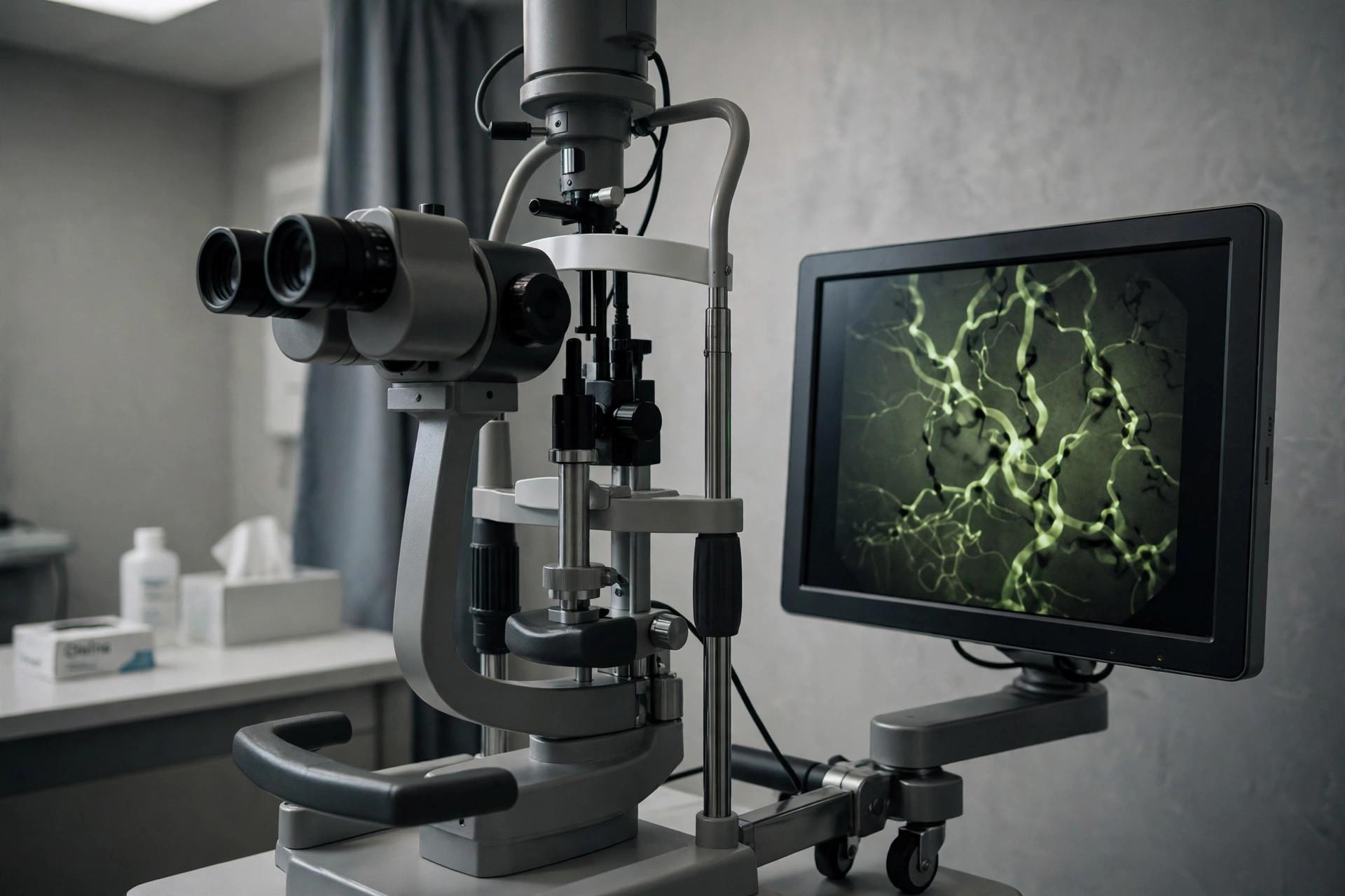
Fluorescein angiography photographs the circulation of the retina: a yellow dye (fluorescein) is injected into an arm vein and a special camera takes a rapid series of pictures as the dye flows through the retinal blood vessels, showing leaks, blockages and abnormal new vessels.
Why it is done
It is used in diabetic retinopathy, macular degeneration assessment, retinal vein occlusions, retinal inflammation (uveitis) and unexplained vision loss, to plan laser and injection treatment.
How to prepare
Tell the team about allergies (especially previous dye reactions), asthma, kidney problems and pregnancy, and list your medicines. Your pupils are dilated, so arrange not to drive home. You may be asked not to eat heavily beforehand, as the dye occasionally causes nausea.
What happens during the procedure
With a cannula in your arm, baseline photographs are taken, the dye is injected over a few seconds, and photographs are taken rapidly for the first minute then at intervals for about 10 to 20 minutes. The flash sequence is bright but painless.
Afterwards — recovery & results
Your skin and urine will look yellow/orange for a day or so as the dye clears — drink fluids. Nausea, if it occurs, passes within minutes. The specialist reviews the images to map treatment; results are usually discussed at the same or a follow-up appointment.
Risks & considerations
Reactions range from mild (nausea, itching, rash) to, very rarely, a severe allergic reaction — the department is equipped to treat reactions and will ask about previous dye exposure. Extravasation (dye leaking under the skin at the injection site) can stain and sting temporarily.
Related guides on MedicalVideos.com
Frequently asked questions
Why does my urine turn orange?
The dye is filtered by the kidneys unchanged — the colour is expected and fades within about 24 hours. It is harmless, though it can amuse children.
How is it different from OCT?
OCT shows retinal structure in cross-section; angiography shows blood flow and leakage in real time. They are complementary and often done together.
Related procedures — Eye Procedures & Tests
Browse the F section of the A–Z list or return to the Procedures & Tests hub.