
- Physical Examination
- Surgical Examination
- Ophthalmology
- Clinical Skills
- Orthopedics
- Surgery Videos
- Laparoscopy
- Pediatrics
- Funny Videos
- Cardiothoracic Surgery
- Nursing Videos
- Plastic Surgery
- Otorhinolaryngology
- Histology and Histopathology
- Neurosurgery
- Dermatology
- Pediatric Surgery
- Urology
- Dentistry
- Oncology and Cancers
- Anatomy Videos
- Health and Fitness
- Radiology
- Anaesthesia
- Physical Therapy
- Pharmacology
- Interventional Radiology
- Cardiology
- Endocrinology
- Gynecology
- Emergency Medicine
- Psychiatry and Psychology
- Childbirth Videos
- General Medical Videos
- Nephrology
- Physiology
- Diet and Food Health
- Diabetes Mellitus
- Neurology
- Women Health
- Osteoporosis
- Gastroenterology
- Pulmonology
- Hematology
- Rheumatology
- Toxicology
- Nuclear Medicine
- Infectious Diseases
- Vascular Disease
- Reproductive Health
- Burns and Wound Healing
- Kidney Zone Podcast
- Other
Breast Cancer Screening (Mammogram)
A mammogram is a low-dose X-ray of the breast that can find breast cancer years before it can be felt. The USPSTF recommends one every two years for women aged 40 to 74.
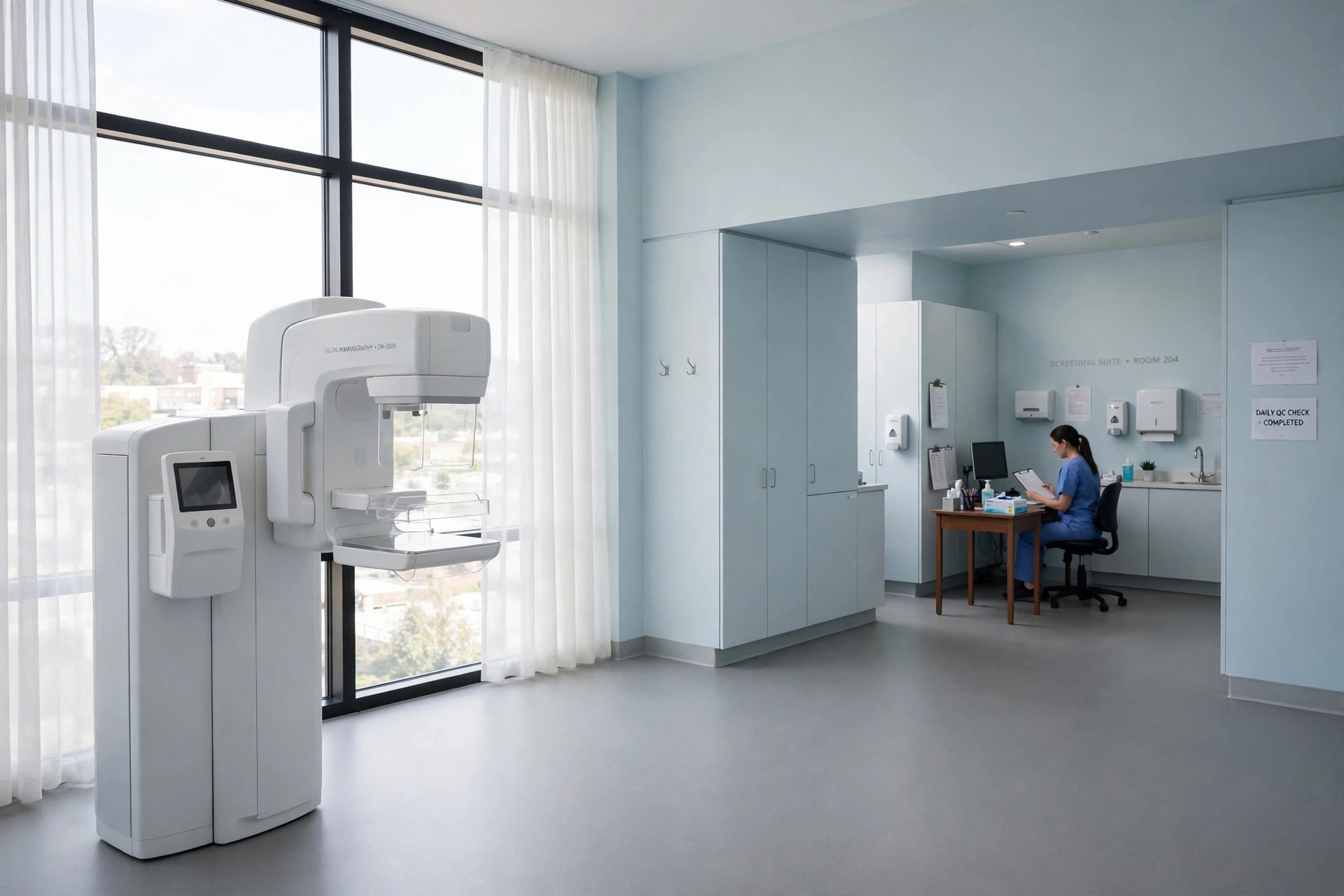
A screening mammogram is an X-ray picture of each breast, taken in people who have no breast symptoms. Its job is to find cancers — and changes that may become cancers — while they are still small and treatment is most likely to work.
The US Preventive Services Task Force (USPSTF) recommends biennial (every-two-years) screening mammography for women aged 40 to 74, with a grade B recommendation. For women aged 75 and older, and for extra ultrasound or MRI scans in women with dense breasts after a normal mammogram, the Task Force found the evidence insufficient (grade I) — those are decisions to make with your own clinician.
What this screening checks
- Lumps or changes too small to feel during a physical examination.
- Areas of abnormal tissue, or tiny calcium deposits (calcifications), that may need a closer look.
- Both standard digital mammography and digital breast tomosynthesis — often called a '3D mammogram' — are effective screening methods, according to the USPSTF.
Who should have it, and how often
Women aged 40 to 74 at average risk should be offered a mammogram every two years. The recommendation applies to people assigned female at birth who are at average risk of breast cancer.
It does not cover people at high risk — for example those with BRCA gene changes, a strong family history, previous breast cancer or high-risk breast lesions, or high-dose radiation to the chest in the past. If that is you, ask your clinician about an earlier, more intensive screening plan instead.
If you find a lump or any new breast change at any age, that is not a screening question — see a clinician promptly for a diagnostic check.
What happens during the test
- You undress from the waist up and wear a gown that opens at the front.
- One breast at a time is placed on a flat plate on the mammography machine.
- A second plate lowers to press the breast firmly for a few seconds while the X-ray is taken. The pressure spreads the tissue so the picture is clear and the radiation dose stays low.
- The position is repeated — usually two pictures of each breast — and the whole appointment is typically over in well under half an hour.
- The images are read by a specialist doctor (radiologist), and results are usually sent to you and your clinician afterwards.
How to prepare
- Do not wear deodorant, antiperspirant, powders or lotions on your chest or underarms that day — they can show up as white spots on the images.
- Wear a top you can remove easily, with separate bottoms.
- If you have had mammograms elsewhere, tell the clinic where, so the new images can be compared with your old ones.
- If your breasts are usually tender before a period, booking the appointment for after your period may be more comfortable.
What the results mean
Most screening mammograms show no sign of cancer. Some women are called back for extra pictures or an ultrasound — a 'recall' — and most recalls turn out not to be cancer; the extra views are often needed because tissue overlapped in the first pictures.
If something still looks suspicious after the extra checks, the next step is usually a biopsy: a small sample of tissue is taken with a needle and examined under a microscope. Only a biopsy can confirm or rule out cancer.
Benefits
- Finding breast cancer early, before it can be felt, when treatment is more likely to succeed.
- Catching some cancers while they are smaller and may need less extensive treatment.
- A regular two-yearly rhythm balances early detection against the downsides of screening.
Limits and possible harms
- False alarms: being called back for more tests that turn out to be normal is common, and can cause real anxiety.
- Overdiagnosis: screening finds some cancers that would never have caused problems in a woman's lifetime, yet are still treated.
- False reassurance: a normal mammogram does not guarantee there is no cancer — keep reporting new symptoms.
- The compression can be uncomfortable, and each mammogram uses a small amount of radiation.
Watch: related videos
Useful links and tools
More in Cancer Screening
- Cervical Cancer Screening (Pap & HPV Tests)
- Colorectal Cancer Screening (Colonoscopy & Stool Tests)
- Lung Cancer Screening (Low-Dose CT)
- Prostate Cancer Screening (PSA Test)
Frequently asked questions
How often should I have a mammogram?
The USPSTF recommends a screening mammogram every two years for women aged 40 to 74 (grade B, 2024). For women 75 and older the evidence is insufficient to set a rule, so that decision is best made with your own clinician, based on your health and preferences.
What does it mean if I am told I have dense breasts?
Dense breasts have more glandular and fibrous tissue and less fatty tissue, and they are very common — nearly half of all women have them. Dense tissue can make cancers harder to see on a mammogram. Whether extra ultrasound or MRI screening helps after a normal mammogram is still uncertain (USPSTF grade I), so discuss it with your clinician.
This guide explains how a screening test usually works and who it is generally offered to. It is general educational information only — not medical advice, and not a recommendation for you personally. Screening decisions depend on your age, history and risk, so talk to your own clinician about which tests you need and when. If you have symptoms now, do not wait for a screening test — seek medical care promptly.