
- Physical Examination
- Surgical Examination
- Ophthalmology
- Clinical Skills
- Orthopedics
- Surgery Videos
- Laparoscopy
- Pediatrics
- Funny Videos
- Cardiothoracic Surgery
- Nursing Videos
- Plastic Surgery
- Otorhinolaryngology
- Histology and Histopathology
- Neurosurgery
- Dermatology
- Pediatric Surgery
- Urology
- Dentistry
- Oncology and Cancers
- Anatomy Videos
- Health and Fitness
- Radiology
- Anaesthesia
- Physical Therapy
- Pharmacology
- Interventional Radiology
- Cardiology
- Endocrinology
- Gynecology
- Emergency Medicine
- Psychiatry and Psychology
- Childbirth Videos
- General Medical Videos
- Nephrology
- Physiology
- Diet and Food Health
- Diabetes Mellitus
- Neurology
- Women Health
- Osteoporosis
- Gastroenterology
- Pulmonology
- Hematology
- Rheumatology
- Toxicology
- Nuclear Medicine
- Infectious Diseases
- Vascular Disease
- Reproductive Health
- Burns and Wound Healing
- Kidney Zone Podcast
- Other
Latest videos
The newest additions to the MedicalVideos.com library — freshly curated medical and surgery videos, explainers, and lectures from educators and institutions worldwide.
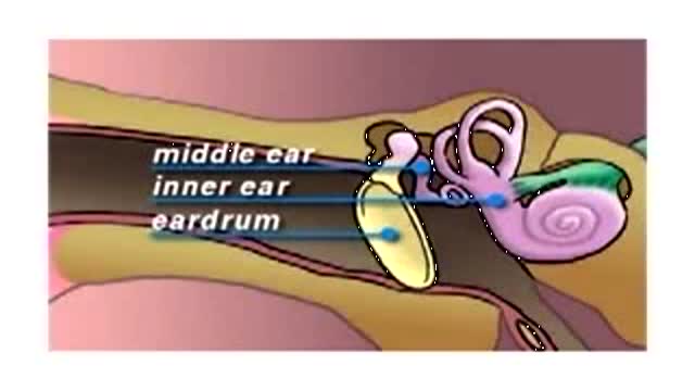

Myringotomy is the surgery to place tubes in the ear. This animated video reviews the anatomy of the ear and what happens after frequent infection. As well as treatment with tubes in the ear or myringotomy.
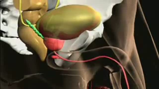
This short narrated lesson explains the anatomy of the prostate gland. The recording describes its size, its position under the bladder, the urethra passing through it, and the seminal vesicles above it. It is a basic anatomy study aid for medical students. The lesson runs just under a minute in total.
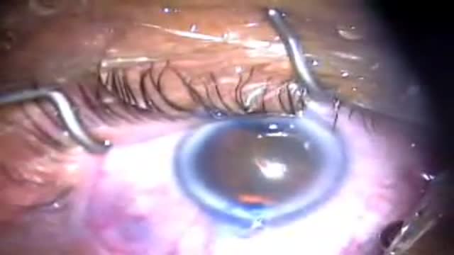
Cataract surgery with dense arcus
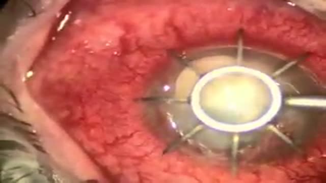
Cornea Transplant
Lap Band Procedure done on a patient with a BMI of 45. Minimal editing and includes narration.
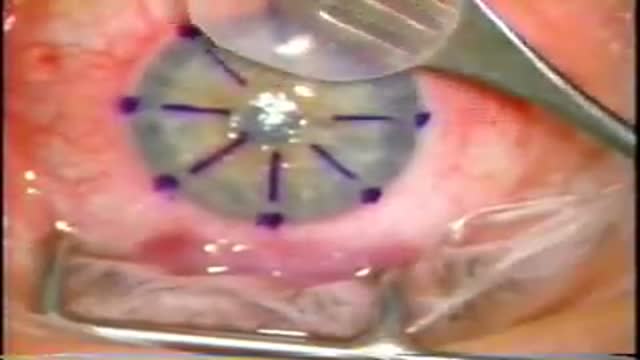
Most corneal transplants performed in the U.S. involve replacing the entire thickness of the diseased cornea with a healthy donor cornea (called penetrating keratoplasty or PK). In partial-thickness corneal transplants (LK), only the anterior (surface) layers of the cornea are removed. The donor cornea is then attached to the host corneal bed, containing only posterior (deeper) layers. LK is less risky, but tends to result in somewhat inferior vision vs. PK and cannot be performed if the disease process (e.g. scar) involves the deeper layers of the cornea.
Radial Keratotomy
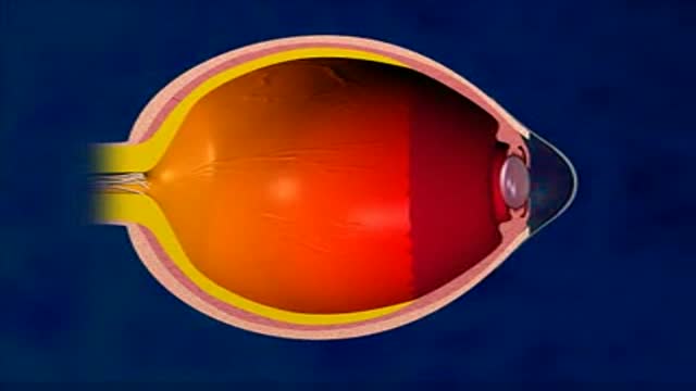
LASIK or Laser-Assisted In Situ Keratomileusis is a surgical procedure intended to reduce a person's dependency on glasses or contact lenses.
LASIK surgery is most commonly performed as a cure for myopia (nearsightedness), but can also be used to cure hyperopia (farsightedness) or astigmatism (corneal irregularities).
LASIK is a procedure that permanently changes the shape of the cornea using a special laser and thus focusing the light rays exactly on the retina.
The steps of the procedure are as follows:
A suction ring is placed on the eye to stabilize and check the eye pressure.
The microkeratome, a cutting instrument, is attached to the suction ring.
The blade of the microkeratome is used to cut a flap in the cornea.
The exposed inner layer of the cornea is then reshaped with an excimer laser.
The corneal flap is returned to its original position.
LASIK is an ambulatory procedure; the patient can walk into the surgery center, have the procedure and walk out again and is awake the whole time. Occasionally, the doctor may administer a mild oral sedative.
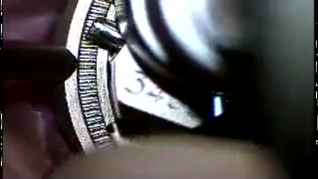
LASIK eye surgery operation
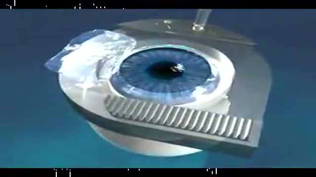
Microkeratome in Lasik
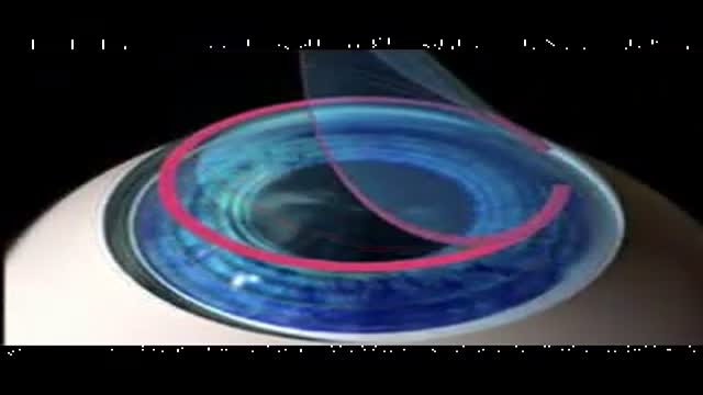
LASIK Surgery Procedure
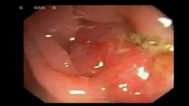
Endoscopic finding in a patient with a typical rectal cancer (adenocarcinoma)
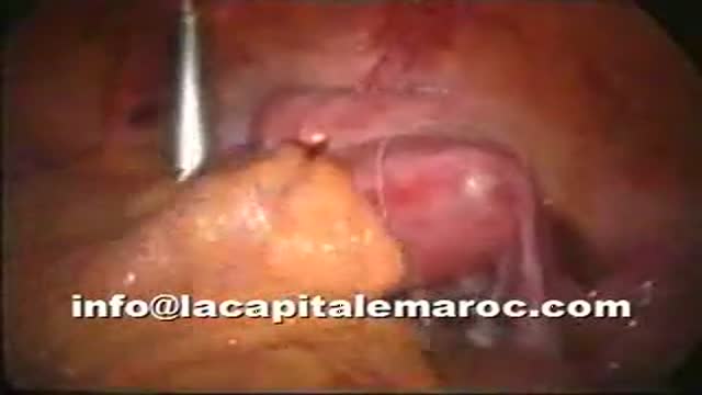
Treatment of bowel injury by IUD

An educational narration introduces internal birth control methods. The recording explains the diaphragm, the vaginal ring, and the implant, how each prevents pregnancy, and their advantages and requirements. It is presented as general information about contraceptive options.
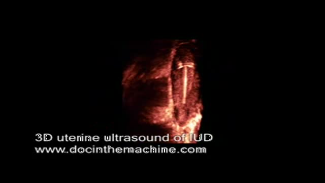
3D ultrasound of IUD in uterus
This light-hearted medical comedy clip features a humorous check-up trick. The short video presents a playful examination scene intended purely for entertainment. It is shared in the funny videos category as a break from the clinical education recordings elsewhere on the site for entertainment.
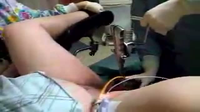
Delivery using foreceps
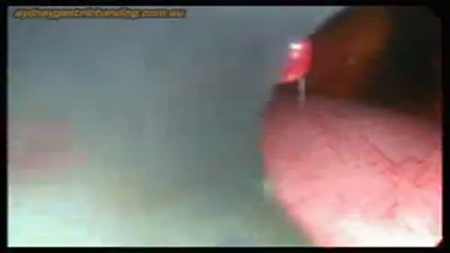
Laparoscopy in acute bowel obstruction following previous surgery is a difficult procedure and avoided by most of the surgeons due to the difficulty in obtaining pneumoperitoneum, port placement, lack of working space, adhesions and risk of bowel injury.
Here is a patient who had a previous laparotomy for trauma with a midline incision from xyphysternum to pubis; after unsuccessful conservative management he underwent a laparoscopy; a prior CT scan showed adhesions in the left side and a distal-mid small bowel obstruction. The pneumoperitoneum was obtained with the Visiport placed in the right lower quadrant; although the abdomen was grossly distended, under significant tension and distended loops of small bowel were occupying most the peritoneal cavity, with muscle relaxation there is usually enough space to perform a thorough inspection of the abdominal cavity. Port placement has to be done with special care as there is no room to push and usually a blunt trocar directed away from the bowel is employed in my practice. The collapsed loops of small bowel point quickly to the site of obstruction -- it is better to avoid manipulating the distended bowel as it is heavy, oedematous and prone to be lacerated with the instruments; once the pathology is identified, in this case the obstructive band, light packing is performed in order to expose the working space and protect the bowel from instruments like scissors or diathermy. In this case the band adhesion was slightly more difficult to separate from the bowel and required a combination of sharp and gentle blunt dissection.
Once the obstruction is release and the transit of contents is confirmed in the collapsed bowel the procedure is terminated. No abdominal drainage is usually necessary.
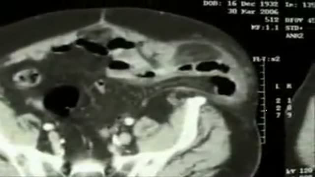
Giant spigelian stranguled hernia with small bowel loop and omental flap inside. The omentum required resection, the bowel appears vital. After the handle of hernia sac and his content has been done, a overlapped prolene repair will be done.
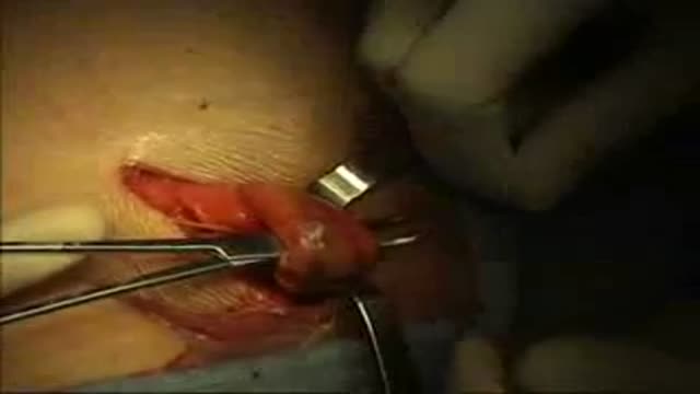
Right indirect (Gilbert II)inguinal hernia has been repared using PHSe prosthetic device
Medical Videos
Free medical and surgery videos for patients, students and professionals — trusted since 2007.
Founded by Dr. Mohamed Ibrahim, MD, PhD — physician-scientist.
Follow us on Facebook · Instagram